Glycosaminoglycans analogs from marine invertebrates: structure, biological effects, and potential as new therapeutics
 Mauro S. G. Pavão
- Programa de Glicobiologia, Laboratório de Bioquímica e Biologia Cellular de Glicoconjugados, Instituto de Bioquímica Médica Leopoldo De Meis, Hospital Universitário Clementino Fraga Filho, Universidade Federal do Rio de Janeiro, Rio de Janeiro, Brazil
In this review, several glycosaminoglycan analogs obtained from different marine invertebrate are reported. The structure, biological activity and mechanism of action of these unique molecules are detailed reviewed and exemplified by experiments in vitro and in vivo. Among the glycans studied are low-sulfated heparin-like polymers from ascidians, containing significant anticoagulant activity and no bleeding effect; dermatan sulfates with significant neurite outgrowth promoting activity and anti-P-selectin from ascidians, and a unique fucosylated chondroitin sulfate from sea cucumbers, possessing anticoagulant activity after oral administration and high anti P- and L-selectin activities. The therapeutic value and safety of these invertebrate glycans have been extensively proved by several experimental animal models of diseases, including thrombosis, inflammation and metastasis. These invertebrate glycans can be obtained in high concentrations from marine organisms that have been used as a food source for decades, and usually obtained from marine farms in sufficient quantities to be used as starting material for new therapeutics.
Introduction
Heparin is a highly sulfated glycosaminoglycan composed by disaccharide units containing a hexuronic acid (α-L-iduronic acid or β-D-glucuronic acid) linked 1,4 to α-D-glucosamine. The heparin molecules consist of a heterogeneous mixture of polymers with a similar backbone, which results from variations of sulfation on the D-glucosamine (N-acethylated, N-sulfated, O-sulfated at C6 and/or C3) and on the uronic acid residue (O-sulfated at C2) (Lindahl et al., 1989).
Because of its unique binding to antithrombin, involving a specific pentasaccharide sequence containing a 3-O-sulfated glucosamine, heparin is endowed of a potent anticoagulant activity (Lindahl et al., 1980, 1989). In fact, based on its ability to inhibit fluid phase coagulation, unfractionated heparin (UF) isolated from porcine and bovine intestinal mucosa has been used clinically for decades. However, the therapeutic use of UH is limited mostly by its potent hemorrhagic effect, implying that patients under heparin therapy have to be closely monitored (Hirsh, 1984). UH also has poor bioavailability, requires multiple daily dosing, and has side effects such as heparin-induced thrombocytopenia (Hirsh and Raschke, 2004; Hirsh et al., 2004). To circumvent these problems, different low-molecular-weight heparins (LMW-Hep) have been produced by degrading UF, using a variety of methods, including chemical depolymerization and enzymatic digestion (Hirsh and Raschke, 2004).
In addition to its anticoagulant effect, mammalian UH has also anti-inflammatory effect in several animal models of inflammation, which is possibly mediated by P- and L-selectins. The recruitment of leukocytes from blood and lymphatic systems into tissues facilitates the response to tissue injury. Adhesion molecules of the selectin family (E, P, and L) mediate the initial events that direct the movement of leukocytes across the endothelium in inflamed tissues by interacting with sialylated, fucosylated carbohydrate antigens related to sialyl Lewisx [Slex, Neu5Acα2,3Galβ 1,4(Fucα1,3)GlcNAcβ-] found at the cell surface (Lasky, 1995; McEver, 1995; Nelson et al., 1995; Butcher and Picker, 1996). It has been reported (Stevenson et al., 2005) that the dose of UH required for the inhibitory effect on the selectins is higher than that required for the anticoagulant action, which increases the hemorrhagic risk and makes the clinical use of UH impractical to treat inflammation. Similarly, the use of LMW-Hep, which has a much lower hemorrhagic effect, is not a good alternative for UH, since it is a poor inhibitor of selectins (Stevenson et al., 2005).
UF and heparin-like oligosaccharides inhibit L- and P-selectin binding to Slex and has been shown to dramatically reduce leukocyte infiltration in thioglycollate-induced peritoneal inflammation in mice (Burg et al., 1997; Wang et al., 2002). In addition, UH has also been used successfully as a therapeutic agent in different animal models of nephropathy. Subcutaneous injection of non-anticoagulant UH reduces glomerulosclerosis in rats (Burg et al., 1997), and ameliorates the progression of renal disease in rats with subtotal renal ablation (Purkerson et al., 1988). In addition, it has also been demonstrated that heparin inhibits macrophage infiltration and TGF-β synthesis in puromycin glomerulosclerosis (Ceol et al., 2003).
The risk of contamination with pathogens is an important aspect to take into account in the therapeutic use of natural products from mammalian origin. For example, the association of mammalian prionic proteins with transmissible spongiform encephalopathy has restricted the use of bovine heparin in Europe, USA and Japan. In these countries, commercial heparin is obtained exclusively from porcine tissues and the risk of contamination with a prionic protein or even a virus is still present.
Therefore, as we consider the molecular mechanism of the anti-inflammatory effect of UF, its side effects, and the possibility of pathogen contamination, it becomes extremely relevant the search for alternative heparin-like compounds, obtained from non-mammalian sources, possessing similar biological activities, but devoid of the undesired side effects.
Unique Invertebrate Glycosaminoglycans
Heparin-Like Glycans
Heparin with a structure similar to that of vertebrate heparin but with lower anticoagulant activity has been identified in the tissues of the ascidian Styela plicata (Chordata-Tunicata) (Cavalcante et al., 2000). An extensive structural analysis of the polymer indicated that the invertebrate heparin is composed mainly of the disaccharide α-L-IdoA(2SO4)-1→4β-D-GlcN(SO4)(6SO4)-1, with a minor contribution (~25%) of the disaccharide α-L-IdoA-1→4β-D-GlcN(SO4)(6SO4)-1. Activated partial thromboplastin time (APTT) assays of the ascidian heparin showed that the polymer has lower anticoagulant activity than mammalian heparin. In addition, it is about 20 times less potent than mammalian heparin in stimulating the inhibition of thrombin by purified ATIII. However, S. plicata heparin activates HCII with approximately the same potency as vertebrate heparin, as indicated by comparison of the IC50 values for inhibition of thrombin by purified HCII. To compare the hemorrhagic effect of the ascidian and mammalian heparin, we used a rat cut-tail bleeding model. S. plicata heparin at a dose of 4 mg/kg body weight, which is above the dose required to prevent thrombus on an animal experimental model did not increase the amount of blood loss in comparison with the saline control. In contrast, mammalian heparin, compared to the control, increased blood loss almost 2-fold (Cardilo-Reis et al., 2006).
In a TNBS-induced colitis model in rats, S. plicata heparin drastically reduced inflammation after subcutaneous administration during a 7-day period (Belmiro et al., 2009), as observed by the normalization of the macroscopic and histological characteristics of the colon. At molecular level, TNF-alpha, TGF-beta, and VEGF were reduced to normal values. Lymphocyte and macrophage recruitment and epithelial cell apoptosis were also decreased after the treatment. A drastic reduction in collagen-mediated fibrosis was also observed. No hemorrhagic events were observed after glycan treatment (Belmiro et al., 2009). These results strongly indicate a potential therapeutic use of the ascidian heparin in the treatment of colonic inflammation with a lower risk of hemorrhage, when compared with mammalian heparin.
The bivalve mollusk Nodipecten nodosus, currently cultivated in different parts of the world with an annual production of about 75,000 tons (http://www.fao.org/fishery/topic/14884/en), has been shown to contain high amounts of a heparan sulfate-like glycosaminoglycan. 1D 1H, 2D COZY and HSQC nuclear magnetic resonance revealed characteristic signals of non-sulfated, 3- or 2-sulfated glucuronic acid, as well as N-sulfated and/or 6-sulfated glucosamine (Gomes et al., 2010). The mollusk glycan possesses an anticoagulant activity of 36 IU mg−1, 5-fold lower than bovine heparin (180 IU mg−1). It inhibits factor Xa (IC50 = 0.835 μg mL−1) and thrombin (IC50 = 9.3 μg mL−1) in the presence of antithrombin. Experiments in vivo, demonstrated that, the mollusk HS inhibited thrombus growth in photochemicaly injured arteries, at the dose of 1 mg Kg−1. No bleeding effect, factor XIIa-mediated kallikrein activity or toxic effect on fibroblast cells were induced by the invertebrate HS at the antithrombotic dose (Gomes et al., 2010).
Oversulfated Dermatans
The ascidians S. plicata and Halocyntia pyriformis, contain dermatan sulfates formed by [α-L-IdoA(2SO4)-1→3β-D-GalNAc(4SO4)] disaccharide units. These oversulfated dermatans have high heparin cofactor II-mediated anticoagulant activity (Pavao et al., 1998). Due to a higher concentration of [α-L-IdoA(2SO4)-1→3β-D-GalNAc(4SO4)]-containing sequences, that bind to the glycosaminoglycan binding site in the inhibitor, their heparin cofactor II activities are at least 10 times higher than that of the mammalian counterpart.
The occurrence of a well-defined relationship between sulfate position on the disaccharides and biological activity can be observed studying dermatan sulfates from different species of ascidians. In the ascidian Phallusia nigra, the dermatan sulfate has the same degree of sulfation of that from S. plicata, but is composed mainly by [α-L-IdoA(2SO4)-1→3β-D-GalNAc(6SO4)] disaccharide units (Pavao et al., 1995). As a result of the different sulfation on the N-acetylgalactosamine (6-sulfated instead of 4-sulfated), P. nigra dermatan sulfate has very low heparin cofactor II-mediated anticoagulant activity. Overall, these results strongly suggest that binding of oversulfated dermatan sulfate polymers to heparin cofactor II requires a specific sulfation pattern on the glycans, composed by [α-L-IdoA(2SO4)-1→3β-D-GalNAc(4SO4)]-enriched sequences.
These unique oversulfated dermatan sulfates from ascidians, with different heparin cofactor II activities, allowed the study of the relationship between heparin cofactor II activity and antithrombotic effect. Thus, after intravascular administration S. plicata dermatan sulfate, with high heparin cofactor II activity, prevents thrombus formation in veins (Vicente et al., 2001). On the other hand, P. nigra dermatan sulfate, with a discernible heparin cofactor II activity, has no antithrombotic effect in the same venous thrombotic model and dose (Vicente et al., 2001). These results indicate that a heparin cofactor II-mediated mechanism is associated with the antithrombotic effect of dermatan sulfate polymers.
The oversulfated dermatan sulfates from the ascidian P. nigra and S. plicata, composed by [α-L-IdoA(2SO4)-1→3β-D-GalNAc(6SO4)] and [α-L-IdoA(2SO4)-1→3β-D-GalNAc(4SO4)] disaccharide unities were used in the study of mouse hippocampal neurons behavior. P. nigra dermatan possesses significant neurite outgrowth-promoting activity, which resulted specific morphological features. The ascidian dermatan sulfate induced a flattened neuronal cell soma and dendrite-like multiple neurites (Hikino et al., 2003). S. plicata dermatan sulfate, composed by [α-L-IdoA(2SO4)-1→3β-D-GalNAc(4SO4)], on the other hand, exhibited only a modest neurite outgrowth-promoting activity (Hikino et al., 2003; Bao et al., 2005).
Heparin has been shown to exhibit P- and/or L-selectin-mediated antimetastatic and antiinflammatory activities. P-selectin-mediated platelet-tumor cell and tumor-cell endothelium interactions facilitate the initial steps of hematogeneous metastasis (Borsig et al., 2001, 2002). The dermatan sulfates from S. plicata and P. nigra, that contain the same disaccharide core structure [α-L-IdoA(2SO4)-1→3β-D-GalNAc]n, but sulfated at carbon 4 or 6 of the GalNAc residues, respectively, and opposed HCII activities are potent inhibitors of P-selectin (Kozlowski and Pavao, 2011; Kozlowski et al., 2011). Both ascidian dermatan sulfates regardless of the position of sulfation on the N-acetylgalactosamine drastically attenuate metastasis of both MC-38 colon carcinoma and B16-BL6 melanoma cells, and the infiltration of inflammatory cells in a thioglycollate peritonitis mouse model (Kozlowski and Pavao, 2011; Kozlowski et al., 2011). Additionally, both ascidians glycosaminoglycans reduced thrombus size in a FeCl3-induced arterial thrombosis model, irrespective of their HCII activities. Interestingly, the arterial thrombi demonstrated a markedly reduced platelet deposition after dermatan sulfate treatment (Kozlowski and Pavao, 2011; Kozlowski et al., 2011), suggesting that the ascidians glycosaminoglycan inhibited P-selectin and thereby the binding of activated platelets during thrombus formation. These results provide evidence that inhibition of P-selectin is a potential therapeutic target in thrombosis, inflammation and metastasis, and ascidian dermatan sulfates may serve as anti-selectin agents.
Fucosylated Chondrotitin Sulfate
A unique natural chondroitin sulfate analog, containing sulfated fucose branches has been identified in several species of sea cucumbers (Vieira and Mourao, 1988; Vieira et al., 1991; Kariya et al., 1997). The Ludwigothurea grisea glycan has a core like that of mammalian chondroitin sulfate but substituted at the 3-position of the glucuronic acid residues with fucose-2,4 disulfated branches (Vieira and Mourao, 1988; Vieira et al., 1991). The fucosylated chondroitin sulfate has high anticoagulant and antithrombotic activities that disappear after removal of the sulfated fucose branches by mild acid hydrolysis (Mourao et al., 1996). Two anticoagulant mechanism have been proposed: activation of thrombin inhibition by heparin cofactor II, and inhibition of factor-Xa and thrombin generation by the tenase and prothrombinase complexes, respectively (Glauser et al., 2008; Buyue and Sheehan, 2009).
Interestingly, thrombosis inhibition in artery by the fucosylated chondroitin sulfate occurs at low doses, and does not modify the plasma anticoagulant activity. On the contrary, in venous thrombosis the antithrombotic activity of the fucosylated chondroitin requires high doses and occurs with an increase in the plasma anticoagulant activity (Zancan and Mourao, 2004). Additionally, daily oral doses of this glycosaminoglycan showed a decrease in thrombus weight on experimental models of venous and arterial thrombosis in experimental animals (Fonseca and Mourao, 2006).
The fucosylated chondroitin sulfate from H. grisea inhibited P- and L-selectin binding to immobilized sialyl Lewisx, a component of leukocyte surface glycoproteins, which is also overexpressed in several tumor cells (Borsig et al., 2007). The glycan also inhibited LS180 carcinoma cell attachment to immobilized P- and L-selectins (Borsig et al., 2007). As a result of its anti-selectin effect, the intact sea cucumber glycan attenuates lung colonization by adenocarcinoma MC-38 cells in an experimental metastasis model in mice, as well as neutrophil recruitment in thioglycollate-induced peritonitis. Removal of the sulfated fucose branches abolishes the inhibitory effect in vitro and in vivo (Borsig et al., 2007). These results suggest that this glycan may be a potential therapeutic drug for blocking metastasis and inflammatory reactions.
Concluding Remarks and Future Perspectives
Glycosaminoglycans analogs with unique structures and pharmacological activities have been described in different marine invertebrates (Table 1). The structure, biological activity and mechanism of action have been extensively studied and the glycans evaluated in pre-clinical experiments in rodent animals with promising results. Heparin-like polymers with low anticoagulant activity, significant antithrombotic and anti-inflammatory activities, but devoid of bleeding effects occur in different species of ascidians and mollusks. Similarly, oversulfated dermatan sulfates containing the same disaccharide core structure of [α-L-IdoA(2SO4)-1→3β-D-GalNAc]n, but differing in the position of sulfation (4-sulfate or 6-sulfate) on the N-acetylgalactosamine, occur in high amounts in ascidians. These polymers have high anti-selectin activity, which results in attenuation of metastasis and leukocyte recruitment. The 2,6-sulfated dermatan sulfate from P. nigra has very low heparin cofactor II activity and antithrombotic effect, different from its 2,4-sulfated high anticoagulant counterpart from S. plicata. The anti-P-selectin activity of these disulfated glycans is involved in low platelet arterial thrombus formation. Moreover a significant neurite outgrowth promoting activity is associated with the di-sulfated ascidian dermatan sulfates. The fucosylated chondroitin sulfate from L.grisea has significant therapeutic effect after oral administration, attenuating metastasis and inflammation due to the high anti-selectin activity of the sulfated fucose branches. So far, no significant toxic effect has been associated with the use of these marine compounds. The marine invertebrates that are the primary source of these potential therapeutic compounds abound in different parts of the world, mainly in western seas, where they are cultivated in large scale. In general, the marine invertebrate glycans occur in higher concentration in the tissue (about 0.5% of the dry weight, comparing to 0.022% from pig intestinal mucosa) and can be easily isolated by procedures similar to those already employed in the preparation of pharmaceutical heparin. Whereas these marine organisms will be a source of new heparin analogs with significant therapeutic effect in thrombosis, inflammation and cancer in the future will depend on the economic pressure of the pharmaceutical industry and the increasing demand for new natural drugs with less undesired side effects to treat specific diseases.
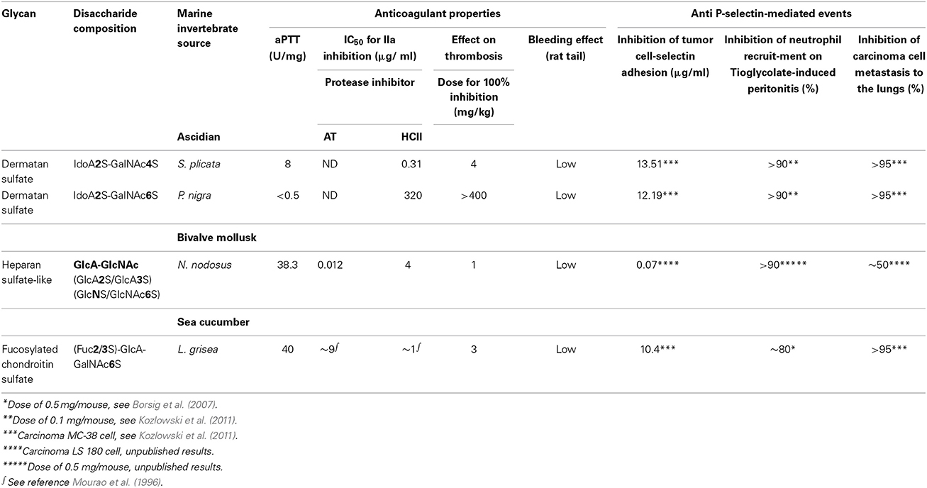
Table 1. Effect of invertebrate glycans on coagulation, inflammation, and metastasis.
Conflict of Interest Statement
The author declares that the research was conducted in the absence of any commercial or financial relationships that could be construed as a potential conflict of interest.
Acknowledgments
This work was supported by grants from CNPq and FAPERJ. Mauro S. G. Pavão is a research fellow from CNPq and FAPERJ.
Abbreviations
HexA, hexuronic acid; GlcA, glucuronic acid; IdoA, iduronic acid; GalNAc, N-acetyl galactosamine; GlcNAc, N-acetyl glucosamine; GlcA2S, 2-O-sulfated glucuronic acid; GlcA3S, 3-O-sulfated glucuronic acid; IdoA2S, 2-O-sulfated iduronic acid; GalNAc4S, 4-O-sulfated N-acetyl galactosamine; GalNAc6S, 6-O-sulfated N-acetyl galactosamine; GalNAc4S,6S, 4-O- and 6-Osulfated N-acetyl galactosamine; GlcNS, N-sulfated glucosamine; GlcNS,6S, N- and 6-sulfated glucosamine; GlcNS,3S,6S, N, 3-O- and 6-Osulfated glucosamine.
References
Bao, X., Pavao, M. S., Dos Santos, J. C., and Sugahara, K. (2005). A functional dermatan sulfate epitope containing iduronate(2-O-sulfate)alpha1-3GalNAc(6-O-sulfate) disaccharide in the mouse brain: demonstration using a novel monoclonal antibody raised against dermatan sulfate of ascidian Ascidia nigra. J. Biol. Chem. 280, 23184–23193. doi: 10.1074/jbc.M503036200
Belmiro, C. L., Castelo-Branco, M. T., Melim, L. M., Schanaider, A., Elia, C., Madi, K., et al. (2009). Unfractionated heparin and new heparin analogues from ascidians (chordate-tunicate) ameliorate colitis in rats. J. Biol. Chem. 284, 11267–11278. doi: 10.1074/jbc.M807211200
Borsig, L., Wang, L., Cavalcante, M. C., Cardilo-Reis, L., Ferreira, P. L., Mourao, P. A., et al. (2007). Selectin blocking activity of a fucosylated chondroitin sulfate glycosaminoglycan from sea cucumber. Effect on tumor metastasis and neutrophil recruitment. J. Biol. Chem. 282, 14984–14991. doi: 10.1074/jbc.M610560200
Borsig, L., Wong, R., Feramisco, J., Nadeau, D. R., Varki, N. M., and Varki, A. (2001). Heparin and cancer revisited: mechanistic connections involving platelets, P-selectin, carcinoma mucins, and tumor metastasis. Proc. Natl. Acad. Sci. U.S.A. 98, 3352–3357. doi: 10.1073/pnas.061615598
Borsig, L., Wong, R., Hynes, R. O., Varki, N. M., and Varki, A. (2002). Synergistic effects of L- and P-selectin in facilitating tumor metastasis can involve non-mucin ligands and implicate leukocytes as enhancers of metastasis. Proc. Natl. Acad. Sci. U.S.A. 99, 2193–2198. doi: 10.1073/pnas.261704098
Burg, M., Ostendorf, T., Mooney, A., Koch, K. M., and Floege, J. (1997). Treatment of experimental mesangioproliferative glomerulonephritis with non-anticoagulant heparin: therapeutic efficacy and safety. Lab. Invest. 76, 505–516.
Butcher, E. C., and Picker, L. J. (1996). Lymphocyte homing and homeostasis. Science 272, 60–66. doi: 10.1126/science.272.5258.60
Buyue, Y., and Sheehan, J. P. (2009). Fucosylated chondroitin sulfate inhibits plasma thrombin generation via targeting of the factor IXa heparin-binding exosite. Blood 114, 3092–3100. doi: 10.1182/blood-2009-02-203661
Cardilo-Reis, L., Cavalcante, M. C., Silveira, C. B., and Pavao, M. S. (2006). In vivo antithrombotic properties of a heparin from the oocyte test cells of the sea squirt Styela plicata(Chordata-Tunicata). Braz. J. Med. Biol. Res. 39, 1409–1415. doi: 10.1590/S0100-879X2006001100004
Cavalcante, M. C., Allodi, S., Valente, A. P., Straus, A. H., Takahashi, H. K., Mourao, P. A., et al. (2000). Occurrence of heparin in the invertebrate styela plicata (Tunicata) is restricted to cell layers facing the outside environment. An ancient role in defense? J. Biol. Chem. 275, 36189–36186. doi: 10.1074/jbc.M005830200
Ceol, M., Vianello, D., Schleicher, E., Anglani, F., Barbanti, M., Bonfante, L., et al. (2003). Heparin reduces glomerular infiltration and TGF-beta protein expression by macrophages in puromycin glomerulosclerosis. J. Nephrol. 16, 210–218.
Fonseca, R. J., and Mourao, P. A. (2006). Fucosylated chondroitin sulfate as a new oral antithrombotic agent. Thromb. Haemost. 96, 822–829. doi: 10.1160/TH06-06-0304
Glauser, B. F., Pereira, M. S., Monteiro, R. Q., and Mourao, P. A. (2008). Serpin-independent anticoagulant activity of a fucosylated chondroitin sulfate. Thromb. Haemost. 100, 420–428. doi: 10.1160/th08-04-0210
Gomes, A. M., Kozlowski, E. O., Pomin, V. H., de Barros, C. M., Zaganeli, J. L., and Pavao, M. S. (2010). Unique extracellular matrix heparan sulfate from the bivalve Nodipecten nodosus (Linnaeus, 1758) safely inhibits arterial thrombosis after photochemically induced endothelial lesion. J. Biol. Chem. 285, 7312–7323. doi: 10.1074/jbc.M109.091546
Hikino, M., Mikami, T., Faissner, A., Vilela-Silva, A. C., Pavao, M. S., and Sugahara, K. (2003). Oversulfated dermatan sulfate exhibits neurite outgrowth-promoting activity toward embryonic mouse hippocampal neurons: implications of dermatan sulfate in neuritogenesis in the brain. J. Biol. Chem. 278, 43744–43754. doi: 10.1074/jbc.M308169200
Hirsh, J., Heddle, N., and Kelton, J. G. (2004). Treatment of heparin-induced thrombocytopenia: a critical review. Arch. Intern. Med. 164, 361–369. doi: 10.1001/archinte.164.4.361
Hirsh, J., and Raschke, R. (2004). Heparin and low-molecular-weight heparin: the seventh ACCP conference on antithrombotic and thrombolytic therapy. Chest 126, 188S–203S. doi: 10.1378/chest.126.3_suppl.188S
Kariya, Y., Watabe, S., Kyogashima, M., Ishihara, M., and Ishii, T. (1997). Structure of fucose branches in the glycosaminoglycan from the body wall of the sea cucumber Stichopus japonicus. Carbohydr. Res. 297, 273–279. doi: 10.1016/S0008-6215(96)00258-3
Kozlowski, E. O., and Pavao, M. S. (2011). Effect of sulfated glycosaminoglycans on tumor invasion and metastasis. Front. Biosci. 3, 1541–1551. doi: 10.2741/244
Kozlowski, E. O., Pavao, M. S., and Borsig, L. (2011). Ascidian dermatan sulfates attenuate metastasis, inflammation and thrombosis by inhibition of P-selectin. J. Thromb. Haemost. 9, 1807–1815. doi: 10.1111/j.1538-7836.2011.04401.x
Lasky, L. A. (1995). Selectin-carbohydrate interactions and the initiation of the inflammatory response. Annu. Rev. Biochem. 64, 113–139. doi: 10.1146/annurev.bi.64.070195.000553
Lindahl, U., Backstrom, G., Thunberg, L., and Leder, I. G. (1980). Evidence for a 3-O-sulfated D-glucosamine residue in the antithrombin-binding sequence of heparin. Proc. Natl. Acad. Sci. U.S.A. 77, 6551–6555.
Lindahl, U., Kusche, M., Lidholt, K., and Oscarsson, L. G. (1989). Biosynthesis of heparin and heparan sulfate. Ann. N.Y. Acad. Sci. 556, 36–50. doi: 10.1111/j.1749-6632.1989.tb22488.x
McEver, R. P. (1995). Regulation of function and expression of P-selectin. Agents Actions Suppl. 47, 117–119.
Mourao, P. A., Pereira, M. S., Pavao, M. S., Mulloy, B., Tollefsen, D. M., Mowinckel, M. C., et al. (1996). Structure and anticoagulant activity of a fucosylated chondroitin sulfate from echinoderm. Sulfated fucose branches on the polysaccharide account for its high anticoagulant action. J. Biol. Chem. 271, 23973–23984. doi: 10.1074/jbc.271.39.23973
Nelson, R. M., Venot, A., Bevilacqua, M. P., Linhardt, R. J., and Stamenkovic, I. (1995). Carbohydrate-protein interactions in vascular biology. Annu. Rev. Cell Dev. Biol. 11, 601–631.
Pavao, M. S., Aiello, K. R., Werneck, C. C., Silva, L. C., Valente, A. P., Mulloy, B., et al. (1998). Highly sulfated dermatan sulfates from Ascidians. Structure versus anticoagulant activity of these glycosaminoglycans. J. Biol. Chem. 273, 27848–27857. doi: 10.1074/jbc.273.43.27848
Pavao, M. S., Mourao, P. A., Mulloy, B., and Tollefsen, D. M. (1995). A unique dermatan sulfate-like glycosaminoglycan from ascidian. Its structure and the effect of its unusual sulfation pattern on anticoagulant activity. J. Biol. Chem. 270, 31027–31036. doi: 10.1074/jbc.270.52.31027
Purkerson, M. L., Tollefsen, D. M., and Klahr, S. (1988). N-desulfated/acetylated heparin ameliorates the progression of renal disease in rats with subtotal renal ablation. J. Clin. Invest. 81, 69–74. doi: 10.1172/JCI113312
Stevenson, J. L., Choi, S. H., and Varki, A. (2005). Differential metastasis inhibition by clinically relevant levels of heparins–correlation with selectin inhibition, not antithrombotic activity. Clin. Cancer Res. 11, 7003–7011. doi: 10.1158/1078-0432.CCR-05-1131
Vicente, C. P., Zancan, P., Peixoto, L. L., Alves-Sa, R., Araujo, F. S., Mourao, P. A., et al. (2001). Unbalanced effects of dermatan sulfates with different sulfation patterns on coagulation, thrombosis and bleeding. Thromb. Haemost. 86, 1215–1220.
Vieira, R. P., and Mourao, P. A. (1988). Occurrence of a unique fucose-branched chondroitin sulfate in the body wall of a sea cucumber. J. Biol. Chem. 263, 18176–18183.
Vieira, R. P., Mulloy, B., and Mourao, P. A. (1991). Structure of a fucose-branched chondroitin sulfate from sea cucumber. Evidence for the presence of 3-O-sulfo-beta-D-glucuronosyl residues. J. Biol. Chem. 266, 13530–13536.
Wang, L., Brown, J. R., Varki, A., and Esko, J. D. (2002). Heparin's anti-inflammatory effects require glucosamine 6-O-sulfation and are mediated by blockade of L- and P-selectins. J. Clin. Invest. 110, 127–136. doi: 10.1172/JCI0214996
Keywords: glycosaminoglycans, heparin, dermatan sulfate, selectins, marine invertebrates
Citation: Pavão MSG (2014) Glycosaminoglycans analogs from marine invertebrates: structure, biological effects, and potential as new therapeutics. Front. Cell. Infect. Microbiol. 4:123. doi: 10.3389/fcimb.2014.00123
Received: 09 June 2014; Accepted: 18 August 2014;
Published online: 10 September 2014.
Edited by:
Eliana Barreto-Bergter, Universidade Federal do Rio de Janeiro, BrazilReviewed by:
Marcio Rodrigues, Oswaldo Cruz Foundation, BrazilBarbara Mulloy, King's College London, UK
Copyright © 2014 Pavão. This is an open-access article distributed under the terms of the Creative Commons Attribution License (CC BY). The use, distribution or reproduction in other forums is permitted, provided the original author(s) or licensor are credited and that the original publication in this journal is cited, in accordance with accepted academic practice. No use, distribution or reproduction is permitted which does not comply with these terms.
*Correspondence: Mauro S. G. Pavão, Programa de Glicobiologia, Laboratório de Bioquímica e Biologia Celular de Glicoconjugados, Instituto de Bioquímica Médica Leopoldo de Meis, Hospital Universitário Clementino Fraga Filho, Universidade Federal do Rio de Janeiro, Rua Prof. Rodolpho P. Rocco 255, 4° andar, Sala 4A-08, Cidade Universitária, Rio de Janeiro, RJ 21941-913, Brazil e-mail: mpavao@hucff.ufrj.br