Article

Growth, Survival and Spore Formation of the Pathogenic Aquatic Oomycete Aphanomyces astaci and Fungus Fusarium avenaceum Are Inhibited by Zanthoxylum rhoifolium Bark Extracts In Vitro

1
Department of Sciences and Technologies, University of Sannio, Via Port’Arsa 11, 82100 Benevento, Italy
2
Department of Pharmacy, University of Salerno, 84084 Fisciano, Italy
3
Department of Molecular Medicine and Medical Biotechnology, Federico II University, Via S. Pansini 5, 80131 Napoli, Italy
4
Department of Environmental and Biological Sciences, University of Eastern Finland, 70210 Kuopio Campus, Finland
5
CEINGE, Advanced Biotechnologies s.c.ar.l., Via Gaetano Salvatore 486, 80145 Napoli, Italy
*
Author to whom correspondence should be addressed.
Submission received: 22 January 2018 / Revised: 9 February 2018 / Accepted: 12 February 2018 / Published: 15 February 2018

Abstract

:
This study aimed to evaluate the in vitro activity of Zanthoxylum rhoifolium bark (Zr-b) extracts against pathogenic aquatic oomycete/fungal isolates that cause different diseases in native European crayfish resulting in an elevated mortality rate and severe economic repercussions. n-hexane, chloroform, chloroform–methanol (9:1) and methanol extracts of Zr-b were used to evaluate the antifungal activity against the strain UEF88662 of Aphanomyces astaci (oomycete) and the strain SMM2 of Fusarium avenaceum (fungus). The anti-oomycete and antifungal activity was quantitatively evaluated by growth, survival and sporulation microbiological assays. The extracts tested demonstrated a dose-dependent inhibitory effect on oomycete and fungal growth and survival, as well as on the production of oomycete and fungal spores. This work presents alternatives for the treatment and prevention of the spreading of Aphanomyces astaci and Fusarium avenaceum, the etiological agents of the diseases crayfish plague and brown spot disease, respectively. The antifungal properties of Zanthoxylum rhoifolium bark extracts warrant further research on their use in the prevention and treatment of both oomycete and fungal diseases. The antifungal properties of Zanthoxylum rhoifolium bark extracts, shown in vitro, indicate the possibility of their use in new therapeutic and prophylactic strategies, providing perspectives for the design of in vivo studies.

1. Introduction

Fungal diseases are often caused by species that are common in the environment. Although most fungi are not dangerous, some types can be harmful to health [1]. Fusarium avenaceum belongs to the phylum Ascomycota, class Sordariomycetes, order Hypocreales, family Nectreaceae. It is found worldwide and commonly isolated from soil and many different species of plants [2]. Many important plant pathogens and mycotoxin producers, including saprophytes and endophytes, belong to the genus Fusarium [3]. These pathogenic fungi are the etiological agent of the melanisation of the esoskeleton and gills of crustaceans such as freshwater crayfish, causing burn spot disease, which is manifested by a series of injuries on the exoskeleton of the crayfish, similar to burns [4,5,6,7,8,9,10]. Recently, epidemics caused by burn spot disease due to Fusarium avenacem have been reported in both mainland Estonian and Saaremaa Island, where the survival of noble crayfish (Astacus astacus) stocks was put at risk [6]. Under aquaculture conditions, the effects of Fusarium avenaceum infections can be even more dramatic, since the entire crops can be lost during the epidemics [6]. In the case of freshwater crayfish aquaculture, prevention of the fungal diseases is important [11]. Recently, it was shown that Fusarium avenaceum can cause erosion of the female signal crayfish swimmerets, affecting their reproductive potential [12].
Aphanomyces astaci (Phylum Chromista, class Oomycetes, order Saprolegniales, family Saprolegniaceae) is the etiological agent of the crayfish plague [13]. Crayfish plague is an infectious disease that affects freshwater crayfish. In susceptible species, the infection can cause the extirpation of entire populations [14]. The crayfish plague is the most common infectious disease among European freshwater crayfish with a very high fatality rate, even resulting in the complete eradication of the hosts during outbreaks [11]. Different species of crayfish show different degrees of sensitivity to infection, and some studies indicate that the resistance towards Aphanomyces astaci infection is increasing among populations [4], while others indicate a considerable between-strain variation among Aphanomyces astaci genotypes, suggesting an environment adaptation of the original North American parasite to the European continent, that can be the cause of the reduced virulence of some oomycete strains [15]. All European crayfish species are highly susceptible to infection. These include the noble crayfish (Astacus astacus) of north-western Europe, the white clawed crayfish (Austropotamobius pallipes) of south-western and western Europe, the stone crayfish (Austropotamobius torrentium) (mountain streams of south-western Europe) and the narrow clawed or Turkish crayfish (Astacus leptodactylus) of eastern Europe and Asia Minor [4,16,17,18,19]. On the other hand, North American crayfish such as the signal crayfish (Pacifastacus leniusculus), the red swamp crayfish (Procambarus clarkii) and Orconectes spp. are often chronic carriers of Aphanomyces astaci with symptoms from minor melanised lesions to tissue erosion and limb losses with the infection also being, under certain circumstances, fatal to these species [20]. Due to human-assisted translocations, North American crayfish acting as carriers of infectious diseases such as Aphanomyces astaci, have caused serious ecological and economic problems for both wild native crayfish stocks and crayfish aquaculture [21,22,23,24]. Indeed, especially in the Fennoscandian countries, crayfish trapping and also farming is, to some extent, a flourishing activity as crayfish consumption is part of the culinary tradition [25,26]. So far, the treatment and prevention of the spread of both oomycete and fungal diseases, causing losses among native European stocks (Astacus astacus, Austropotamobius pallipes, Austropotamobius torrentium, Astacus leptodactylus, Astacus pachypus), have been scarcely studied [27,28,29,30,31].
In recent decades, the research of plant-derived natural bioactive compounds with therapeutic effects has increased significantly [32]. Although some phytogenic compounds have traditionally been used as complementary or alternative drugs to improve human health or cure human diseases, phytogenic compounds, such as essential oils, botanicals and herbal extracts, have been shown to possess positive effects also on animal growth and health. Their antimicrobial, antiviral, antifungal and antioxidative properties make them good candidates as alternatives to conventional antimicrobial agents not only for the therapy of important human diseases, but also for the prevention and treatment of diseases which represents a serious scourge among animals and invertebrates [33].
The genus Zanthoxylum (Rutaceae) includes about 250 species distributed throughout the world; among these, the Zanthoxylum rhoifolium species, native to South America, has long been used in traditional Brazilian medicine to treat various health problems [34] and in French Guiana as antimalarial treatment [35]. These plants are known to be a rich source of natural alkaloids and lignans, which are extracted from the bark [36]. Some of these alkaloids, including benzofenantridine and fluoroquinolones, exhibit a variety of biological activities: anticancer [37], antimicrobial [38], anti-inflammatory [39] and antifungal [40,41] activities. Moreover, recently, Zanthoxylum rhoifolium extracts showed a fungistatic effect on different fungal plant pathogens, such as, Botrytis cinerea, Sclerotinia sclerotiorum, Alternaria alternata, Colletotrichum gloeosporioides and Clonostachys rosea [36].
In this context, we studied the effects of extracts of the bark of Zanthoxylum rhoifolium on the growth, survival and spore formation of an oomycete and a fungus that cause severe diseases in crayfish: Aphanomyces astaci and Fusarium avenaceum.

2. Results

2.1. Chemical Constituents of Chloroform–Methanol (9:1) Zr-b Extract

All Zanthoxylum rhoifolium bark (Zr-b) extracts tested in this study showed antioomycete and antifungal activity; the chloroform–methanol (9:1) Zr-b extract in particular, purified by RP-HPLC, determined the wider inhibition zone (in millimeters) by the agar-diffusion method (see below) against crayfish isolates of Aphanomyces astaci (UEF88662 strain) and Fusarium avenaceum (SMM2 strain). The phytochemical investigation revealed the presence of 3-OMe-benzoyl-1-β-d-(6-galloyl)-glucopyranoside, hesperidin (PubChem CID: 10621), isorhamnetin-3-0-rutinoside (PubChem CID: 5481665), coralydine (PubChem CID: 92233282), and N,O-dimethylthaicanine. The most abundant chemicals in the chloroform–methanol (9:1) extract were N,O-dimethylthaicanine and coralydine, (3.3 mg/g and 1.4 mg/g, respectively), followed by hesperidin (0.5 mg/g), 3-OMe-benzoyl-1-β-d-(6-galloyl)-glucopyranoside (0.4 mg/g) and isorhamnetin-3-0-rutinoside (0.3 mg/g). Altogether, the identified chemical constituents sum up to 5.9 mg/g of chloroform–methanol (9:1) extract.

2.2. In Vitro Anti Oomycete and Antifungal Activity of Zr-b Extracts

The antioomycete and antifungal activity of all the Zr-b extracts was assayed against crayfish isolates of Aphanomyces astaci (UEF88662 strain) and Fusarium avenaceum (SMM2 strain), respectively (Figure 1).
The diameters of the inhibition zones, obtained with 6 mg/disc of the Zr-b extracts against the two isolates, are reported in Table 1 and Figure 2.
The results of in vitro assays indicate that all the Zr-b extracts exhibited a clear antioomycete and antifungal activity, with the formation of wide inhibition zones against Fusarium avenaceum and Aphanomyces astaci, although the widest area of inhibition was obtained with the chloroform–methanol extract. Tioconazole (TCZ) (1.4 mg/disc), used as a positive control, exerted the expected antifungal activity against Fusarium avenaceum with an inhibition zone of 40.1 ± 7.2 mm, but also induced an inhibition zone of 20.0 ± 4.1 mm against Aphanomyces astaci. The extraction buffers, used as negative controls, showed no effects toward the tested microorganisms. The results indicate that the SMM2 Fusarium avenaceum strain was more sensitive to all the Zr-b extracts tested and to Tioconazole, compared to the UEF88662 Aphanomyces astaci strain. This result was also confirmed by quantitative assays.

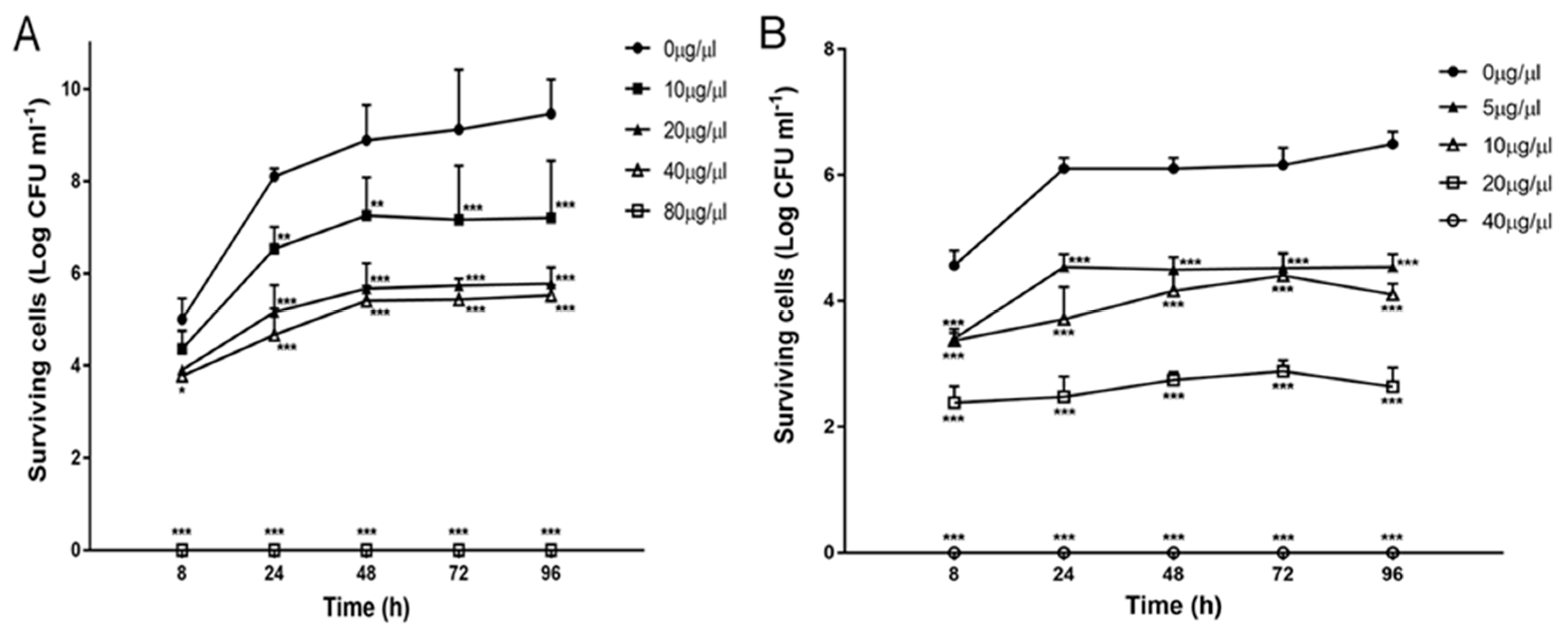
2.3. Effect of Zr-b Extracts on Oomycete and Fungal Growth and Survival

The antioomycete and antifungal activities of all the Zr-b extracts have been confirmed by quantitative assays. All Zr-b extracts showed both oomycestatic and fungistatic effects other than oomyceticidal and fungicidal effects (Table 2). The growth of UEF88662 Aphanomyces astaci isolate was inhibited by n-hexane, chloroform, chloroform–methanol (9:1) and methanol Zr-b extracts at a concentration of 40 μg/µL. The growth of SMM2 Fusarium avenaceum isolate was inhibited by lower concentrations of n-hexane, chloroform, chloroform–methanol (9:1) and methanol Zr-b extracts equal to 5 μg/µL. The minimum inhibitory concentration (MIC) values for the TCZ were 1.5 μg/µL and 1.0 μg/µL for Aphanomyces astaci and Fusarium avenaceum, respectively.
Furthermore, the minimum oomycetidal concentration (MOC) of n-hexane, chloroform, chloroform–methanol (9:1) and methanol Zr-b extracts was 80 μg/µL for UEF88662 Aphanomyces astaci strain. The minimum fungicidal concentration (MFC) of n-hexane, chloroform, chloroform–methanol (9:1) and methanol Zr-b extracts was 40 μg/µL for SMM2 Fusarium avenaceum strain. Both the oomycete and fungal isolates used in this study were sensitive to TCZ, with a MOC value of 15 μg/µL for Aphanomyces astaci and MFC value of 1.5 μg/µL for Fusarium avenaceum.

2.4. Effect of the Chloroform–Methanol (9:1) Zr-b Extract on Oomycete and Fungal Fitness

The fitness of the pathogenic aquatic oomycete and fungal isolates was monitored in the presence of different concentrations (0–80 µg/µL) of the chloroform–methanol (9:1) Zr-b extract (Figure 3A,B); however, the growth curves were not reported because the spectrophotometric determination was distorted by the turbidity of the extracts. The figures show that the extract interferes with both the oomycete and fungal survival of UEF88662 Aphanomyces astaci strain and SMM2 Fusarium avenaceum strain in a dose-dependent manner. Indeed, the number of viable cells was reduced proportionally with the increasing concentration of the extract during the 96 h of observation. Fusarium avenaceum was more sensitive to the chloroform–methanol (9:1) Zr-b extract compared to Aphanomyces astaci.

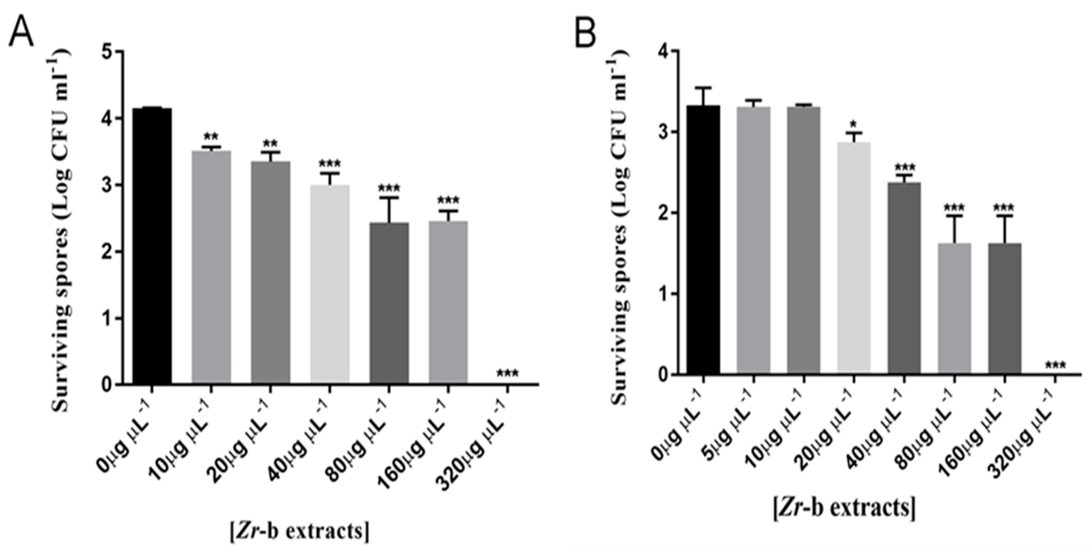
2.5. Effect of the Chloroform–Methanol (9:1) Zr-b Extract on Oomycete and Fungal Sporulation and Spore Germination

After the evaluation and quantification of the inhibitory effect of the chloroform–methanol (9:1) Zr-b extract on the vegetative form of UEF88662 Aphanomyces astaci strain and SMM2 Fusarium avenaceum strain, we evaluated the ability of the chloroform–methanol extract to inhibit the production of spores from both strains. The sporulation inhibition assay show that the inhibitory effect of Zr-b extracts on both oomycete and fungal spore production was dose-dependent, with complete inhibition of the sporulation at a concentration of 320 μg/µL (Figure 4A,B).
To verify the sporicidal effect of the Zr-b extract, a spore germination inhibition assay was performed in vitro with 320 µg/µL chloroform–methanol (9:1) Zr-b extract. After the appropriate incubation, no development of viable colonies of Aphanomyces astaci and Fusarium avenaceum was observed on media supplemented with the Zr-b extract (Figure 5)

3. Discussion

The results obtained from the present study show that the Zanthoxylum rhoifolium bark extracts possess both oomycetidal and antifungal activity and in vitro can antagonize the growth of both Aphanomyces astaci and Fusarium avenaceum isolates. Zanthoxylum has been studied for several types of biological actions including the antifungal activity [42].
All Zr-b extracts were effective against Aphanomyces astaci and Fusarium avenaceum, counteracting their growth in the agar-diffusion assay; the chloroform–methanol (9:1) extract in particular exhibited a wider inhibition zone than n-hexane, chloroform and methanol extracts, suggesting that the efficacy of the organic solvents to extract the active principles is more or less equivalent. Extracts from medicinal plants have been extensively used from ancient times against fungal infections for treating various disease conditions in humans and in agriculture [43]. Ethanolic extracts of the bark of Zanthoxylum fagara, Zanthoxylum elephantiasis and Zanthoxylum martinicense showed antifungal activity [44], as well as ether, chloroform and methanol extracts of Zanthoxylum budrunga bark [45]. The Zanthoxylum armatum essential oil has been shown to inhibit the mycelial growth of the fungus Bipolaris sorokiniana [46]. Biological activity against fungi has been reported also in the bark of Zanthoxylum usambarense [47] and Zanthoxylum americanum [9]. The fungicidal activity is likely due to the presence of alkaloids, the major components, along with flavonoids, within the Zanthoxylum genus [48]. The alkaloidal extract of the bark of Zanthoxylum chiloperone exhibited antifungal activity against Candida albicans, Aspergillus fumigatus and Trichophyton mentagrophytes [49]. Moreover, a synergistic action against fluconazole-resistant Candida albicans clinical strains was observed with concomitant use of berberine and fluconazole [50]. Because of their good antifungal properties and synergistic action, berberine and its derivatives have been suggested to represent a new class of antifungal agents with low host toxicity [51]. Interestingly, we found that the most abundant bioactive molecules in the bark of Zanthoxylum rhoifolium were N,O-dimethylthaicanine and coralydine, two protoberberine-derived alkaloids. Protoberberine alkaloids display a great variety of biological and pharmacological activities. Such activities include the inhibition of DNA synthesis, protein biosynthesis, the inhibition of membrane permeability, and the uncoupling of oxidative phosphorylation. These processes likely explain the allelochemical and toxic effects observed against bacteria, fungi, other plants, insects, and vertebrates [52].
Other compounds found in the present study in Zr-b were the flavonoids 3-OMe-benzoyl-1-β-d-(6-galloyl)-glucopyranoside, hesperidin (aflavonoid glycoside) and isorhamnetin-3-0-rutinoside (an O-methylated flavonol), already reported in Zanthoxylum schinifolium by Li et al. [48]. Flavonoids have been reported to possess many biological properties including antimicrobial [53] and antifungal [54] activities. Owing to the widespread ability of flavonoids to inhibit spore germination in plant pathogens, they have been proposed to be useful against human fungal pathogens [55] and their activity against the opportunistic pathogen Candida albicans has been shown [56,57]. Galangin, a flavonol commonly found in bee propolis samples [58], has been shown to have inhibitory activity against Aspergillus tamari and Aspergillus flavus, fungi responsible for massive diseases in immunosuppressed patients [59,60].
The MIC values of the Zanthoxylum rhoifolium extracts against Aphanomyces astaci and Fusarium avenaceum isolates showed that the extracts were oomycestatic and fungistatic at lower concentrations while becoming oomycetidal and fungicidal at higher concentrations. It is worth noting that the quantitative microbiological tests highlighted the higher antioomycete and antifungal activity of the chloroform–methanol (9:1) Zr-b extract in comparison to Tioconazole, used as a positive control. Indeed, according to the HPLC quantification, the amount of alkaloids and flavonoids in the chloroform–methanol extract was 5.9 mg/g. This allows us to estimate a MIC of 0.24 and 0.003 µg/µL for Aphanomyces astaci and Fusarium avenaceum, respectively; and a MOC of 0.48 for Aphanomyces astaci and an MFC of 0.24 µg/µL for Fusarium avenaceum, concentrations well below the MIC, MOC and MFC of Tioconazole. Numerous natural substances possess both antimicrobial and antifungal activity more powerful than the pharmaceutical products of chemical synthesis [51].
We hypothesize that the reason for such high antioomycete and antifungal activities, shown by the chloroform–methanol (9:1) Zr-b extract, can reside in the synergistic effect of the different phytochemicals present in the extract. The crude extract of Spirulina spp., showed improved antifungal activity against Fusarium graminacearum, compared to the single phenolic acids. The improvement occurred also when the crude extract was mixed with the purified standard gallic acid [61]. It is interesting to note that the research has been focusing on novel natural products to substitute the chemical synthesis products that often show high toxicity and the prolonged use is accompanied by the drawback of the onset of resistant strains, that can be minimized by the use of blends of natural molecules [62].
In vitro, the chloroform–methanol (9:1) Zr-b extract is able to effectively inhibit the spore production from Aphanomyces astaci and Fusarium avenaceum isolates, with sporicidal activity to high concentrations. As expected, the minimum sporicidal concentration of Zr-b extracts is four-fold higher than the MOC for Aphanomyces astaci and eight-fold higher than the MFC for Fusarium avenaceum, being generally the mycotic spores more resistant than the vegetative forms to the effects of natural and synthetic antimycotics which often have no or very low influence on the spores [63,64].
These properties are very important as the sporulation is crucial to the potential spreading of these disease agents. Although many chemicals had antifungal properties, some of them are very toxic (such as formaldehyde, malachite green, etc.) and prohibited in aquaculture, while for others (such as hydrogen peroxide, acetic acid, povidone iodine, etc.) the effective concentrations are higher than the levels recommended by the U.S. Food and Drug Administration, and therefore they may cause adverse effects on animal health, and in general may be injurious to aquatic biota [65].
To conclude, the results of the microbiological tests performed in this study on the evaluation of the inhibitory effects of the Zr-b extracts on the survival and sporulation of Aphanomyces astaci and Fusarium avenaceum, suggest that these extracts could be used as means to prevent the spreading of the tested oomycete and fungal disease agents and encourage the promotion of their use as alternatives to chemicals in the prevention and treatment of diseases, that represent a real scourge, especially in crayfish aquaculture. Additionally, the present study constitutes a basis for further investigations on molecular mechanisms of antioomycete and antifungal activities of the Zr-b extracts and for the design of in vivo studies. The extracts of the plant used in this study could be useful in the treatment and prevention of infections caused by these two pathogens. However, further studies are required to determine the cost, applicability, and safety of these extracts as potential antioomycetes and antifungals.

4. Materials and Methods

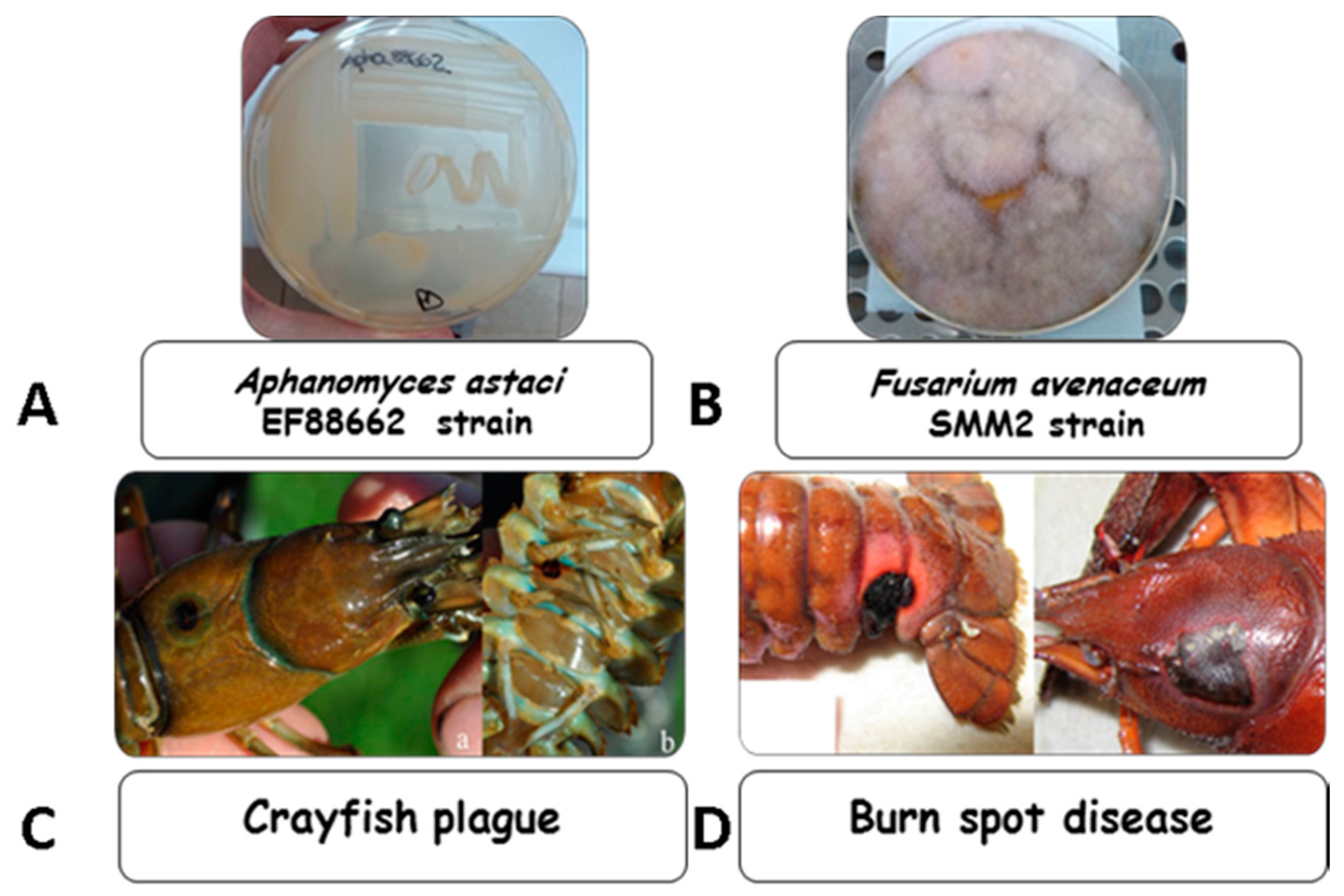
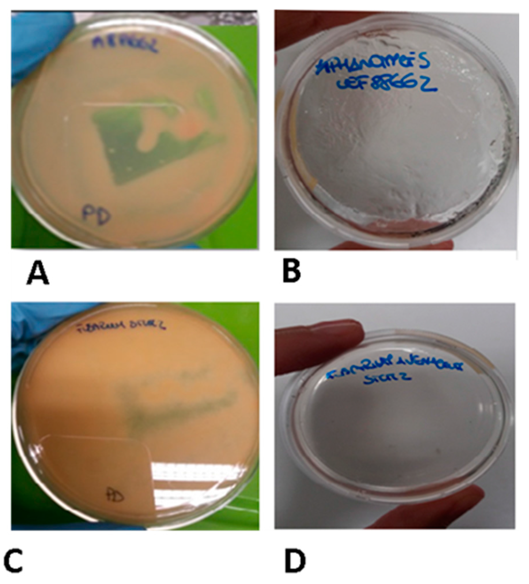
4.1. Microorganisms and Growth Conditions

The strains UEF88662 of Aphanomyces astaci and SMM2 of Fusarium avenaceum (crayfish isolates) were provided by the Department of Biology Kuopio Campus, University of Eastern Finland. The microorganisms were cultured in Potato-Dextrose (PD) broth/agar (CONDA, Torrejón de Ardoz, Spain), a medium developed for cultivation of yeasts and molds, at 20 °C. Both strains were maintained at 4 °C on agar media. The isolates were stored frozen at −80 °C in PD broth supplemented with 10% glycerol (v/v) (Carlo Erba Reagents, Milan, Italy) until use and the working cultures were activated in the PD broth at 20 °C for 24–48 h.

4.2. Plant Material

The bark of Zanthoxylum rhoifolium Lam was collected in Venezuela and identified by Eng. Juan Carmona, Universidad de Los Andes, Merida, Venezuela. A voucher specimen (No. 607) was deposited to Jardin de Plantas Medicinales de la Facultad de Farmacia y Bioanalisis, Merida.

4.3. Extraction and Characterization of Zanthoxylum rhoifolium bark

An amount of 810 g of Zr-b was sequentially extracted with solvents of increasing polarity yielding n-hexane, chloroform, chloroform–methanol (9:1) and methanol extracts, in the amounts of 25.6 g, 16.08 g, 50.48 g, and 94.12 g, respectively (3.14%, 2.06%, 6.19% and 11.55% with respect to the dry plant material). The chloroform–methanol (9:1) extract was partitioned between n-butanol and H2O to remove the sugar portion and subjected (2.5 g) to Column Chromatography (CC) over Sephadex LH-20 in MeOH to give seven major fractions (A–G) grouped by Thin Layer Chromatography (TLC). TLC was performed on precoated Kieselgel 60 F254 plates (Merck). Only the most abundant fractions (B and E) were subjected to further purification. Fraction B (466.3 mg) was purified by reverse-phase high performance liquid chromatography on a Shimadzu LC-20AT series pumping system equipped with a Shimadzu RID10A refractive index detector and a Shimadzu injector, using a C18μ-Bondapak column (30 cm × 7.8 mm, 10 μm, Waters-Milford) at a flow rate of 2 mL/min with MeOH–H2O (1:4) as eluent to afford coralydine (3.5 mg, tR (Retention time) = 40 min) and N,O-dimethylthaicanine (8.2 mg, tR = 50 min). Fraction E (51.9 mg) was purified by RP-HPLC with MeOH–H2O (10:15) as eluent to afford 3-OMe-benzoyl-1-β-d-(6-galloyl)- (1.0 mg, tR = 8 min), hesperedin (1.3 mg, tR = 30 min), and isorhamnetin-3-0-rutinoside (0.8 mg, tR = 70 min). The structures of the isolated compounds were established by NMR experiments which were performed on a Bruker DRX-600 spectrometer (Bruker BioSpin GmBH, Rheinstetten, Germany) equipped with a Bruker 5 mm TCI CryoProbeat 300 K. All 2D NMR spectra were acquired in methanol-d4 (99.95%, Sigma-Aldrich Srl, Milan, Italy), and standard pulse sequences and phase cycling were used for DQF-COSY, HSQC, and HMBC spectra.

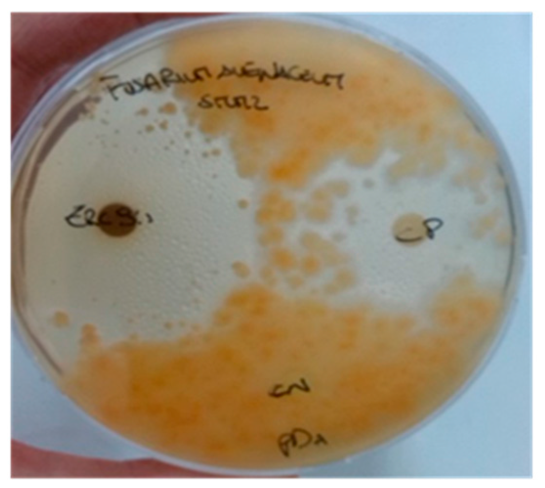
4.4. In Vitro Antioomycetidal and Antifungal Activity Assay of Zr-b Extracts with the Agar-Diffusion Method

In order to evaluate the inhibitory spectrum of Zr-b extracts, against test microorganisms, a variation of the agar-diffusion method was performed [66]. Briefly, the oomycete and fungal strains were grown in PD broth to an optical density of 0.5 at 600 nm; an aliquot of 200 µL of oomycete and fungal suspension, appropriately homogenized to generate fragmented hyphae, was spread on the surface of agar media. Paper disks (6 mm in diameter, Oxoid, S.p.a., Rodano, Milano, Italy), impregnated with 20 µL of Zr-b extracts, were positioned on media using sterile forceps. Different concentrations of Zr-b extracts (1–6 mg/disc) were used to evaluate both antioomycetidal and antifungal activities. TCZ (Pfizer Italia Srl, Latina, Italy), at a concentration of 1.4 mg/disc was used as positive control. The extraction buffers, n-hexane, chloroform, methanol and chloroform–methanol (9:1), were used as negative controls. Plates were incubated at 20 °C for 2–5 days. The size (expressed in mm) of the inhibition zones (including disc diameter) around the disc was measured. The antioomycetidal and antifungal activities were expressed as the diameter of the inhibition zones produced by the Zr-b extracts against the test microorganisms. The experiments were repeated three times.

4.5. Quantitative Evaluation of the Antioomycetidal and Antifungal Activities of Zr-b Extracts

The susceptibility of both oomycete and fungal strains to different concentrations of Zr-b extracts was determined by the dilution tube method, with 1 × 10⁵ CFU/mL as standard inoculums [67]. Zr-b extracts were added in a series of tubes achieving final concentrations of 0, 5, 10, 20, 40, 80, and 160 µg/µL, and incubated at 20 °C for 2–5 days. The isolates were also tested with TCZ as positive control and with the extraction buffer as negative control. After the incubation, an aliquot of the suspension of hyphae fragments from each tube was used to determine the optical density at 600 nm, while an aliquot was spread on the surfaces of PD agar plates in duplicate. Plates were then incubated at 20 °C for 2–5 days, and a colony count was performed. The MIC was assigned to the lowest concentration of Zr-b extract which prevented fungal growth. The MOC and the MFC were defined as the minimum extract concentration that killed 99% of oomycete and fungi respectively from the initial inoculums. The experiments were repeated three times.

4.6. Oomycete and Fungal Fitness Evaluation of the Chloroform–Methanol (9:1) Zr-b Extract

To verify the effect of Zr-b extracts on the fitness of Aphanomyces astaci and Fusarum avenaceum, the assays of oomycete and fungal growth and assays of oomycete and fungal survival were performed in the presence of increasing concentrations of chloroform–methanol (9:1) Zr-b extracts. To evaluate the growth of each strain, during the observation period (96 h), aliquots of serial dilutions of the oomycete and fungal suspensions were used to determine the optical density at 600 nm. To evaluate the survival of each strain, during the observation period (96 h), aliquots of serial dilutions of the oomycete and fungal suspensions were spread on PD agar, and the plates were incubated at 20 °C for 2–5 days. Thereafter, the count of oomycete and fungal colonies on agar was carried out. All experiments were performed in triplicate with three independent cultures.

4.7. Assays of Oomycete and Fungal Sporulation with the Chloroform–Methanol (9:1) Zr-b Extract

In order to evaluate the inhibitory effect of the chloroform–methanol (9:1) Zr-b extract on the spore production of Aphanomyces astaci and Fusarium avenaceum, sporulation inhibition assays were performed in the presence of increasing concentrations of the extract. To induce the production of spores, a variation of the procedure described by Makkonen et al. was performed [68], both for Aphanomyces astaci and for Fusarium avenaceum. Briefly, the oomycete and the fungus were grown for 2–5 days on PD agar and a few millimeters of agar layer covered with mycelia was transferred in 1 mL of PD broth. After 1 week of incubation at 20 °C, a filtration was carried out with sterile gauze in order to remove the agar layer, and 1 mL of sterile distilled water was added. After 24 h of incubation at 20 °C, a count of oomycete and fungal spores was carried out under an optical microscope (Nikon Eclipse E600, NITAL Spa, Turin, Italy) with a Burker chamber. For the sporulation inhibition assays, the Zr-b extracts were added to a final concentration of 0, 5, 10, 20, 40, 80, 160, and 320 µg/µL, initially in growth medium and subsequently in sterile distilled water during the incubation of the mycelia in nutritional stress. All experiments were performed in triplicate.

4.8. Assays of Spore Germination with the Chloroform–Methanol (9:1) Zr-b Extract

To verify and confirm the efficacy on the plate of the minimum sporicidal concentration value obtained from the previous experiment, we designed an assay of zoospore and spore germination inhibition. The experiments were performed with 320 µg/µL chloroform–methanol (9:1) Zr-b extract, a concentration that completely abolished the in vitro oomycete and fungal sporulation. Briefly, 1 mL of PD broth, containing 2 × 10⁴ zoospores of Aphanomyces astaci and spores of Fusarium avenaceum, was spread on PD agar extract-free (as a positive control) and on PD agar supplemented with 320 µg/µL of chloroform–methanol (9:1) Zr-b extracts. After 1 week of incubation at 20 °C, the oomycete and fungal growth was verified. The experiment was performed in triplicate.

4.9. Statistical Analysis

Data are reported as mean ± standard deviation (SD). Data were analyzed and graphically reported by using “GraphPad Prism 4” software, validating the statistical significance of the two-way ANOVA test with a Bonferroni correction. In all cases, p values lower than 0.05 were considered statistically significant.

Acknowledgments

This study was supported, in part, by FRA (Fondi Ricerca di Ateneo) 2014–2015, University of Sannio.

Author Contributions

C.P. and P.S. conceived and designed the experiments; N.D.T. prepared the Zr-b extracts; D.S., E.S., R.C., C.P. and E.S performed the experiments; J.J. and J.M. provided the Aphanomyces astaci and Fusarium avenaceum strains; C.P. and M.P. wrote the paper.

Conflicts of Interest

The authors declare no conflict of interest.

Abbreviations

Zr-bZanthoxylum rhoifolium bark
PDPotato-Dextrose
CCColumn Chromatography
TLCThin Layer Chromatography
NMRNuclear magnetic resonance
RP-HPLCReversed-phase high-performance liquid chromatography
TCZTioconazole (PubChem CID: 5482)
MICminimum inhibitory concentration
MOCminimum oomycetidal concentration
MFCminimum fungicidal concentration.

References

  1. Latijnhouwers, M.; de Wit, P.J.G.M.; Govers, F. Oomycetes and fungi: Similar weaponry to attack plants. Trends Microbiol. 2003, 11, 462–469. [Google Scholar] [CrossRef] [PubMed]
  2. Uhlig, S.; Jestoi, M.; Parikka, P. Fusarium avenaceum-The North European situation. Int. J. Food Microbiol. 2007, 119, 17–24. [Google Scholar] [CrossRef] [PubMed]
  3. Nalim, F.A.; Elmer, W.H.; McGovern, R.J.; Geiser, D.M. Multilocus Phylogenetic Diversity of Fusarium avenaceum Pathogenic on Lisianthus. Phytopathology 2009, 99, 462–468. [Google Scholar] [CrossRef] [PubMed]
  4. Alderman, D.J. Geographical spread of bacterial and fungal diseases of crustaceans. Rev. Sci. Tech. 1996, 15, 603–632. [Google Scholar] [CrossRef] [PubMed]
  5. Chinain, M.; Vey, A. Experimental study of Fusarium solani: Infections in Astacus leptodactylus and Pacifastacus leniusculus (Crustacea, Decapoda). Dis. Aquat. Org. 1988, 5, 215–223. [Google Scholar] [CrossRef]
  6. Makkonen, J.; Jussila, J.; Koistinen, L.; Paaver, T.; Hurt, M.; Kokko, H. Fusarium avenaceum causes burn spot disease syndrome in noble crayfish (Astacus astacus). J. Invertebr. Pathol. 2013, 113, 184–190. [Google Scholar] [CrossRef] [PubMed]
  7. Vey, A. Infection fongines chez l’écrevisse Astacus leptodactylus Esch. Freshw. Crayfish 1978, 4, 403–410. [Google Scholar]
  8. Edgerton, B.F. Hazard analysis exotic pathogens of potential threat to european freshwater crayfish. Bull. Fr. Pêche Piscic. 2002, 367, 813–820. [Google Scholar] [CrossRef]
  9. Quaglio, F.; Morolli, C.; Galuppi, R.; Tampieri, M.P.; Marcer, F.; Rotundo, G. Pathological investigation on crayfish (Procambarus clarkii, Girard 1852) from canals in Padana Plain. Freshw. Crayfish 2006, 15, 365–375. [Google Scholar]
  10. Dörr, A.J.M.; Rodolfi, M.; Elia, A.C.; Scalici, M.; Garzoli, L.; Picco, A.M. Mycoflora on the cuticle of the invasive crayfish Procambarus clarkii. Fund. Appl. Limnol. 2012, 180, 77–84. [Google Scholar] [CrossRef]
  11. Edgerton, B.F.; Henttonen, P.; Jussila, J.; Mannonen, A.; Paasonen, P.; Taugbøl, T.; Edsman, L.; Souty-Grosset, C. Understanding the causes of disease in European freshwater crayfish. Conserv. Biol. 2004, 18, 1466–1474. [Google Scholar] [CrossRef]
  12. Edsman, L.; Nyström, P.; Sandström, A.; Stenberg, M.; Kokko, H.; Tiitinen, V.; Makkonen, J.; Jussila, J. Eroded swimmeret syndrome in female crayfish Pacifastacus leniusculus associated with Aphanomyces astaci and Fusarium spp. infections. Dis. Aquat. Org. 2014, 112, 219–228. [Google Scholar] [CrossRef] [PubMed]
  13. Diéguez-Uribeondo, J.; García, M.A.; Cerenius, L.; Kozubíková, E.; Ballesteros, I.; Windels, C.; Weiland, J.; Kator, H.; Söderhäll, K.; Martín, M.P. Phylogenetic relationships among plant and animal parasites, and saprotrophs in Aphanomyces (Oomycetes). Fungal Genet. Biol. 2009, 46, 365–376. [Google Scholar] [CrossRef] [PubMed]
  14. Souty-Grosset, C.; Holdich, D.M.; Noël, P.Y.; Reynolds, J.; Haffner, P. Atlas of crayfish in Europe, Muséum National d’Histoire Naturelle, Paris. Patrim. Nat. 2006, 64, 187. [Google Scholar]
  15. Makkonen, J.; Kokko, H.; Vainikka, A.; Kortet, R.; Jussila, J. Dose-dependent mortality of the noble crayfish (Astacus astacus) to different strains of the crayfish plague (Aphanomyces astaci). J. Invertebr. Pathol. 2014, 115, 86–91. [Google Scholar] [CrossRef] [PubMed]
  16. Rahe, R.; Soylu, E. Identification of the pathogenic fungus causing destruction to Turkish crayfish stocks (Astacus leptodactylus). J. Invertebr. Pathol. 1989, 54, 10–15. [Google Scholar] [CrossRef]
  17. Unestam, T. Resistance to the crayfish plague in some American, Japanese and European crayfishes. Rep. Inst. Freshw. Res. 1969, 4, 202–209. [Google Scholar]
  18. Unestam, T. Defense reactions and susceptibility of Australian and New Guinea freshwater crayfish to European-crayfish-plague fungus. Aust. J. Exp. Biol. Med. Sci. 1976, 53, 349–359. [Google Scholar] [CrossRef] [PubMed]
  19. Unestam, T.; Weiss, D.W. The host-parasite relationship between fresh-water crayfish and the crayfish disease fungus Aphanomyces astaci: Responses to infection by a susceptible and a resistant species. J. Gen. Microbiol. 1970, 60, 77–90. [Google Scholar] [CrossRef] [PubMed]
  20. Aydin, H.; Kokko, H.; Makkonen, J.; Kortet, R.; Kukkonen, H.; Jussila, J. The signal crayfish is vulnerable to both the As and the PsI-isolates of the crayfish plague. Knowl. Manag. Aquat. Ecosyst. 2014, 413, 03. [Google Scholar] [CrossRef]
  21. Alderman, D.J.; Holdich, D.; Reeve, I. Signal crayfish as crayfish plague vectors in Britain. Aquaculture 1990, 86, 3–6. [Google Scholar] [CrossRef]
  22. Alderman, D.J.; Polglase, J.L.; Frayling, M.; Hogger, J. Crayfish plague in Britain. J. Fish Dis. 1984, 7, 401–405. [Google Scholar] [CrossRef]
  23. Bohman, P.; Nordwall, F.; Edsman, L. The effect of the large-scale introduction of signal crayfish on the spread of crayfish plague in Sweden. Bull. Fr. Pêche Piscic. 2006, 380–381, 1291–1302. [Google Scholar] [CrossRef]
  24. Holdich, D.M.; Reynolds, J.D.; Souty-Grosset, C.; Sibley, P.J. A review of the ever increasing threat to European crayfish from non-indigenous crayfish species. Knowl. Manag. Aquat. Ecosyst. 2009, 394–395, 11. [Google Scholar] [CrossRef]
  25. Ackefors, H. The culture and capture crayfish fisheries in Europe. World Aquac. Mag. 1998, 29, 18–67. [Google Scholar]
  26. Jussila, J.; Mannonen, A. Crayfisheries in Finland, a short overview. Bull. Fr. Pêche Piscic. 2004, 372–373, 263–273. [Google Scholar] [CrossRef]
  27. Jussila, J.; Makkonen, J.; Kokko, H. Peracetic acid (PAA) treatment is an effective disinfectant against crayfish plague (Aphanomyces astaci) spores in aquaculture. Aquaculture 2011, 320, 37–42. [Google Scholar] [CrossRef]
  28. Jussila, J.; Makkonen, J.; Vainikka, A.; Kortet, R.; Kokko, H. Latent crayfish plague (Aphanomyces astaci) infection in a robust wild noble crayfish (Astacus astacus) population. Aquaculture 2011, 321, 17–20. [Google Scholar] [CrossRef]
  29. Gherardi, F.; Aquiloni, L.; Diéguez-Uribeondo, J.; Tricarico, E. Managing invasive crayfish: Is there a hope? Aquat. Sci. 2011, 73, 185–200. [Google Scholar] [CrossRef]
  30. Rantamäki, J.; Cerenius, L.; Söderhäll, K. Prevention of transmission of the crayfish plague fungus (Aphanomyces astaci) to the freshwater crayfish Astacus astacus by treatment with MgCl2. Aquaculture 1992, 104, 11–18. [Google Scholar] [CrossRef]
  31. Lilley, J.H.; Inglis, V. Comparative effects of various antibiotics, fungicides and disinfectants on Aphanomyces invaderis and other saprolegniaceous fungi. Aquac. Res. 1997, 28, 461–469. [Google Scholar] [CrossRef]
  32. Yang, C.; Chowdhury, M.A.K.; Hou, Y.; Gong, J. Phytogenic Compounds as Alternatives to In-Feed Antibiotics: Potentials and Challenges in Application. Pathogens 2015, 4, 137–156. [Google Scholar] [CrossRef] [PubMed]
  33. Makkar, H.P.S.; Francis, G.; Becker, K. Bioactivity of phytochemicals in some lesser-known plants and their effects and potential applications in livestock and aquaculture production systems. Animal 2007, 1, 1371–1391. [Google Scholar] [CrossRef] [PubMed]
  34. Pereira, S.S.; Lopes, L.S.; Marques, R.B.; Figueiredo, K.A.; Costa, D.A.; Chaves, M.H.; Almeida, F.R. Antinociceptive effect of Zanthoxylum rhoifolium Lam. (Rutaceae) in models of acute pain in rodents. J. Ethnopharmacol. 2010, 129, 227–231. [Google Scholar] [CrossRef] [PubMed]
  35. Jullian, V.; Bourdy, G.; Georges, S.; Maurel, S.; Sauvain, M. Validation of use of a traditional antimalarial remedy from French Guiana, Zanthoxilum rhoifolium Lam. J. Ethnopharmacol. 2006, 106, 348–352. [Google Scholar] [CrossRef] [PubMed]
  36. Carotenuto, G.; Carrieri, R.; Tarantino, P.; Alfieri, M.; Leone, A.; De Tommasi, N.; Lahoz, E. Fungistatic activity of Zanthoxylum rhoifolium Lam. bark extracts against fungal plant pathogens and investigation on mechanism of action in Botrytis cinerea. Nat. Prod. Res. 2015, 29, 2251–2255. [Google Scholar] [CrossRef] [PubMed]
  37. Messmer, W.M.; Tin-Wa, M.; Fong, H.H.S.; Bevelle, C.; Farnswort, N.R.; Abraham, D.J.; Trojánek, J. Fagaronine, a new tumor inhibitor isolated from Fagara zanthoxyloids Lam. (Rutaceae). J. Pharm. Sci. 1972, 61, 1858–1859. [Google Scholar] [CrossRef] [PubMed]
  38. De A Gonzaga, W.; Weber, A.D.; Giacomelli, S.R.; Dalcol, I.I.; Hoelzel, S.C.; Morel, A.F. Antibacterial alkaloids from Zanthoxylum rhoifolium. Planta Med. 2003, 69, 371–374. [Google Scholar] [CrossRef] [PubMed]
  39. Hu, J.; Zhang, W.D.; Liu, R.H.; Zhang, C.; Shen, Y.H.; Li, H.L.; Liang, M.J.; Xu, X.K. Benzophenanthridine alkaloids from Zanthoxylum nitidum (Roxb.) DC, and their analgesic and anti-inflammatory activities. Chem. Biodivers. 2006, 3, 990–995. [Google Scholar] [CrossRef] [PubMed]
  40. Bafi-Yeboa, N.F.A.; Arnason, J.T.; Baker, J.; Smith, M.L. Antifungal constituents of Northern prickly ash, Zanthoxylum americanum Mill. Phytomedicine 2005, 12, 370–377. [Google Scholar] [CrossRef] [PubMed]
  41. Gong, Y.; Huang, Y.; Zhou, L.; Shi, X.; Guo, Z.; Wang, M.; Jiang, W. Chemical Composition and Antifungal Activity of the Fruit Oil of Zanthoxylum bungeanum Maxim. (Rutaceae) from China. J. Essent. Oil Res. 2009, 21, 174–178. [Google Scholar] [CrossRef]
  42. Negi, J.S.; Bisht, V.K.; Bhandari, A.K.; Singh, P.; Sundriyal, R.C. Chemical constituents and biological activities of the genus Zanthoxylum: A review. Afr. J. Pure Appl. Chem. 2011, 5, 412–416. [Google Scholar]
  43. Abad, M.J.; Ansuategui, M.; Bermejo, P. Active antifungal substances from natural sources. ARKIVOC 2007, 2007, 116–145. [Google Scholar]
  44. Dieguez-Hurtado, R.; Garrido-Garrido, G.; Prieto-Gonzalez, S.; Iznaga, Y.; Gonzalez, L.; Molina-Torres, J.; Curini, M.; Epifano, F.; Marcotullio, M.C. Antifungal activity of some Cuban Zanthoxylum species. Fitoterapia 2003, 74, 384–386. [Google Scholar] [CrossRef]
  45. Islam, A.; Sayeed, A.; Bhuiyan, M.S.A.; Mosaddik, M.A.G. In vitro Antimicrobial effect of three Terpenes, Isolated from the Bark of Zanthoxylum budrunga. Pak. J. Biol. Sci. 2001, 4, 711–713. [Google Scholar]
  46. Manandhar, A.; Tiwari, R.D. Antifungal efficacy of Zanthoxylum oil against Bipolaris sorokiniana (Sacc.) Shoem. Ecoprint 2005, 12, 91–93. [Google Scholar] [CrossRef]
  47. He, W.; Van Puyvelde, L.; De Kimpe, N.; Verbruggen, L.; Anthonissen, K.; Van der Flaas, M.; Bosselaers, J.; Mathenge, S.G.; Mudida, F.P. Chemical Constituents and Biological Activities of Zanthoxylum usambarense. Phytother. Res. 2002, 16, 66–70. [Google Scholar] [CrossRef] [PubMed]
  48. Li, Y.; Zhang, W.; Zheng, D.; Zhou, Z.; Yu, W.; Zhang, L.; Feng, L.; Liang, X.; Guan, W.; Zhou, J.; et al. Genomic evolution of Saccharomyces cerevisiae under Chinese rice wine fermentation. Genome Biol. Evol. 2014, 6, 2516–2526. [Google Scholar] [CrossRef] [PubMed]
  49. Thouvenel, C.; Gantier, J.C.; Duret, P.; Fourneau, C.; Hocquemiller, R.; Ferreira, M.E.; de Arias, A.R.; Fournet, A. Antifungal compounds from Zanthoxylum chiloperone var. angustifolium. Phytother. Res. 2003, 17, 678–680. [Google Scholar] [CrossRef] [PubMed]
  50. Quan, H.; Cao, Y.Y.; Xu, Z.; Zhao, J.X.; Gao, P.H.; Qin, X.F.; Jiang, Y.Y. Potent in vitro synergism of fluconazole and berberine chloride against clinical isolates of Candida albicans resistant to fluconazole. Antimicrob. Agents Chemother. 2006, 50, 1096–1099. [Google Scholar] [CrossRef] [PubMed]
  51. Sheng, C.; Zhang, W. New Lead Structures in Antifungal Drug Discovery. Curr. Med. Chem. 2011, 18, 733–766. [Google Scholar] [CrossRef] [PubMed]
  52. Grycová, L.; Dostál, J.; Marek, R. Quaternary protoberberine alkaloids. Phytochemistry 2007, 68, 150–175. [Google Scholar] [CrossRef] [PubMed]
  53. Cushnie, T.P.T.; Lamb, A.J. Antimicrobial activity of flavonoids. Int. J. Antimicrob. Agents 2005, 26, 343–356. [Google Scholar] [CrossRef] [PubMed]
  54. Galeotti, F.; Barile, E.; Curir, P.; Dolci, M.; Lanzotti, V. Flavonoids from carnation (Dianthus caryophyllus) and their antifungal activity. Phytochem. Lett. 2008, 1, 44–48. [Google Scholar] [CrossRef]
  55. Harborne, J.B.; Williams, C.A. Advances in flavonoid research since 1992. Phytochemistry 2000, 55, 481–504. [Google Scholar] [CrossRef]
  56. Valsaraj, R.; Pushpangadan, P.; Smitt, U.W.; Adsersen, A.; Christensen, S.B.; Sittie, A.; Nyman, U.; Nielsen, C.; Olsen, C.E. New anti-HIV-1, antimalarial, and antifungal compounds from Terminalia bellerica. J. Nat. Prod. 1997, 60, 739–742. [Google Scholar] [CrossRef] [PubMed]
  57. Wachter, G.A.; Hoffmann, J.J.; Furbacher, T.; Blake, M.E.; Timmermann, B.N. Antibacterial and antifungal flavanones from Eysenhardtia texana. Phytochemistry 1999, 52, 1469–1471. [Google Scholar] [CrossRef]
  58. Fearnley, J. Bee Propolis; Souvenir Press Ltd.: London, UK, 2001; ISBN 9780285635227. [Google Scholar]
  59. Zheng, W.F.; Tan, R.X.; Yang, L.; Liu, Z.L. Two flavones from Artemisia giraldii and their antimicrobial activity. Planta Med. 1996, 62, 16–162. [Google Scholar] [CrossRef] [PubMed]
  60. Afolayan, A.J.; Meyer, J.J. The antimicrobial activity of 3,5,7-trihydroxyflavone isolated from the shoots of Helichrysum aureonitens. J. Ethnopharmacol. 1997, 57, 177–1781. [Google Scholar] [CrossRef]
  61. Pagnussatt, F.A.; Del Ponte, E.M.; Garda-Buffon, J.; Badiale-Furlong, E. Inhibition of Fusarium graminearum growth and mycotoxin production by phenolic extract from Spirulina sp. Pest Biochem. Physiol. 2014, 108, 21–26. [Google Scholar] [CrossRef] [PubMed]
  62. Lewis, R.E.; Kontoyiannis, D.P. Rationale for combination antifungal therapy. Pharmacotherapy 2001, 21, 149–164. [Google Scholar] [CrossRef]
  63. Maxim, D.; Bucşa, L.; Moza, M.I.; Chachula, O. Preliminary antifungal investigation of ten biocides against moulds from two different church frescoes. Ann. RSCB XVII 2012, 17, 139–146. [Google Scholar]
  64. Mironescu, M.; Georgescu, C.; Oprean, L. Comparative sporicidal effects of volatile oils. J. Agroaliment. Process. Technol. 2009, 15, 361–365. [Google Scholar]
  65. Häll, L.; Unestam, K. The effect of fungicides on survival of the crayfish plague fungus Aphanomyces astaci (Oomycetes) growing on fish scales. Mycopathologia 1980, 72, 131–134. [Google Scholar] [CrossRef] [PubMed]
  66. Bauer, A.W.; Kirby, W.M.M.; Sherris, J.C.; Turck, M. Antibiotic susceptibility testing by a standardized single disk method. Am. J. Clin. Pathol. 1966, 36, 493–496. [Google Scholar] [CrossRef]
  67. Varaldo, P.E. Antimicrobial resistance and susceptibility testing: An evergreen topic. J. Antimicrob. Chemother. 2002, 50, 1–4. [Google Scholar] [CrossRef] [PubMed]
  68. Makkonen, J.; Jussila, J.; Kortet, R.; Vainikka, A.; Kokko, H. Differing virulence of Aphanomyces astaci isolates and elevated resistance of noble crayfish Astacus astacus against crayfish plague. Dis. Aquat. Organ. 2012, 102, 129–136. [Google Scholar] [CrossRef] [PubMed]
Figure 1. In the upper panel, Aphanomyces astaci strain 88662 (A) and Fusarium avenaceum strain SMM2 (B) grown on Potato-Dextrose (PD) agar (CONDA), a selective medium for the growth of yeasts and molds. In the lower panel, the injuries on the exoskeleton of a crayfish caused by Aphanomyces astaci, the etiological agent of “crayfish plague” (C) and Fusarium avenaceum, the etiological agent of the “burn spot disease” (D).
Fishes 03 00012 g001
Figure 2. Example of the antifungal activity of the chloroform–methanol Zr-b extract on Fusarium avenaceum growth, determined by the agar diffusion method. The inhibition zone on the left is obtained with the chloroform–methanol Zr-b extract, and on the right with Tioconazole.
Fishes 03 00012 g002
Figure 3. Effect of the Zr-b extract on the survival of Aphanomyces astaci UEF88662 and Fusarium avenaceum SMM2 strains. Chloroform–methanol (9:1) Zr-b extract concentrations are reported in µg/µL. The extracts were used at a concentration of 0 (●), 10 (■), 20 (▲), 40 (∆) and 80 (□) µg/µL for Aphanomyces astaci (A), and at a concentration of 0 (●), 5(▲), 10 (∆), 20 (□) and 40 (○) µg/µL for Fusarium avenaceum (B). The experiments were performed in triplicate and statistical significance was examined by the two-way ANOVA test with a Bonferroni correction. Results are indicated as means ± standard deviations (SDs). Asterisks indicate statistical significance (* p < 0.05; ** p < 0.001; *** p < 0.0001).
Fishes 03 00012 g003
Figure 4. The inhibitory effect of the Zr-b extract on the sporulation of Aphanomyces astaci UEF88662 (A) and Fusarium avenaceum SMM2 strains (B). The chloroform–methanol (9:1) Zr-b extract concentrations are reported in µg/µL. The experiments were performed in triplicate and statistical significance was examined by the two-way ANOVA test with a Bonferroni correction. Values for surviving spores are reported as mean ± SDs. Asterisks indicate statistical significance (* p < 0.05; ** p < 0.001; *** p < 0.0001).
Fishes 03 00012 g004
Figure 5. Sporicidal effect of the chloroform–methanol Zr-b extract, in an in vitro spore germination inhibition assay. No viable colonies of Aphanomyces astaci (B) and Fusarium avenaceum (D) were observed on media supplemented with the chloroform–methanol Zr-b extract (right panel). Left panel: growth control on PD agar of Aphanomyces astaci (A) and Fusarium avenaceum (C) without the Zr-b extract.
Fishes 03 00012 g005
Table 1. Anti-oomycete and antifungal activity of different Zanthoxylum rhoifolium bark (Zr-b) extracts determined by the agar diffusion method a.
Oomycete/Fungal Strainn-Hexane
(6 mg disc−1)
Chloroform
(6 mg disc−1)
Chloroform–Methanol 9:1
(6 mg disc−1)
Methanol
(6 mg disc−1)
Tioconazole
(1.4 mg disc−1)
Aphanomyces astaci
UEF88662
10.2 ± 0.911.4 ± 1.417.5 ± 3.510.2 ± 0.320.0 ± 4.1
Fusarium avenaceum
SMM2
13.1 ± 1.016.5 ± 2.147.5 ± 3.512.5 ± 3.540.1 ± 7.2
a The inhibition zone (mm) is reported as mean ± standard deviation (SD). The extraction buffers were used as negative controls and showed no effects on tested microorganisms.
Table 2. Quantitative evaluation of the anti-oomycete and antifungal activity of different Zr-b extracts.
Oomycete/Fungal Strainn-HexaneChloroformChloroform–Methanol 9:1MethanolTioconazole
MICMFC/MOCMICMFC/MOCMICMFC/MOCMICMFC/MOCMICMFC/MOC
Aphanomyces astaci
UEF88662
40804080408040801.515
Fusarium avenaceum
SMM2
54054054054011.5
MIC: minimum inhibitory concentration; MFC: minimum fungicidal concentration; MOC: minimum oomycetidal concentration; MIC, MFC and MOC are reported as μg/μL.

