Review

Carotenoid Cleavage Oxygenases from Microbes and Photosynthetic Organisms: Features and Functions

by
Oussama Ahrazem
1,
Lourdes Gómez-Gómez
1,
María J. Rodrigo
2,
Javier Avalos
3 and
María Carmen Limón
3,*
1
Instituto Botánico, Departamento de Ciencia y Tecnología Agroforestal y Genética, Facultad de Farmacia, Universidad de Castilla-La Mancha, Campus Universitario s/n, 02071 Albacete, Spain
2
Instituto de Agroquímica y Tecnología de Alimentos (IATA-CSIC), Departamento de Ciencia de los Alimentos, Calle Catedrático Agustín Escardino 7, 46980 Paterna, Spain
3
Departamento de Genética, Facultad de Biología, Universidad de Sevilla, Avenida Reina Mercedes 6, 41012 Sevilla, Spain
*
Author to whom correspondence should be addressed.
Int. J. Mol. Sci. 2016, 17(11), 1781; https://0-doi-org.brum.beds.ac.uk/10.3390/ijms17111781
Submission received: 5 September 2016 / Revised: 7 October 2016 / Accepted: 8 October 2016 / Published: 26 October 2016
(This article belongs to the Section Bioactives and Nutraceuticals)

Abstract

:
Apocarotenoids are carotenoid-derived compounds widespread in all major taxonomic groups, where they play important roles in different physiological processes. In addition, apocarotenoids include compounds with high economic value in food and cosmetics industries. Apocarotenoid biosynthesis starts with the action of carotenoid cleavage dioxygenases (CCDs), a family of non-heme iron enzymes that catalyze the oxidative cleavage of carbon–carbon double bonds in carotenoid backbones through a similar molecular mechanism, generating aldehyde or ketone groups in the cleaving ends. From the identification of the first CCD enzyme in plants, an increasing number of CCDs have been identified in many other species, including microorganisms, proving to be a ubiquitously distributed and evolutionarily conserved enzymatic family. This review focuses on CCDs from plants, algae, fungi, and bacteria, describing recent progress in their functions and regulatory mechanisms in relation to the different roles played by the apocarotenoids in these organisms.

Graphical Abstract

1. Introduction

Carotenoids are a large group of terpenoid fat-soluble pigments widely distributed in nature. They are abundantly present in plants, where they are masked by the green color of chlorophyll, but they are responsible for the yellow, orange, or reddish colors of many fruits and flowers. They provide color also to many microorganisms and to some animals, including birds, fishes, and crustaceans. Chemically, the carotenoids are C40 polyene compounds with a chain of conjugated double bonds, which creates a chromophore that absorbs light in the UV and blue range of the spectrum. Carotenoids are produced by algae and plants as well as by many fungi and bacteria [1,2,3]. Animals are mostly unable to synthesize carotenoids, but an outstanding exception was found in certain aphids, explained by recent carotenoid biosynthetic gene horizontal transfer from fungi [4,5]. Carotenoids exert important biological functions in most living organisms, usually related with their photoprotective and light-absorbing properties. Their functions are especially relevant in plants, algae, cyanobacteria, and anoxygenic prototrophic bacteria [4,6,7], where they are indispensable in photosynthesis because of their light harvesting and protecting roles [8]. Further, carotenoids act as precursors of a vast group of bioactive compounds, the apocarotenoids, with very diverse biological functions [9].
The electron-rich polyene chain of carotenoids makes them susceptible to oxidative breakdown, leading to the generation of unspecific apocarotenoid products by random cleavage, carried out by carotenoid-unrelated enzymes such as lipoxygenases or peroxidases. However, apocarotenoids are usually the result of a biologically active process, resulting from the action of specific carotenoid cleavage dioxygenases (CCDs), frequently referred to as oxygenases (CCOs), a family of non-heme iron-type enzymes that cleave double bonds in the conjugated carbon chain of carotenoids [10]. Although the first carotenoid cleaving activity was reported in animal tissues [11], the first CCD was identified in the plant Arabidopsis thaliana [12], initiating the discovery of a large series of CCD enzymes in many other species. CCDs typically catalyze the cleavage of non-aromatic double bonds by dioxygen to form aldehyde or ketone products. Some CCDs act specifically on apocarotenoid substrates, and these enzymes are known as apocarotenoid cleavage oxygenases (ACOs). In addition to carotenogenic organisms, represented by plants, algae, fungi, and bacteria, CCDs are also widespread in animals, using them to cleave carotenoids acquired through the diet.
This review covers the different CCD families identified hitherto in microorganisms and in photosynthetic species. In the microbial sections, the name CCDs will be generically used to include all types of oxygenases, and the nomenclature ACO will be reserved for the apocarotenoid specific oxygenases. In the plant section, we will refer to the CCD1, 2, 4, 7, and 8 enzyme subfamilies. The members of the nine-cis-epoxycarotenoid dioxygenases (NCED) subfamily responsible for the specific cleavage of 9-cis-epoxycarotenoids and involved in the production of abscisic acid (ABA) are not included in this review since it has been the subject of numerous studies and their activities and functions are well known [12,13,14,15,16,17,18].

2. Bacterial Carotenoid Oxygenases

Biological roles of CCDs in bacteria are not well established. In cyanobacteria, apocarotenoids act as photoprotective and accessory pigments in thylakoid membranes. Cyanobacteria are known also to be responsible for undesired flavors in drinking water and fish from aquiculture. They produce different odor compounds such as fermentation products and apocarotenoids, the former of which is also found in microbially produced dairy food. The apocarotenoids include derivatives of β-ionone, which generally exhibit pleasant odors found in many flowers and are widely used by industry. Apocarotenoids are produced from carotenoids by CCDs, the first of which was described in Microcystis PPC 7806 [19]. A very different function is found, however, in some archaea and eubacteria, where these enzymes are essential for the biosynthesis of retinal, the chromophore for rhodopsins, or similar pumps [20,21,22]. In fungi, a similar function has been also described (see fungal section).

2.1. Structural Studies

The first crystal structure of a CCD was determined for an apocarotenoid cleavage oxygenase (ACO) from Synechocystis sp. PCC 6803 [23]. The spatial organization resembles a propeller with seven blades, conserved in all described CCDs and, in fact, a structural signature for all of them. Five blades (I to V) are made of four antiparallel β strands, and two blades (VI and VII) consist of 5 strands (Figure 1) [24].
The active center is located on the top of the enzyme, close to the propeller axis. CCDs contain a Fe2+ ion as a cofactor that is indispensable for the cleavage activity. Its putative role is to activate oxygen involved in the enzymatic reaction. The Fe2+ is coordinated by four His residues, which are conserved in the CCD family. There is a second coordination center formed by three Glu residues interacting through hydrogen bonds to three of the His residues. The requirement for these amino acids has been demonstrated via mutagenesis [25,26,27].
Another characteristic of CCDs is a large tunnel perpendicular to the propeller axis that enters the protein, passes through the active center, and exits the protein parallel to the propeller axis. The access to the tunnel is important for the entrance of the substrate and is located in a large hydrophobic patch that allows for the localization of the enzyme in the cell membrane. This long tunnel consists of hydrophobic residues (Phe, Val, Leu) and a few aromatic residues (Tyr, Trp, His), forming “van der Waals” forces allowing a correct orientation of the substrate [24]. The hydrophobic and aromatic residues play an important role in isomerase activity, demonstrated through mutagenesis experiments [24].
The propeller-forming β-strands are conserved between ACO (Synechocystis) and NOV2 (Novosphingobium aromaticivorans). However, there are structural differences in the entrance loop and the dome of both enzymes. The differences in the residues of these structural domains are seemingly involved in their different substrate specificities. In fact, the Synechocystis model suggests differences in the substrate requirement compared with the NOV model. In ACO, besides the substrate tunnel, there are two other tunnels made mainly by hydrophobic residues that connect the active site to a hydrophilic mouth. The reaction products are directed to the cytosol through the mouth of the exit tunnel.

2.2. Substrate Specificity

Studies on bacterial CCDs usually focused the attention on the purification of different enzymes and the determination of their specificity through their incubation with diverse carotenoid substrates. Frequently, the enzymes exhibit a high specificity, cleaving at a certain position of the polyene chain, while others are less specific. The available information is summarized below.

2.2.1. Apocarotenoid Cleavage Oxygenases (ACO)

ACO enzymes cleave exclusively at C15–C15′ double bonds of apo-β-carotenals. The best-known ACOs are Diox1 from Synechocystis sp. PCC 6803 and NosACO, from Nostoc sp. PCC 7120 (Table 1). Diox1 cleaves at the C15–C15′ double bond of certain all-trans-apocarotenoids, such as apo-β-carotenals, apo-β-carotenols, apo-8′-lycopenal, and apo-lycopenol [28,29]. The enzyme is rather unspecific, since it accepts apo-β-carotenals, apo-β-carotenols, and their 3-hydroxy derivatives, with lengths ranging from C25 (12′-apo) to C35 (4′-apo), to generate a C20 retinal (or 3-hydroxy-retinal) and a dialdehyde product (between C5 to C15) (Table 1). However, larger substrates, such as β-carotene (C40), are not cleaved [29].
NosACO, also named NSC2 [30], is one of the three CCDs of the cyanobacterium Nostoc sp. (strain PCC 7120). The three enzymes exhibit different substrate preferences, as expected from cooperative functions among them [31]. In vitro, NosACO cleaves monocyclic or acyclic carotenoids at C15–C15′ double bonds to generate retinal. In vivo, bleaching activity of NosACO was also observed on β-carotene, zeaxanthin, torulene, lycopene, and diapocarotenedial [32,33] (Table 1).

2.2.2. CCDs with Symmetrical Mode of Action

A β-carotene oxygenase from Microcystis, not identified yet, is able to carry out two symmetrical cleavages on β-carotene and zeaxanthin at the C7–C8 and C7′–C8′ double bonds [34]. It has been suggested that one molecule of crocetindial and two molecules of β-cyclocitral are released from each molecule of β-carotene [19]. On the other hand, one molecule of crocetindial and two molecules of hydroxyl-β-cyclocitral would be released from a molecule of zeaxanthin.
Carotenoid oxygenases are responsible for the generation of some bacterial aromatic compounds, as those produced by cyanobacterial species of the genera Calotrix and Plectonema. For instance, Plectonema notatum PCC 6306 and Plectonema sp. PCC 7410 produced 6-methyl-5-hepten-2-one, β-ionone, 2,6,6-trimethylcyclohexanone, β-cyclocitral, 2-hydroxy-2,6,6-trimethylcyclo-hexan-1-one, 6-methyl-5-hepten-2-ol, dihydroactinidiolide, and β-ionone-5,6-epoxide. Moreover, Plectonema notatum PCC 6306 and Plectonema sp. PCC 7410 produced cyclogeraniol, 4-oxo-β-ionone, and dihydro-β-ionol. Based on Microcystis data, carotenoid oxygenases may be also present in biofilms with Phormidium sp., Rivularia sp., and Tolypothrix distorta [35].

2.2.3. Carotenoid Oxygenases Cleaving at Different Positions

NSC1, also known as NosCCD, is a soluble CCD from Nostoc sp. PCC 7120 that shares 44% homology with AtCCD1 from Arabidopsis thaliana, but only 26% identity with NSC3, also from Nostoc sp. When purified, NSC1 was incubated with β-apo-8′-carotenal in vitro, HPLC chromatograms showed a peak corresponding to 8,10′-apocarotenal, and a second product was identified by GC-MS as the volatile β-ionone [33]. If the incubation was kept for 5 h, the product 8,10′-apocarotenal was not accumulated proportionally to the disappearance of the substrate. Moreover, when the incubation was prolonged to 14 h, the 8,10′-apocarotenal disappeared, and isomers of 8,10′-apocarotenal and additional products resulting from the cleavage of β-apo-8′-carotenal at its C7–C8 and C9′–C10′ bonds, were identified [33]. In any case, the main target were the C9–C10 double bonds and cleavage at other positions releases only minor products that may be a mechanism of eliminating competing species. In summary, in vitro NSC1 cleaves at C9–C10 and C9′–C10′ double bonds in bicyclic carotenoids and at C9–C10 and C7′–C8′ double bonds in monocyclic carotenoids. Interestingly, β,β-carotene cleavage products such as β-ionone, methylheptenone, and geranylacetone are known to inhibit growth of some cyanobacteria [35].
NSC3, also named NosDiox2, cleaves β-apo-8′-carotenals at C13–C14, C13′–C14′, and C15–C15′ double bonds in vitro; in vivo, NSC3 cleaved C30 compounds, such as 4,4′-diaponeurosporene at the C13–C14′ double bond and 4,4′-diaponeurosporen-4′-al at the C9′–C10′ double bond. Using C40 carotenoids as substrate, NSC3 cleaved torulene at C15–C15′ double bond and seemingly also 3,4,3′,4′-tetrahydrolycopene, but the cleaving site was not identified in that case; however, no products were detected using lycopene [31]. The explanation could be that the fully conjugated double bonds of the carotenoid backbone could facilitate the entry and the binding to the active pocket of the enzyme [23]. In vitro, 4,4′-diaponeurosporene and 4,4′-diaponeurosporen-4′-al were cleaved at more sites than in vivo. Discrepancies were observed in torulene cleavage by NSC3, which might be due to the differences among Escherichia coli strains and protein solubilisation [33]. Apocarotenals and apocarotendials were shown to have anticancer effects, suggesting that NSC3 could be used biotechnologically to produce novel bioactive compounds [31].
Several mycobacterial species are known to synthesize carotenoids; however, Mycobacterium tuberculosis does not contain carotenogenic genes, which were probably lost during evolution. Nevertheless, two ORFs coding for putative carotenoid cleavage oxygenases, Rv0654 and Rv0913c, were found in the genome of this species. Rv0654, named MtCCO from M. tuberculosis carotenoid cleavage oxygenase [36], shows a 44% similarity with the Nostoc CCO [30] and contains the four conserved His residues involved in binding the Fe2+ cofactor.
Expression of MtCCO in E. coli as a GST fusion protein allowed its biochemical characterization in vitro. β-Apo-10′-carotenal was mainly cleaved by this enzyme at the C13–C14 double bond and less frequently at the C15–C15′double bond, while 3-OH-β-apo-11-carotenal was equally cleaved at both double bonds. MtCCO cleaved also C30 compounds efficiently, but exhibited only a weak activity on C25 compounds and no activity on shorter molecules, indicating that the substrate of this enzyme must have a minimal length of 25 carbons [36]. MtCCO also showed higher affinity for unsubstituted apocarotenoids, but the conversion was faster on those hydroxylated. Additionally, the enzyme cleaved C40 carotenoid substrates both symmetrically at the C15–C15′ and asymmetrically at the C13–C14 or C13′–C14′ double bonds, albeit the symmetrical and the asymmetrical cleavages were not equally targeted among the tested substrates (β-carotene, zeaxanthin, and lutein). For instance, there was a preference for a symmetrical cleavage when the β-ionone ring has a 3-OH radical (see zeaxanthin and lutein in Table 2). On the other hand, the acyclic lycopene was not cleaved by MtCCO in vitro, but it was converted to apo-13-lycopenone and apo-15′-lycopenal (acycloretinal) in lycopene-accumulating E. coli cells [36].
The enzyme NACOX1 from Novosphingobium aromaticivoransi exhibited cleavage activity at the C13–C14 double bound of carotenoids with a β-ionone ring giving β-apo-13-carotenone as resulting product [37].
Blast searches with the Nostoc NosCCD sequence in the genomes of the marine proteobacteria Sphingopyxis alaskensis and Plesiocystis pacifica retrieved two putative CCD genes whose identities with NosCCD ranged from 27% to 38%. After expression in carotenoid-expressing E. coli, only one of the two enzymes encoded by the putative CCD genes from both species, named respectively SaCCO and PpCCO, exhibited carotenoid cleavage activity [38]. Purified SaCCO cleaved apo-8′-carotenal in vitro and released apo-12′-carotenal and apo-10′-carotenal (Table 2); however, lycopene was poorly cleaved, and no activity was found against β-carotene or zeaxanthin. On the other hand, PpCCO cleaved zeaxanthin at the C13′–C14′ double bond, producing apo-13′-zeaxanthinone and apo-14′-zeaxanthinal, and at the C11′–C12′ double bond, releasing apo-12′-zeaxanthinal (Table 2).
Similar products were observed after cantaxanthin, astaxanthin, and nostoxanthin were cleaved by PpCCO at the C13′–C14′ position, producing apo-13′-cantaxanthinone and apo-14′-cantaxanthinal (Table 2), apo-13′-astaxanthinone and apo-14′-astaxanthinal (Table 2), and apo-14′-nostoxanthinal (Table 3), respectively. When PpCCO cleaves cantaxanthin and nostoxanthin at their C11′–C12′ bonds, apo-12′-cantaxanthinal (Table 2) and apo-12′-nostoxanthinal (Table 3) are released, respectively.

3. Fungal Carotenoid Oxygenases

Carotenoid production is a frequent trait in fungi. Best-known examples are the production of β-carotene in different taxonomic groups, neurosporaxanthin in ascomycetes, and astaxanthin in basidiomycetes. In some fungi, as Phycomyces blakesleeanus, Mucor circinelloides, Neurospora crassa, Fusarium fujikuroi, and Xanthophyllomyces dendrorhous, the genetic and biochemical basis of their carotenoid production has received considerable attention, and the functions and regulation of all the structural genes have been thoroughly investigated [2]. In addition, some fungi are excellent carotenoid producers, and they have been adopted as biotechnological carotenoid sources [39,40]. In contrast to photosynthetic organisms, in the cases investigated, the presence of carotenoids is dispensable, and the mutants unable to make carotenoids are viable. In some fungal models, such as N. crassa, such mutants have been widely used as easily traceable genetic markers.
In fungi, carotenoid biosynthesis derives from the mevalonate pathway, as indicates the labeling of carotenoids with radioactive mevalonate [41,42]. The biosynthetic pathways are similar to those of photosynthetic species, except that a single desaturase is responsible for all desaturation steps from phytoene, and the cyclase and phytoene synthase activities depend on a single bifunctional gene. Several CCD enzymes have been identified in fungi participating in late steps of their carotenoid pathways. They are related to the production of three different compounds: retinal, neurosporaxanthin, and trisporic acids (Figure 2).

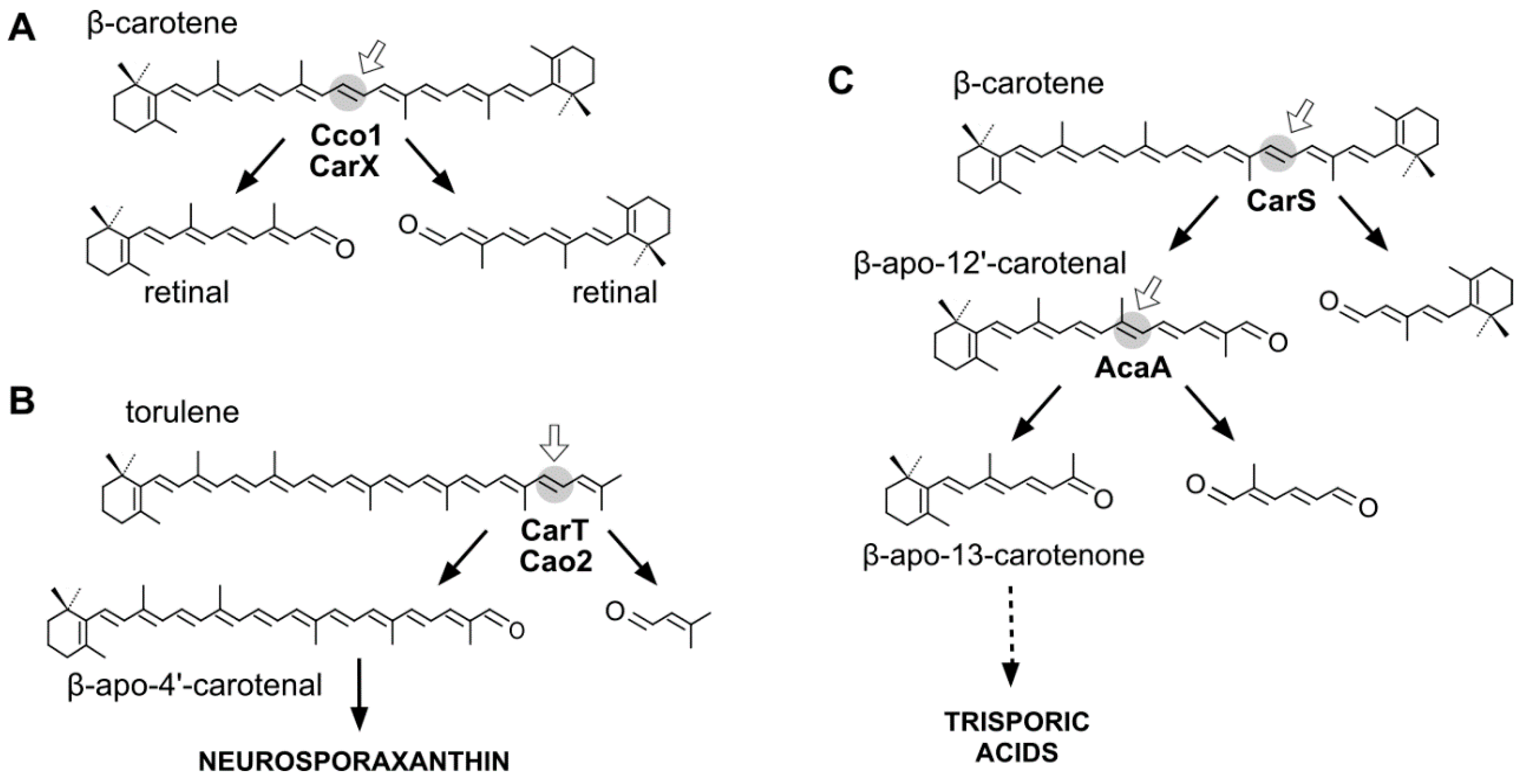
3.1. Fungal CCDs Involved in Retinal Production

Rhodopsins are membrane photoreceptors using retinal as a chromophore. The tertiary structure of these proteins is highly conserved and consists of seven transmembrane helices with a lysine residue to which an all-trans-retinal chromophore is covalently bound. Rhodopsins are found in all major taxonomic groups, including fungi [43]. Very few fungal rhodopsins have been functionally investigated, among them NOP-1 in N. crassa [44], Ops in Leptosphaeria maculans [45,46], and CarO [47], and OpsA in F. fujikuroi [48]. The participation of retinal in the activity of these proteins has only been demonstrated for NOP-1 [49] and CarO [50].
Animals obtain retinal from β-carotene through its oxidative cleavage by a class of CCD enzymes [51]. A similar CCD, encoded by the gene carX [52], is used by F. fujikuroi to produce retinal from β-carotene (Figure 2) [53]. The gene carX is located in a coregulated cluster with the genes needed to produce β-carotene, carRA, and carB, and with the rhodopsin gene carO. Unexpectedly, despite the biochemical similarities between their carotenoid pathways, no carX ortholog has been identified in N. crassa. The only possible candidate for such activity in the genome of this fungus, the CCD CAO-1, was not active on carotenoid substrates, but cleaved efficiently the stilbene resveratrol [54]. A retinal-forming CCD enzyme, called Cco1, has also been described in the basidiomycete Ustilago maydis, producing minor amounts of β-carotene [55]. In this organism, retinal could be detected in cell extracts of the wild type and strains overexpressing cco1, but not in those of null cco1 mutants, and the in vivo β-carotene levels correlated inversely with the Cco1 activity. Retinal production seems to be particularly relevant in this fungus, as suggested by the occurrence of three genes in its genome for presumptive photoactive rhodopsins [55].

3.2. Fungal CCDs Involved in Trisporic Acid Production

Trisporic acids, a class of fungal sexual hormones belonging to the trisporoids family, stand out among the compounds derived from β-carotene for their biological relevance [56,57]. Depending on small variations in their chemical structures, these hormones are distributed in five groups, named A, B, C, D, and E [56,58,59]. The synthesis, investigated in detail in B. trispora and other related species, requires the participation of two CCD enzymes. In fact, thorough chemical analyses revealed the generation of an unexpected apocarotenoid complexity in these fungi: B. trispora produces at least three groups of β-carotene derivatives—C18 trisporoids, C15 cyclofarnesoids, and C7 methylhexanoids [60]—and the same compounds were found in P. blakesleeanus cultures [61]. In the latter case, the origin from β-carotene was confirmed by their absence in a mutant unable to produce β-carotene.
The first CCD enzymes known in fungi were Tsp3 and Tsp4 from Rhizopus oryzae, identified through the analysis of its genome and involved in trisporic acid biosynthesis [62]. This discovery led to identification of the tsp3 ortholog in B. trispora, whose connection with trisporic acids was reinforced by its induced expression by sexual interaction and its capacity to cleave β-carotene in a tsp3-expressing E. coli strain. Further evidence was obtained from the chemical structures of the apocarotenoids produced by P. blakesleeanus [63], which were consistent with the cleavage of β-carotene at the C11′–C12′ and C12–C13 double bonds (Figure 2).
The carS mutants of P. blakesleeanus, which accumulate large amounts of β-carotene, were found to be affected in a CCD enzyme [64], orthologous of Tsp3. The function of the gene, corroborated by the finding of relevant mutations in six different carS mutants, was confirmed by the capacity of the CarS protein to generate β-apo-12′-carotenal (Figure 2) in carS-expressing E. coli cells [65]. The identification of CarS as a CCD was unexpected, since the β-carotene over-accumulation phenotype of carS mutants suggested that the gene encoded a regulatory protein. This finding leads to the reinterpretation of the carotene overproduction of the carS mutation as a result of a blockage of the pathway or of the lack of a negative-acting apocarotenoid signal, which could be responsible for a formerly proposed feed-back regulation [66]. In support of the blockage hypothesis, the large increase in β-carotene content in these mutants is not sufficiently explained by the minor changes that could be found in the transcript levels of the structural genes carB and carRA [67]. However, the function of CarS as a CCD enzyme does not discard an additional regulatory role, as suggested by the unexpected albino phenotype of some double carS mutants [68]. Moreover, the carS mutants exhibit increased enzymatic activities in vitro [42], a result more coherent with a putative regulatory hypothesis.
A second CCD enzyme, AcaA, has been characterized in P. blakeseleeanus. AcaA cleaves β-apo-12′-carotenal (C25) to generate β-apo-13-carotenone (C18) [65]. The available information indicates that CarS and AcaA act sequentially to produce apocarotenoids in this fungus. First, CarS cleaves β-carotene at the C11′–C12′ double bond to generate the C25 and C15 apocarotenals, and AcaA cleaves the resulting C25-product at the C13–C14 double bond afterwards (Figure 2). The resulting C18 product is the origin of the trisporic acids and other trisporoids, while the former CarS C15 product is used for the synthesis of cyclofarnesoids.
The genome of P. blakesleeanus contains three additional genes for presumptive CCDs. Only one of them contains the expected histidine residues to support CCD cleavage activity, but its function has not been investigated. The genes and enzymes for later steps of trisporoid metabolism are currently under study, but they are presumably CCD-unrelated enzymes: they include at least a 4-dihydromethyltrisporate dehydrogenase [69,70] and a 4-dihydrotrisporin-dehydrogenase [71], both identified in the P. blasleeanus relative Mucor mucedo.

3.3. Fungal CCDs Involved in Neurosporaxanthin Production

Neurosporaxanthin is a C35 carotenoid derived from the oxidative cleavage of its C40 precursor torulene. The first enzymatic reactions in the neurosporaxanthin biosynthetic pathway are similar to those for β-carotene production in other fungi, but in this case five desaturations instead of four and only one cyclization are introduced into the C40 carotene skeleton, leading to torulene (Figure 2). The analysis of the proteomes of N. crassa and F. fujikuroi and the study of mutants blocked in different steps of the pathway led to the identification of the enzymes involved in the late oxidative reactions. The genomes of both species contain two CCD-encoding genes. One of them, called carT in F. fujikuroi, was found to catalyze the cleavage of torulene to produce β-apo-12′-carotenal. This function was corroborated by the finding of a mutated carT allele in a reddish torulene -accumulating mutant and the ability of the wild-type carT allele to restore neurosporaxanthin production when it was introduced in the mutant [53,72]. CarT activity was also confirmed by targeted mutation in Gibberella zeae, a teleomorph of Fusarium graminearum [73]. The same function was achieved in N. crassa by its ortholog, cao-2, as demonstrated the accumulation of torulene in a mutant for this gene and the finding of cao-2 mutations in two torulene-producing mutants of this fungus [74].
As found for β-carotene production in Mucorales, the synthesis of neurosporaxanthin is induced by the light in N. crassa [75] and Fusarium sp. [76]. This photoresponse is achieved through an outstanding increase in mRNA levels for most of the structural genes, including cao-2 [74] in N. crassa, and carT [72] in F. fujikuroi. Moreover, the expression of carT is enhanced in carotenoid-overproducing mutants.
The CarT and CAO-2 enzymes, catalyzing the asymmetrical cleavage of torulene at its acyclic end to remove a C5 segment, represent a novel CCD subgroup. The reaction is highly specific, as shown by the incapacity of CAO-2 to cleave the torulene precursor γ-carotene, indicating the need for five desaturations in the substrate molecule. Subsequently, the product β-apo-12′-carotenal is converted to neurosporaxanthin by the aldehyde dehydrogenase YLO-1 in N. crassa [77] and by its ortholog CarD in F. fujikuroi [78].

4. Plant CCDs

The oxidative cleavage of carotenoids in plants leads to the production of a range of apocarotenoids compounds that serve critical functions including photoprotection, photosynthesis, pigmentation, and signaling [79,80]. Plant CCDs constitute the most abundant group identified so far. These CCDs have been classified into two large families based on whether they are involved or not in the production of abscisic acid (ABA), a hormone involved in drought stress responses and in bad and seed dormancy [81]. Those involved in ABA production are the nine-cis-epoxy-carotenoid-dioxygenases (NCEDs), which cleave 9-cis-violaxanthin and 9-cis-neoxanthin to xanthoxin, the precursor of ABA. In fact, the first CCD cloned from any organism was the Vp14 gene from maize that catalyzed the first committed step in ABA biosynthesis [81]. After the identification of Vp14, other CCD enzymes were isolated, including β-carotene oxygenase (BCO) in mammals involved in the synthesis of vitamin A, CCDs from microorganisms (mentioned in former sections), and other plant CCDs. In plants, five CCD groups have been identified so far–CCD1, CCD2, CCD4, CCD7, and CCD8. However, transcriptome and large-scale genomic sequencing projects in many different plant species are allowing the identification of new species-specific CCDs with yet unknown functions. In fact, biochemical studies have been reported for a limited number of plant CCDs, and the roles they play in different organisms are not fully understood.

4.1. CCD1 and CCD2 Subfamilies

AtCCD1 from A. thaliana was the first carotenoid cleavage dioxygenase isolated not involved in ABA biosynthesis (Figure 1) [82]. The first cleavage activity reported for CCD1 enzymes was on the C9–C10 (C9′–C10′) double bonds in C40 carotenoids, producing a colored C14 dialdehyde and two scent C13 products [82]. Homologues of this enzyme have been identified in many other plant species [83,84,85,86,87,88,89,90,91], and the studies performed on these CCD1 enzymes suggested additional cleavage activities on acyclic, monocyclic, and bicyclic carotenoids, such as ζ-carotene, lycopene, phytofluene, β-carotene, δ-carotene, zeaxanthin, lutein, and violaxanthin, and on different apocarotenoids, catalyzing the symmetric cleavage at C9–C10 (C9′–C10′), C5–C6 (C5′–C6′), C7–C8 (C7′–C8′), and C13–C14 (C13′–C14′) (Figure 3) [85,92,93,94,95]. The enzyme cleaved symmetrically at C9–C10 (C9′–C10′) of acyclic and cyclic trans-carotenoids and did not cleave adjacently to a -cis double bond or an allenic bond found in some carotenoids [82]. Therefore, an asymmetric cleavage is observed in such cases. The C5–C6 or C5′–C6′ activity of CCD1 enzymes on lycopene (or both), leading to the formation of the C8 ketone 6-methyl-5-hepten-2-one (MHO), was reported for the first time in tomato, maize, and A. thaliana [92] and was later detected in Cucumis melo [84], Rosa damascena [96], Oryza sativa [97], and Vitis vinifera [98]. The cleavage of C7–C8 and C7′–C8′ double bonds of linear and monocyclic carotenoids constitutes a novel recognition site for the CCD1 plant subfamily. So far only detected by in vitro assays for the rice CCD1 enzyme [85], this activity allowed for the formation of C10-aldehyde geranial, suggesting an alternative pathway for the geranial formation in plants.

4.1.1. Mode of Action and Functional Implications

Initially, the CCD1 enzymes were suggested to be involved in the biosynthesis of apocarotenoid volatiles, such as geranylacetone, pseudoionone, and β-ionone [79], which possess an extremely low threshold for human perception [99]. Lowering of CCD1 expression in tomato or petunia led to significant reductions in the emission rates of β-ionone and geranylacetone, but did not lead to significant changes in the carotenoid concentration [90], suggesting additional roles for the CCD1 enzymes. In addition, the expression of CCD1 during tomato, strawberry, melon, and grape development, or in saffron flowers, did not mirror the emission of apocarotenoid volatiles [83,84,86,89,90]. Furthermore, experiments conducted to identify loci affecting volatile composition in tomato did not reveal the implication of CCD1 in the emissions of apocarotenoid volatiles [100]. These are produced from C40 carotenoids localized in plastids, and the CCD1 enzymes lack plastid targeting signals and are cytoplasmatic [87,101], suggesting that CCD1 enzymes mainly act in planta as scavengers of carotenoid degradation products of different chain lengths rather than primary cleavers of intact carotenoids [80,85,93,94,98,102].
The key roles of carotenoids in photosynthesis, photomorphogenesis, and plant development suggest that their biosynthesis and degradation is coordinately regulated with processes such as plastid biogenesis, flowering, and fruit development [103]. In addition, the link of carotenoid biosynthesis with those of gibberellin, ABA, and strigolactone phytohormones implies that changes in the composition or content of carotenoids might bring about physiological or biochemical shiftings in plants [4]. In 2008, a role of CCD1 in the production of strigolactones (SLs) during the arbuscular mycorrhiza symbiosis in roots was described [94]. CCD1 cleaves the C27 apocarotenoid derivatives produced by the activity of CCD7, which is also involved in the biosynthesis of SLs [104], producing C13 α-ionol and C14 mycorradicin, which are indicators of a well-established and functional symbiosis [105], reducing SL production and avoiding over-colonization [106].
CCD1 could be also important in plant stress responses. Enhanced CCD1 expression during berry development has been associated with the increased osmotic stress that occurs during ripening, resulting in leaky membranes and concomitant chloroplast degradation [98]. In addition, CCD1 expression is induced during leaf senescence [89,107], suggesting that the enzyme-mediated degradation of carotenoids, apocarotenoids, or both is catalyzed by cytosolic CCD1 enzymes. Furthermore, plant–insect interactions, extreme temperatures, high irradiance, or ultraviolet (UV) stress induce the production of β-ionone and β-cyclocitral among other apocarotenoid volatiles [108,109]. CCD1 expression has been detected in all tested tissues in different plant species [89,90,91,95,98,110,111], but its expression is stimulated by abiotic stress or ABA treatment [112,113].

4.1.2. CCD1 Gene Family and Genomic Organization

The CCD1 family is present in some plant species as a multigene family (Table S1), and studies on maize and tomato CCD1 enzymes have suggested varying expression profiles and different specificities in their activity towards different substrates and in their double bond preferences [90,92,93,102,114]. In most of the analyzed species, there are two genes encoding for CCD1 enzymes, and in many cases the genes are present in tandem in the same chromosome (Table S1). The identities among the paralogous CCD1 genes range between 90% and 99%. All the CCD1 genes show several introns in their sequence [115], these positions being conserved [115]. Moreover, the presence of such introns allows for the generation of different splice variants (Table S1). These variants may differ as well in the untranslated regions, with implications in transcript stability, localization, translation, or a combination thereof. It is likely that the truncated proteins from the CCD1 mRNA isoforms may act as dominant-negative regulators. In fact, several reports provide evidences of a dominant regulatory role for truncated proteins generated by splice variants in plants [116]. AtCCD1 has been suggested to act as a dimer [82], and the presence of these variants could be part of a regulatory mechanism for CCD1 activity.
The phylogenetic analysis of the CCD1 proteins reveals several main clusters that fit with the phylogenetic relationships in embryophytes (Figure S1). In the angiosperm group, there are three sub-clusters corresponding to monocots, dicots (excluding crucifers), and crucifers. The latter sub-cluster of Brassicaceae species is more related to the monocot group than to the dicot group, suggesting a specific function for CCD1 in these plants.

4.1.3. Mode of Action of CCD2

Recently, a close CCD subfamily related to the CCD1 family has been identified in Crocus species [117,118]. This subfamily, named as CCD2 (Figure 1), is involved in the formation of the apocarotenoid crocetin, which accumulates in stigma tissue at high levels [119]. Crocetin is derived by bio-oxidative cleavage of zeaxanthin by a C7–C8 and C7′–C8′ cleavage reaction catalyzed by CsCCD2 [117]. The expression of CsCCD2 is restricted to the stigma in saffron, and it mirrors the levels of crocetin in this tissue during its development [89,117]. The same behavior is observed for CaCCD2, a CCD2 homologue isolated from the spring crocus C. ancyrensis [118]. Both CCD2 enzymes are plastidic, a major difference with the CCD1 subfamily [118]. No other CCD2 homologues have been identified in other organisms, presumably due to the uncommon ability to synthesize crocetin in plants [120] or bacteria [121]. Besides zeaxanthin, CCD2 is able to recognize and cleave lutein and 3-OH-β-apocarotenals at the C7–C8 position, but it does not cleave β-carotene, lycopene, or β-cryptoxanthin [117].

4.1.4. Structure of CCD1 and CCD2 Enzymes

Based on the crystal structure of VP14 [122], structural models have been built for CCD1 and CCD2. VP14 is a globular protein that showed a characteristic hydrophobic patch formed by the antiparallel α-helices presumably involved in membrane interaction [28]. CCD1 and CCD2 enzymes display from 58% to 39% overall identity with VP14. Specially conserved are the structural elements of the CCD enzymes. Three regions have been identified in VP14, implicated in the recognition of the bond to be cleaved [122]. The first one is Val478, which is a substitute for a Phe residue in CCD1 and CCD2 enzymes. This substitution is present in many others CCDs. The second region is a loop formed by residues 499 to 503 in VP14. This loop is not present in CCD1 and CCD2 enzymes, in which conserved Leu170 in Vp14 was replaced by Trp. In Vp14, the loop and the leucine residue form a pocket where the second ring of the carotenoid accommodates in such a way that the C11–C12 double bond of the substrate should be close to the Fe2+. The third region, located in the active center, comprises three Phe residues in Vp14 (Phe171, Phe411, and Phe589) and is conserved in all NCED and CCD1 enzymes; however, in CCD2, only one Phe residue is conserved.

4.1.5. Regulation of CCD1 and CCD2 Enzymes

As stated before, CsCCD2 expression is regulated during the development of the stigma and the enzyme is localized in chromoplasts [123] where catalyzes crocetin biosynthesis. Crocetin is oxidated and glucosylated thereafter [124], generating crocins that accumulate in the vacuole. The mechanisms involved in CsCCD2 expression and activity have recently been elucidated [123]. In this study, the authors showed that high temperatures repressed CsCCD2 expression, while low temperatures have an opposite effect by upregulating its expression. In saffron plants, the highest expression levels of CsCCD2 are related to the lowest temperatures. Several classes of cis-regulatory elements were identified in the promoter of CsCCD2. The regulation of carotenoid biosynthesis by light and dark conditions has been investigated in red pepper [125], in tomato leaves [126], and in citrus, where the expression levels of the carotenogenic genes are affected by circadian rhythms [127,128]. Additionally, a new mechanism of regulation by alternative splicing has been observed for CsCCD2, [123]. The obtained data agree with the predicted function of alternative splicing in genes with a circadian clock expression pattern, where oscillations of protein expression levels require rapid adjustments in mRNA levels over the course of a day [129].

4.2. The CCD4 Subfamily

The CCD4 subfamily is present in all flowering plants, usually composed by several members (Figure 1) [115,130]. Their functions have been directly linked to coloration and production of aroma volatiles in flower and fruit tissues [89,98,131,132,133]. More recently, CCD4 activity has been reported in seeds, leaves, and roots associated with carotenoid turnover [107,134] or with the production of novel apocarotenoid-derived signals [135]. The first member of this subfamily was identified in A. thaliana [18]; however, its role in carotenoid catabolism was initially described for CCD4a from Chrysantemum morifolium, whose activity is responsible for the absence of carotenoid accumulation in petals of white flowers [136].
All CCD4 proteins contain a plastid target peptide; moreover, suborganellar studies in different plant species localized these enzymes in the plastoglobuli together with other carotenoid biosynthetic enzymes, which allows direct access to their substrates [131,137,138,139,140,141]. In both chloro- and chromoplasts, CCD4 is one of the major components of plastoglobuli proteomes [137,140], indicating a role for this enzyme in the regulation of carotenoid metabolism in plastid types with marked differences in functionality and carotenoid composition [142]. Moreover, CCD4 protein level is strongly reduced in Arabidopsis mutants defective in the ABC1K1 and ABC1K3 plastoglobuli kinases, suggesting phosphorylation as a post-transcriptional modification necessary for CCD4 protein stability and supporting main activity and localization in plastoglobuli [143].

4.2.1. Enzyme Structure and Functional Implications

As found for other CCDs, molecular modeling of CCD4 enzymes from the structure of the 9-cis-epoxycarotenoid dioxygenase from maize (VP14/NCED) revealed a well-conserved structure among the CCD family [130,133]. The analyzed CCD4s display two main functional domains: the helical domain composed of two antiparallel α-helices, which might penetrate in the hydrophobic core of the plastoglobule where carotenoids accumulate [130,138], and the β-propeller structure that forms a long central tunnel, where Fe2+ is coordinated with four conserved His in the active center [130,133]. However, other critical amino acids for NCED activity are not always conserved among all CCD4 members [130,133]. As was the case in CCD1 and CCD2 enzymes, a large number of CCD4 proteins display a Phe residue instead of the conserved Val478 found in NCED, with the exception of CCD4 from alfalfa (CCD4a and b), grape (CCD4e), potato (CCD4b), poplar (CCD4c), and citrus (CCD4b1), where it is substituted by Leu; however, in citrus CCD4c, Val478 is replaced by Met. Other critical residues in VP14 are three Phe at positions 171, 411, and 589, from which only Phe589 is conserved in all CCD4, while Phe411 is highly variable, and Phe171, which also takes part of the motif FDG, is partially conserved and usually substituted by Leu. Another important region for VP14 activity is a loop located on the back side of the substrate pocket, involving residues 499–503 and Leu170. In contrast with CCD1 and CCD2, residues in this loop are highly conserved in the CCD4 subfamily; however, the citrus CCD4b1 is a relevant exception, with the Glu499 and Pro500 reported in VP14 substituted by Lys and Glu, respectively. The Leu170 in VP14 is also well conserved in all CCD4s; however, in CCD4b1, CCD4b, and CCD4e from citrus, potato, and poplar, it is replaced by Phe and in citrus CCD4c by Pro. Although it is difficult to determine exactly the impact of these residue changes in the enzyme activity, it is likely that they may modulate the carotenoid substrate specificity, the cleavage position, or both. Interestingly, functional assays performed with citrus CCD4b1, which accumulates several non-conservative amino acid substitutions, show an unusual cleavage position, having as preferential substrates the hydroxylated β-ionone ring of the xanthophylls. Future studies on comparative 3D modeling of the different members of this subfamily will provide clues as to substrate specificity or cleavage position in carotenoid backbone, which may help to understand their role in vivo.

4.2.2. Gene Family and Genomic Organization

The number of genes for CCD4 enzymes is very variable among different species, with at least two genes identified in most plants [115,144]. The presence of one single CCD4 gene has been reported only in a limited number of plant species with an available genome sequence, such as A. thaliana, Carica papaya (papaya), Prunus persica (peach), and Sorghum bicolor (sorghum) [115,130,144]. By contrast, the largest number of CCD4 genes has been found in Citrus sp. (a, b1, b2, c and d), Populus trichocarpa (a–e), and Chrysanthemum morifolium (a-1–a-4 and b), with five members and with at least one of them being a putative pseudogene [115,133,145,146]. Four members have been identified in Vitis vinifera (grapevine) (a, b, c, and e) or Brassica napus (rapeseed) (A1, A8, C1, and C3), while three are present in Theobroma cacao (cacao) [98,130]. Another common genomic feature of CCD4 genes is the low presence of introns, most of them being intron-less or containing a single intron [115,144]. Moreover, intron size and position are quite variable among different species. A comprehensive comparative analysis of the genomic structure of CCD4 from diverse plants showed that introns are located in three different sites and only those located at the 3′ end of the gene maintain a conserved position [115]. Exceptionally, only a few CCD4 genes contain more than one intron, such as Osmathus fragans CCD4 and Solanum lycopersicum CCD4a and b, each with two introns, Citrus sp. CCD4d with two or four introns, and an uncharacterized CCD4 from apple (Malus domestica) with 85% identity at the protein level to apple CCD4a [96] with six introns.
The degree of protein sequence identity among CCD4 members is quite variable, ranging from 30% to 98%, with typical values around 50%–70% [115,130,133,147,148]. In multigene CCD4 families, a high degree of homology (85%–98% identity at the protein level) is usually found between the different members, frequently located in tandem on the same chromosome (i.e., tomato CCD4a and b, citrus CCD4b1 and b2, chrysamthemum CCD4a-1 to a-4, or Populus CCD4c to e) [115,133]. As expected, the highest variability is found at the plastid target peptide sequences, while a higher conservation is found in the motifs described as essential for CCD activity [6,28,149]. The moderate conservation within the different CCD4 members of the same family (i.e., citrus CCD4b1 versus CCD4c or grapevine CCD4a/b versus CCD4c/e), and the location in different clusters in the phylogenetic analysis (Figure S2) suggest a divergent evolution and functionality of these members.

4.2.3. CCD4 Activity: Carotenoid Substrates and Cleavage Products

In contrast to other CCD subfamilies, such as NCED or CCD7/8, functional assays of CCD4 indicate that the members are rather heterogeneous in respect of carotenoid substrates and cleavage positions (Table 4). The functional characterization of CCD4 has been an active field of research in the last decade by following three basic strategies: (i) in vivo assays of CCD4-defective plant mutants or CCD4-overexpressing or repressing transgenic plants; (ii) in vivo assays of bacteria over-accumulating different carotenoids and co-expressing CCD4; and (iii) in vitro enzymatic assays performed with recombinant CCD4. Table 4 summarizes the information currently available on CCD4 enzyme activity.
The first comparative study of substrate preferences and cleavage positions of CCD4 enzymes was obtained from in vitro and in vivo assays performed with five CCD4s isolated from Arabidopsis, rose (R. damascene), osmanthus (O. fragans), apple (M. domestica), and chrysanthemum (C. morifolium) [96]. Most of these CCD4 cleaved β-carotene at C9–C10 and C9′–C10′ positions, rendering C13 β-ionone, while no activity was detected on hydroxylated xanthophylls [96]. Further research using Arabidopsis ccd4 mutants or carotenoid over-accumulating lines revealed that epoxy-xanthophylls (mainly violaxanthin) are likely the substrates in Arabidopsis leaves [107,134] to render C13 apocarotenoids (cleavage at C9–C10 or C9′–C10′ double bonds) that undergo further glycosylation [134]. Interestingly, changes in carotenoid composition and apocarotenoid profile in ccd4 Arabidopsis mutants, during seed maturation or in roots of hyperaccumulating β-carotene plants, suggest that CCD4 also accepts β-carotene as substrate [107,134]. One of the two CCD4s from potato, in this work designated as CCD4a (Figure S2), was functionally characterized. Analysis of the carotenoid profile in tubers from RNAi CCD4 potato lines suggested violaxanthin as the main substrate [150]. However, in-depth biochemical characterization of potato CCD4a using in vitro and in vivo assays points to β-carotene as its main substrate, cleaved at C9–C10 or C9′–C10′ positions [148]. In saffron, in vivo assays showed that CCD4a and b cleave β-carotene and zeaxanthin most likely at the C9–C10 and C9′–C10′ double bonds, while CCD4c cleaves other xanthophylls such as lutein [89,130,151]. In vivo assays of grapevine CCD4a and b indicate that these enzymes, in contrast to all CCD4s characterized so far, do not cleave cyclic carotenoids, with the exception of β-carotene, cleaving linear carotenes neurosporene, lycopene, and β-carotene—only CCD4b preferentially at C9–C10 and C9′–10′ positions [98]. The carotenoid profile from rape (Brassica napus) flowers suggests that the member CCD4_C3 cleaves α-carotene or δ-carotene at the C9–C10 position to render α-ionone [152]. Interestingly, in vitro and in vivo assays show that Citrus sp. CCD4b1/CCD4 accepts β-ring hydroxylated xanthophylls as preferential substrates, but also accepts β-carotene or α-carotene. In contrast to the other plant CCD4s, citrus CCD4b1 and CCD4 cleaves the carotenoid backbone at C7–C8 or C7′–C8′ double bonds, resembling CCD2 activity, but only at one side of the molecule [131,133].
In summary, in vitro and in vivo assays for most of the CCD4 enzymes point to β-carotene and a C9–C10 or C9′–C10′ double bond cleavage as a favorite substrate and positions, respectively, although some relevant exceptions, such as citrus or grape CCD4, have been identified regarding the substrate or cleavage site. In particular, the exclusive cleavage activity reported for citrus CCD4b1 and CCD4, together with its restricted expression pattern, may indicate that this enzyme belongs to a novel class of CCD more functionally related to the recently characterized CCD2 from saffron [117,118]. In planta data, derived from the analysis of apocarotenoid profiles, the alteration in carotenoid complement in ccd4 mutants and overexpressed or repressed CCD4 plants, or both, point to xanthophylls as the preferential substrates in vivo [130,133,134,145]. Nevertheless, in some species and tissues, such as potato tubers, chrysanthemum petals, and peach fruit, β-carotene cannot be ruled out as the primary target since alterations in the xanthophylls composition in defective or overexpressing CCD4 plants can be explained by a reduction or increase in β-carotene content as the precursor of β-xanthophylls [136,150,153].
These differences in CCD4 substrate preferences may also reflect the differences in substrate availability or accessibility depending on the types of assay. In the in vitro and in vivo assays, the enzyme has direct access to the potential substrates; this clearly differs from the in planta situation, where carotenoids are integrated in the suborganellar structures of the plastids, and CCD4 is preferentially associated with plastoglobuli, where xanthophylls are synthesized and accumulated. The CCD4 activity on linear carotenes is usually very weak either in vivo or in vitro, with the exception of grapevine CCD4 [98]. Recently, the phenotypic characteristics of the Arabidopsis double mutant clb5 ccd4 (defective in ZDS and CCD4) suggests that, under this particular scenario of hyper-accumulation of linear upstream carotenes, CCD4 generates an apocarotenoid derived from phytofluene, ζ-carotene, or both, indicating an activity on the backbone of these carotenes [135]. None of the CCD4 characterized so far catalyzes the cleavage of phytoene, in agreement with the hypothesis that a double bond in the carotenoid structure must be adjacent to the cleaved double bond [92,98].
These differences in CCD4 substrate preferences may also reflect the differences in substrate availability or accessibility depending on the types of assay. In the in vitro and in vivo assays, the enzyme has direct access to the potential substrates; this clearly differs from the in planta situation, where carotenoids are integrated in the suborganellar structures of the plastids, and CCD4 is preferentially associated with plastoglobuli, where xanthophylls are synthesized and accumulated. The CCD4 activity on linear carotenes is usually very weak either in vivo or in vitro, with the exception of grapevine CCD4 [98]. Recently, the phenotypic characteristics of the Arabidopsis double mutant clb5 ccd4 (defective in ZDS and CCD4) suggests that, under this particular scenario of hyper-accumulation of linear upstream carotenes, CCD4 generates an apocarotenoid derived from phytofluene, ζ-carotene, or both, indicating an activity on the backbone of these carotenes [135]. None of the CCD4 characterized so far catalyzes the cleavage of phytoene, in agreement with the hypothesis that a double bond in the carotenoid structure must be adjacent to the cleaved double bond [92,98]. In summary, in vitro and in vivo assays for most of the CCD4 enzymes point to β-carotene and C9–C10 or C9′–C10′ double bond cleavage as the favorite substrate and positions, respectively, although some relevant exceptions, such as citrus or grape CCD4, have been identified regarding the substrate or cleavage site. In particular, the exclusive cleavage activity reported for citrus CCD4b1 and CCD4, together with its restricted expression pattern, may indicate that this enzyme belongs to a novel class of CCD more functionally related to the recently characterized CCD2 from saffron [117,118]. In planta data, derived from the analysis of apocarotenoid profiles, the alteration in carotenoid complement in ccd4 mutants and overexpressed/repressed CCD4 plants, or both, point to xanthophylls as the preferential substrates in vivo [130,133,134,145]. Nevertheless, in some species and tissues such as potato tubers, chrysanthemum petals, or peach fruit, β-carotene cannot be ruled out as the primary target since alterations in the xanthophylls composition in defective or overexpressing CCD4 plants can be explained by a reduction or increase in β-carotene content as the precursor of β-xanthophylls [136,150,153]. These differences in CCD4 substrate preferences may also reflect the differences in substrate availability or accessibility depending on the type of assay. In the in vitro and in vivo assays, the enzyme has direct access to the potential substrates; this clearly differs from the in planta situation, where carotenoids are integrated in the suborganellar structures of the plastids, and CCD4 is preferentially associated with plastoglobuli, where xanthophylls are synthesized and accumulated. The CCD4 activity on linear carotenes is usually very weak either in vivo or in vitro, with the exception of grapevine CCD4 [98]. Recently, the phenotypic characteristics of the Arabidopsis double mutant clb5 ccd4 (defective in ZDS and CCD4) suggests that, under this particular scenario of hyper-accumulation of linear upstream carotenes, CCD4 generates an apocarotenoid derived from phytofluene, ζ-carotene, or both, indicating an activity on the backbone of these carotenes [135]. None of the CCD4 characterized so far catalyzes the cleavage of phytoene, in agreement with the hypothesis that a double bond in the carotenoid structure must be adjacent to the cleaved double bond [92,98].

4.2.4. Expression Pattern in Plant Tissues

CCD4 genes are predominantly expressed in flower and fruit tissues, suggesting specific roles in these organs [130,147]. Usually at least one CCD4 member is highly or exclusively expressed in flowers, and its function has been related to the production of nor-isoprenoid aroma volatiles to attract pollinators [96,133,146,148,154,155]. In chrysantemum, Osmanthus, and cabbage, the expression of a specific CCD4 gene in petals is responsible for carotenoid degradation and a lack of flower coloration [136,145,152]. In chrysanthemum, CCD4a is represented by a small multigene family, and the white to yellow color gradation in petals among different cultivars can be explained by different combinations and levels of expression of specific CCD4a genes [145]. In the flower petals of lily (Lilium brownii var. colchesteri), CCD4 transcript levels are associated with a loss of carotenoid content during anthesis, resulting in flower whitening [156]. In saffron, the expression of CCD4a/b correlates with the production of the nor-isoprenoid β-ionone in flower tissues, while CCD4c is restricted to stigmas and shows a highly regulated expression pattern during flower development [89,130]. In other species, such as sweet orange, tomato, and potato, at least one CCD4 gene is expressed in flowers, but their involvement in carotenoid degradation has not been elucidated [133,148,150]. Expression of CCD4 genes has also been reported in many fruits and its role has been associated with the biosynthesis of nor-isoprenoid volatiles or apocarotenoids pigments, which may attract animals to facilitate seed dispersion. In grape, CCD4a and b genes are expressed in the berry, but only CCD4b shows a fruit-specific expression profile, highly upregulated during ripening, linking its activity to nor-isoprenoid volatiles production [98]. In summer squash (Cucurbita pepo) varieties with white, green, and yellow-orange coloration, the expression of two CCD4 genes (a, b) correlates inversely with carotenoid accumulation in the peel and flesh; therefore, both genes are good candidates for regulating fruit color [111,157]. One of the best-characterized examples of the CCD4 role in fruit pigmentation has been described in peach (Prunus persica). White-fleshed peach cultivars contain at least 10 times less carotenoid concentration and higher nor-isoprenoid carotenoid-derived volatiles than the yellow ones [158]. Recently, several studies have established that alterations in the CCD4 gene are directly related to carotenoid content in yellow peach varieties. In yellow cultivars, CCD4 gene expression is significantly reduced, the formation of a truncated CCD4 protein avoids carotenoids degradation in fruit mesocarp, or both [159,160,161,162].
In mandarins and oranges, the specific expression of the CCD4b1/CCD4 gene in the fruit peel during ripening is responsible for the asymmetric cleavage of β-cryptoxanthin and zeaxanthin to generate the C30 apocarotenoids 3-OH-β-8-apocarotenal (β-citraurin) and β-apo-8-apocarotenal [131,133], respectively. It is interesting to note that, due to the intense orange-red color of these C30 apocarotenoids, the citrus CCD4 activity enhances fruit pigmentation, in contrast to the other CCD4s described so far whose activity is associated with color loss. In other species, such as bitter melon, goji berry, and tomato, the expression of at least one CCD4 gene has been reported in ripened fruits, but no relationship has been established with carotenoid content [148,155,163]. In potato tubers, compared with the yellow ones, CCD4 activity regulates coloration, as indicated by the elevated CCD4 expression in mature white-fleshed tubers [150]. Moreover, in CCD4 RNAi potato lines, total carotenoid content was enhanced in tubers (up to 5.6-fold) and flowers, but not in other plant organs [150]. The function of this enzyme in tubers is probably associated with stress response since the potato tubers from RNAi lines showed diverse developmental alterations linked to heat stress phenotypes [150]. Recently, it has been proposed that an apocarotenoid derived from β-carotene in potato tubers could act as a signaling molecule in stress responses [153].
In other plant species, CCD4 expression is also modulated by specific abiotic stresses. Saffron CCD4a and b are expressed in leaf tissues upon dehydration or heat stress as well as in senescence leaves [89]. Moreover, CCD4c is upregulated in flower stigmas subjected to different stress treatments, such as osmotic, wounding, and cold or heat stresses [130]. In B. rapa and B. oleracea seedlings, CCD4 transcript levels increase in response to different abiotic stresses as well as to exogenous treatments with SLs and ABA phytohormones, suggesting their involvement in plant stress resistance response [152]. In other processes involving cellular responses to stress, such as dark-induced leaf senescence or seed maturation and desiccation, a major role of CCD4 in carotenoid degradation has been proposed [107]. These processes are associated with severe alterations in chloroplast structures, causing disassembly of photosystems and light-harvesting complexes and the release of carotenoids, in which CCD4 activity may play a crucial role in modulating turnover of β,β-carotenoids. By contrast, high-light stress in Arabidopsis leaves causes a strong downregulation in CCD4 expression [164], which may prevent cleavage of xanthophylls with a photoprotective function [134]. A role of CCD4 maintaining carotenoid homeostasis has also been reported in Arabidopsis overexpressing PSY [134]. In these plants, CCD4 activity is essential for the formation of apocarotenoid (C13) glycosides (derived from epoxidized xanthophylls) in leaves and long-chain apocarotenoids (derived from β-carotene) in roots. Moreover, a lethal phenotype was observed in PSY-overexpressing seedlings defective in CCD4 activity, supporting the key role of CCD4 as a first step in detoxifying excess of carotenoids into apocarotenoid glycosides [134]. The CCD4 activity is also involved in the formation of an uncharacterized apocarotenoid, most likely derived from ζ-carotene, phytofluene, or both, participating in a novel signaling process that regulates early chloroplasts and leaf development [135].
In summary, the presence of different CCD4 members with tissue- and organ-specific expression patterns in many plant species indicates a high functional diversification within this subfamily. In the last few years, we have gained much knowledge on the physiological, biochemical, and molecular regulation of CCD4 enzymes and their presumed involvements in different processes. In one case, the function is concerned with the production of apocarotenoids in flowers and fruits to attract pollinators and seed dispersers, which has eco-physiological and reproductive implications. In another case, the role is directly involved in carotenoid turnover, related to specific stress conditions and alterations of the carotenoid pathway. A third function was recently identified with the involvement of CCD4 activity in the formation of an apocarotenoid-derived signal that regulates early chloroplasts and leaf development. Future research on different CCD4 members in multigene families, including structural, functional, and spatial studies as well potential interactions with other enzymes of the carotenoid pathway, will help towards a better understanding as to how carotenoid content is modulated in different plant organs.

4.3. The CCD7 and CCD8 Subfamilies

The first report on CCD7 and CCD8 enzymes in plants derives from the max4 mutant of Arabidopsis, affected in the AtCCD8 gene (Figure 1). The mutants exhibit an increase in lateral branching, which reminiscent of the phenotype of the max3 mutant, which shows an increase in lateral branching as a result of a mutation in the AtCCD7 gene, indicating that both AtCCD8 and AtCCD7 are involved in this developmental process [165,166]. The biochemical characterization of AtCCD7 and AtCCD8 [167] demonstrated that AtCCD7 catalyzes a C9–C10 cleavage of β-carotene to produce 10′-apo-β-carotenal (C27) and β-ionone (C13). The AtCCD8 protein is able to catalyze a secondary cleavage of 10′-apo-β-carotenal at the C13–C14 position to produce 13-apo-β-carotenone (C18). However, it has been shown that CCD7 is also active on many other carotenoid substrates [166,167]. In contrast to CCD1, which has a cytoplasmatic location, CCD7 and CCD8 are localized in plastids, as CCD4 and CCD2 are, the predominant sites for carotenoid accumulation [87].
The CCD7 and CCD8 enzymes are involved in the biosynthesis of a relatively novel class of apocarotenoid hormones, the strigolactones (SLs), which control lateral shoot growth and which appear to be well conserved among the studied plant species. Branching mutants from Arabidopsis, petunia, pea, and rice lacking CCD7 or CCD8 have reduced SL concentrations, and applications of synthetic SLs to the mutant restores the wild-type branching phenotype [87,168,169]. In tomato, a reduction of CCD7 expression increases branching [170]. This is the case of the tomato mutant Sl-ORT1, which is deficient in SLs and has reduced CCD7 expression [171]. These enzymes have been identified in all high plant genomes sequenced up to date. SLs are derived from carotenoids, thus belonging to the class of the apocarotenoids that function as signaling molecules. This is exemplified by their role promoting germination of the parasitic plants Striga and Orobanche [172] or their involvement in symbiotic interaction with arbuscular mycorrhizal fungi [173]. CCD7 and CCD8 are involved in sequential cleavage reactions needed for the synthesis of SLs. These metabolites share a common C19 structure consisting of a tricyclic lactone connected via an enol ether bridge to a second lactone (Figure 4). The first steps of SL synthesis involve isomerization and dioxygenase-mediated cleavage of a carotenoid precursor via plastidic isomerase action by D27, followed by the cleavage activity of CCD7 and CCD8. MAX1 encodes a cytP450 enzyme predicted to act downstream of CCD7 and CCD8, and is required for the synthesis of active SLs. SL signaling requires the hormone-dependent interaction of the α/β hydrolase protein DWARF 14 (D14), a likely SL receptor, with DWARF 3 (D3), an F-box component of the Skp-Cullin-F-box (SCF) E3 ubiquitin ligase complex. The third component in the signaling pathway is D53, which shares predicted features with the class I Clp ATPase proteins and can form a complex with D14 and D3. SLs induce D53 degradation by the proteasome and abrogate its activity in promoting axillary bud outgrowth [174]. Analysis of mutants, such as D27, D17/CCD7, and D10/CCD8 from rice, showed that all these genes are involved in the biosynthesis of SLs. However, mutants lacking one or more of these genes are able to produce SL metabolites, which indicate an alternative minor pathway for SL biosynthesis [175].
The first identified function of SLs in higher plants was as a stimulatory signal in seed germination of root parasitic weeds, such as the witchweed Striga spp. and broomrapes (Orobanche and Phelipanche spp.) [176]. The first germination stimulant identified for Striga was called strigol, a member of SLs isolated from cotton (Gossypium hirsutum L.) root exudates [177]. Many other compounds with similar structures and with germination stimulatory properties were discovered in root exudates of host plant species [178]. Orobanche and Phelipanche spp. are obligate non-photosynthetic holoparasites that depend fully on their host for nutritional needs. It is not clear how these parasites perceive SLs. Recently, it has been suggested that MAX2, AtD14, or both, which are sensitive to SLs, might be involved in this process [179]. In rice, the obstruction of the carotenoid biosynthesis in different steps of the pathway, including those leading to CCD7 substrates, led to reduced SLs production and secretion into the rhizosphere, resulting in decreased Striga germination and consequently lower Striga infection [180].
The analysis of genomes from several plant species showed a single copy for the CCD7 gene. However, in most of the analyzed genomes, CCD8 is present as several copies [144]. In some cases, as in apple (M. domestica) and camelina (Camelina sativa), CCD8 genes are organized in tandem in the same chromosome (Tables S2 and S3). CCD7 and CCD8, as other members of CCDs genes, contain introns, with the exception of the NCED related group, which is intron-less. The CCD7 genes are characterized by the presence of 4–7 introns, four of which are well conserved among different plant species. The number of introns in CCD8 sequences ranges between 2 and 6, with 5 being the most frequent number among the species investigated. The identities among the CCD7 and CCD8 paralogous genes range between 36% and 100% and between 45% and 100%, respectively. The phylogenetic analysis of CCD7 proteins shows three clusters distinguishing the Embryophyte, Grass, and Eudicot phyla. However, a similar analysis with CCD8 proteins did not so clearly match this taxonomical classification (Figure S3).
In contrast to the high functional conservation of the CCD7 and CCD8 enzymes, their genes present different expression patterns. Previous works in some plants, e.g., in rice, indicated that CCD7 and CCD8 genes are found to be upregulated in root [181]. In the columella cap of primary and lateral roots, CCD8 is highly expressed [182,183], and exogenous treatment with auxin 1-naphthaleneacetic acid (NAA) led to an overexpression of CCD8 expression in the primary root and cortical tissue. The expression of CCD8 in roots tissues from Arabidopsis [87], petunia [184], pea [185], kiwi [186], and tomato [187] exceeds shoot expression. However, in saffron and chrysanthemum, the opposite behavior was detected [188]; in rose, no transcripts are found in roots [189]. High expression of CCD7 in root from Arabidopsis is observed, [166], but more recently even higher transcripts were observed in seeds and in the stem vascular tissue [190]. Different patterns of CCD7 expression was found in rice [191], petunia [192], and tomato [170]. High expression was detected in vascular bundle tissue in rice, while CCD7 was mainly expressed in internodes and nodes in petunia, and CCD7 expression was low in green tissue in tomato in comparison to root and shoots. These differences suggest different mechanisms of the SL-regulation of the shoot branching of the root and shoot in different species.
In addition to regulating shoot architecture and branching, SLs are involved in many other processes, including root growth, root hair elongation, lateral root formation, adventitious rooting, stem elongation, leaf expansion, leaf senescence, secondary growth, and drought and salinity responses [183,193,194,195,196]. CCD7 and CCD8 from soybean are involved in abiotic stress physiology, and their expression levels were greatly influenced by exogenous ABA treatment [112]. CCD7 from Lotus japonicus affects reproduction by reducing the number of flowers, fruits, and seeds and modulates leaf senescence and abscission [197]. CCD8 from kiwifruit modifies branch development and slows up leaf senescence [186]. An analogous association was detected in the rice tillering dwarf mutant D3, and the mutant MAX2/ORE9 from Arabidopsis [198]. Nevertheless, the delayed leaf senescence was not observed in the rms4 mutant from pea [199], suggesting that the connection between leaf senescence and branch development has followed a convergent evolution in plant species.
Recent experimental evidence has revealed that auxin influences pea mycorrhizal symbiosis by regulating the production of SL plant hormones. During the symbiosis between the auxin deficient bsh mutant and the mycorrhizal fungus Glomus intraradices, the low auxin production correlated with reduced mycorrhizal colonization, SL levels, and biosynthetic gene expression, including CCD7 and CCD8 from pea [200]. In Crocus sativus, CCD7 and CCD8 were identified in saffron corms and stigmas, leading to attribute novel roles to SLs in this plant [201]. In corms, SLs act synergically with auxin to arrest the outgrowth of the axillary buds. In stigma tissue, transcripts were detected at a higher level than in vascular tissues, leaves, and roots. The abundance of both transcripts in immature orange stigmas suggests that these enzymes and SLs might regulate procambial activity and the development of new vascular tissues connecting leaves with the mother corm. A similar function was already suggested for SLs in flower development in petunia [184].
There has been great progress over the past years in characterizing plant CCD enzymes and their apocarotenoid products. As more functions are unraveled for apocarotenoids in plants, more additional roles will be assigned to CCD7 and CCD8. Further characterization of these genes may provide hints about the origin of parasitism, as well as new approaches for controlling parasitic plants and understanding important physiological processes of the underlying metabolic pathways.

5. CCDs in Algae

Algae are classified throughout many divisions of the kingdom Plantae. Their sizes range from single cells of picophytoplankton to seaweeds. Algae can synthesize many kinds of carotenoids that are absent in higher plants and therefore have been proposed as excellent chemotaxonomic markers [202,203]. Only few studies have been conducted with CCDs in algae. The best-known apocarotenoid in algae is retinal [204,205]. Most unicellular flagellate algae are phototactic [206]. They have developed an eyespot, which have been most studied in Chlamydomonas reinhardtii. The eye of Chlamydomonas comprises the optical system and at least five different rhodopsin photoreceptors. Two of these receptors are rhodopsin-ion channel hybrids switching between closed and open states by photoisomerization of an attached retinal chromophore [207]. The proteome of the eyespot apparatus in C. reinhardtii includes putative carotenoid cleavage dioxygenases homologous to Synechocystis sp. PCC 6803 ACOX_SYNY3 [208], which forms retinal from diverse apo-carotenoids in vitro [29]. However, the phylogenetic analysis of the CCD sequences present in the databases from algae did not show the presence of such homologues; instead, only CCD7 and CCD8 homologues have been identified (Table S2, Figure S4). As mentioned in the former section, SLs comprise an important class of apocarotenoid derivatives acting as hormones in plants [209] and have also been found in several algae [210]. A recent study showed that freshwater green algae belonging to the Charales, some of the closest freshwater green algal relatives of land plants, produce and exude SLs. The same study showed the presence of CCD7 and CCD8 homologues in different green algae taxa (Table S2, Figure S4) [211]. The authors suggested that SLs could play a role in rhizoid elongation in algae and thus increase their anchorage ability.
Other important apocarotenoids in algae are the carotenoid-derived volatiles, which are released by diverse algae taxa and influence aquatic odors [212]. Ulva prolifera, Ulva linza, Monostroma nitidum, Ulothrix fimbriata, and Porphyra tenera produce volatile apocarotenoids in a high proportion. These C13 apocarotenoids or their derivatives exhibit growth-regulating properties in algae [213]. In addition, they may play ecological roles in providing competitive advantages, e.g., by inhibiting the growth of surrounding phytoplankton [214]. Functional carotenoid cleavage-like enzymes are expected to contribute to the formation of volatile apocarotenoids in these macro-algae [215]. No specific CCD has been isolated and identified as responsible for the production of these apocarotenoids, but the presence of homologous sequences closely related to CCD1 and CCD4 subfamilies (Table S2, Figure S4) suggests that theses CCDs could be good candidates to mediate the formation of these apocarotenoids volatiles.

6. Conclusions

In nearly 20 years, the research on CCDs enzymes has evolved quickly, with the cloning of many different CCDs from plants and microorganisms. The increasing number of biochemical analyses for newly identified CCDs evidences the functional diversification of these enzymes in different species, adapted to a diversity of substrates and cleaving sites in a large variety of organisms. More recently, the knowledge on new putative CCDs is increasing enormously with the breakthrough of genome sequencing projects and transcriptome analyses of many additional species. The experimental basis for their future characterization has been solidly established, and the functional diversity found so far allows anticipating that new carotenoid or apocarotenoid substrates for novel CCD enzymatic reactions will be discovered in the next years. The biological purposes of such reactions will be predictably more elusive. Although the function of several novel CCDs has been resolved by in vitro analysis, we are far from understanding the activities and function of these enzymes in vivo in the different organisms, and more efforts will be needed to learn about how these CCDs are regulated at metabolic, transcriptional, and translational levels. The functions for newly identified CCDs and apocarotenoid products will be a future challenge, and effective research will require integrated multidisciplinary approaches to advance in this fascinating field of research.

Supplementary Materials

Supplementary materials can be found at www.mdpi.com/1422-0067/17/11/1781/s1.

Acknowledgments

This work was supported by the Ibero-American network for the study of carotenoids as food ingredients (IBERCAROT, CYTED 112RT0445), the Spanish Government, Ministry of Science and Technology (projects AGL2015-70218, BIO2013-44239-R, AGL2012-34573, BIO2012-39716, and BIO2009-11131), the Andalusian Government (projects P07-CVI-02813 and CTS-6638), and the Generalitat Valenciana (PROMETEOII/2014/027).

Author Contributions

All authors reviewed the literature and wrote this manuscript.

Conflicts of Interest

The authors declare no conflict of interest.

References

  1. Britton, G.; Liaaen-Jensen, S.; Pfander, H. Biosynthesis and Metabolism; Birkhäuser Verlag: Basel, Switzerland, 1998. [Google Scholar]
  2. Avalos, J.; Díaz-Sánchez, V.; García-Martínez, J.; Castrillo, M.; Ruger-Herreros, M.; Limón, M.C. Carotenoids. In Biosynthesis and Molecular Genetics of Fungal Secondary Metabolites; Martín, J.F., García-Estrada, C., Zeilinger, S., Eds.; Springer Science-Business Media: New York, NY, USA, 2014; pp. 149–185. [Google Scholar]
  3. Cazzonelli, C.I. Carotenoids in nature: Insights from plants and beyond. Funct. Plant Biol. 2011, 38, 833–847. [Google Scholar] [CrossRef]
  4. Nisar, N.; Li, L.; Lu, S.; Khin, N.C.; Pogson, B.J. Carotenoid metabolism in plants. Mol. Plant 2015, 8, 68–82. [Google Scholar] [CrossRef] [PubMed]
  5. Moran, N.A.; Jarvik, T. Lateral transfer of genes from fungi underlies carotenoid production in aphids. Science 2010, 328, 624–627. [Google Scholar] [CrossRef] [PubMed]
  6. Walter, M.H.; Strack, D. Carotenoids and their cleavage products: Biosynthesis and functions. Nat. Prod. Rep. 2011, 28, 663–692. [Google Scholar] [CrossRef] [PubMed]
  7. Armstrong, G. Genetics of eubacterial carotenoid biosynthesis: A colorful tale. Annu. Rev. Microbiol. 1997, 51, 629–659. [Google Scholar] [CrossRef] [PubMed]
  8. Mirkovic, T.; Ostroumov, E.E.; Anna, J.M.; van Grondelle, R.; Shoeles, G.; Scholes, G.D. Light absorption and energy transfer in the antenna complexes of photosynthetic organisms. Chem. Rev. 2016. [Google Scholar] [CrossRef] [PubMed]
  9. Britton, G. Functions of carotenoid metabolites and breakdown products. In Carotenoids; Britton, G., Liaaen-Jensen, S., Pfander, H., Eds.; Birkhäuser: Basel, Switzerland, 2008. [Google Scholar]
  10. Marasco, E.K.; Schmidt-Dannert, C. Identification of bacterial carotenoid cleavage dioxygenase homologues that cleave the interphenyl α,β-double bond of stilbene derivatives via a monooxygenase reaction. Chembiochem 2008, 9, 1450–1461. [Google Scholar] [CrossRef] [PubMed]
  11. Goodman, D.S.; Huang, H.S. Biosynthesis of vitamin A with rat intestinal enzymes. Science 1965, 149, 879–880. [Google Scholar] [CrossRef] [PubMed]
  12. Tan, B.C.; Schwartz, S.H.; Zeevaart, J.A.; McCarty, D.R. Genetic control of abscisic acid biosynthesis in maize. Proc. Natl. Acad. Sci. USA 1997, 94, 12235–12240. [Google Scholar] [CrossRef] [PubMed]
  13. Priya, R.; Siva, R. Analysis of phylogenetic and functional diverge in plant nine-cis-epoxycarotenoid dioxygenase gene family. J. Plant Res. 2015, 128, 519–534. [Google Scholar] [CrossRef] [PubMed]
  14. Finkelstein, R. Abscisic acid synthesis and response. Arabidopsis Book 2013, 11. [Google Scholar] [CrossRef] [PubMed]
  15. Seo, M.; Kanno, Y.; Frey, A.; North, H.M.; Marion-Poll, A. Dissection of Arabidopsis NCED9 promoter regulatory regions reveals a role for ABA synthesized in embryos in the regulation of GA-dependent seed germination. Plant Sci. 2016, 246, 91–97. [Google Scholar] [CrossRef] [PubMed]
  16. Lefebvre, V.; North, H.; Frey, A.; Sotta, B.; Seo, M.; Okamoto, M.; Nambara, E.; Marion-Poll, A. Functional analysis of Arabidopsis NCED6 and NCED9 genes indicates that aba synthesized in the endosperm is involved in the induction of seed dormancy. Plant J. 2006, 45, 309–319. [Google Scholar] [CrossRef] [PubMed]
  17. Martínez-Andújar, C.; Ordiz, M.I.; Huang, Z.; Nonogaki, M.; Beachy, R.N.; Nonogaki, H. Induction of 9-cis-epoxycarotenoid dioxygenase in Arabidopsis thaliana seeds enhances seed dormancy. Proc. Natl. Acad. Sci. USA 2011, 108, 17225–17229. [Google Scholar] [CrossRef] [PubMed]
  18. Tan, B.C.; Joseph, L.M.; Deng, W.T.; Liu, L.; Li, Q.B.; Cline, K.; McCarty, D.R. Molecular characterization of the Arabidopsis 9-cis-epoxycarotenoid dioxygenase gene family. Plant J. 2003, 35, 44–56. [Google Scholar] [CrossRef] [PubMed]
  19. Jüttner, F.; Höflacher, B. Evidence of β-carotene 7,8(7′,8′) oxygenase (β-cyclocitral, crocetindial generating) in Microcystis. Arch. Microbiol. 1985, 141, 337–343. [Google Scholar] [CrossRef]
  20. Balashov, S.P.; Lanyi, J.K. Xanthorhodopsin: Proton pump with a carotenoid antenna. Cell. Mol. Life Sci. 2007, 64, 2323–2328. [Google Scholar] [CrossRef] [PubMed]
  21. Grote, M.; O’Malley, M.A. Enlightening the life sciences: The history of halobacterial and microbial rhodopsin research. FEMS Microbiol. Rev. 2011, 35, 1082–1099. [Google Scholar] [CrossRef] [PubMed]
  22. Jung, K.-H.; Trivedi, V.D.; Spudich, J.L. Demonstration of a sensory rhodopsin in eubacteria. Mol. Microbiol. 2003, 47, 1513–1522. [Google Scholar] [CrossRef] [PubMed]
  23. Kloer, D.P.; Ruch, S.; Al-Babili, S.; Beyer, P.; Schulz, G.E. The structure of a retinal-forming carotenoid oxygenase. Science 2005, 308, 267–269. [Google Scholar] [CrossRef] [PubMed]
  24. Sui, X.; Kiser, P.D.; Lintig, J.V.; Palczewski, K. Structural basis of carotenoid cleavage: From bacteria to mammals. Arch. Biochem. Biophys. 2013, 539, 203–213. [Google Scholar] [CrossRef] [PubMed]
  25. Poliakov, E.; Gentleman, S.; Cunningham, F.X.; Miller-Ihli, N.J.; Redmond, T.M. Key role of conserved histidines in recombinant mouse β-carotene 15,15′-monooxygenase-1 activity. J. Biol. Chem. 2005, 280, 29217–29223. [Google Scholar] [CrossRef] [PubMed]
  26. Redmond, T.M.; Poliakov, E.; Yu, S.; Tsai, J.-Y.; Lu, Z.; Gentleman, S. Mutation of key residues of RPE65 abolishes its enzymatic role as isomerohydrolase in the visual cycle. Proc. Natl. Acad. Sci. USA 2005, 102, 13658–13663. [Google Scholar] [CrossRef] [PubMed]
  27. Takahashi, Y.; Moiseyev, G.; Chen, Y.; Ma, J.-X. Identification of conserved histidines and glutamic acid as key residues for isomerohydrolase activity of RPE65, an enzyme of the visual cycle in the retinal pigment epithelium. FEBS Lett. 2005, 579, 5414–5418. [Google Scholar] [CrossRef] [PubMed]
  28. Kloer, D.P.; Schulz, G.E. Structural and biological aspects of carotenoid cleavage. Cell. Mol. Life Sci. 2006, 63, 2291–2303. [Google Scholar] [CrossRef] [PubMed]
  29. Ruch, S.; Beyer, P.; Ernst, H.; Al-Babili, S. Retinal biosynthesis in eubacteria: In vitro characterization of a novel carotenoid oxygenase from Synechocystis sp. PCC 6803. Mol. Microbiol. 2005, 55, 1015–1024. [Google Scholar] [CrossRef] [PubMed]
  30. Scherzinger, D.; Al-Babili, S. In vitro characterization of a carotenoid cleavage dioxygenase from Nostoc sp. PCC 7120 reveals a novel cleavage pattern, cytosolic localization and induction by highlight. Mol. Microbiol. 2008, 69, 231–244. [Google Scholar] [CrossRef] [PubMed]
  31. Heo, J.; Kim, S.H.; Lee, P.C. New insight into the cleavage reaction of Nostoc sp. strain PCC 7120 carotenoid cleavage dioxygenase in natural and nonnatural carotenoids. Appl. Environ. Microbiol. 2013, 79, 3336–3345. [Google Scholar] [CrossRef] [PubMed]
  32. Scherzinger, D.; Ruch, S.; Kloer, D.P.; Wilde, A.; Al-Babili, S. Retinal is formed from apo-carotenoids in Nostoc sp. PCC7120: In vitro characterization of an apo-carotenoid oxygenase. Biochem. J. 2006, 398, 361–369. [Google Scholar] [CrossRef] [PubMed]
  33. Marasco, E.K.; Vay, K.; Schmidt-Dannert, C. Identification of carotenoid cleavage dioxygenases from Nostoc sp. PCC 7120 with different cleavage activities. J. Biol. Chem. 2006, 281, 31583–31593. [Google Scholar] [CrossRef] [PubMed]
  34. Rodríguez-Bustamante, E.; Sánchez, S. Microbial production of C13-norisoprenoids and other aroma compounds via carotenoid cleavage. Crit. Rev. Microbiol. 2007, 33, 211–230. [Google Scholar] [CrossRef] [PubMed]
  35. Höckelmann, C.; Jüttner, F. Off-flavours in water: Hydroxyketones and β-ionone derivatives as new odor compounds of freshwater cyanobacteria. Flavour. Fragr. J. 2005, 20, 387–394. [Google Scholar] [CrossRef]
  36. Scherzinger, D.; Scheffer, E.; Bär, C.; Ernst, H.; Al-Babili, S. The Mycobacterium tuberculosis ORF Rv0654 encodes a carotenoid oxygenase mediating central and excentric cleavage of conventional and aromatic carotenoids. FEBS J. 2010, 277, 4662–4673. [Google Scholar] [CrossRef] [PubMed]
  37. Kim, Y.-S.; Seo, E.-S.; Oh, D.-K. Characterization of an apo-carotenoid 13,14-dioxygenase from Novosphingobium aromaticivorans that converts β-apo-80-carotenal to β-apo-13-carotenone. Biotechnol. Lett. 2012, 34, 1851–1856. [Google Scholar] [CrossRef] [PubMed]
  38. Hoffmann, J.; Bóna-Lovász, J.; Beuttler, H.; Altenbuchner, J. In vivo and in vitro studies on the carotenoid cleavage oxygenases from Sphingopyxis alaskensis RB2256 and Plesiocystis pacifica SIR-1 revealed their substrate specificities and non-retinal-forming cleavage activities. FEBS J. 2012, 279, 3911–3924. [Google Scholar] [CrossRef] [PubMed]
  39. Avalos, J.; Cerdá-Olmedo, E. Fungal carotenoid production. In Handbook of Fungal Biotechnology, 2nd ed.; Arora, D.K., Ed.; Marcel Dekker, Inc.: New York, NY, USA, 2004. [Google Scholar]
  40. Sandmann, G.; Misawa, N. Fungal carotenoids. In Industrial Applications; Osiewacz, H.D., Ed.; Springer: Berlin/Heidelberg, Germany, 2002. [Google Scholar]
  41. Avalos, J.; Mackenzie, A.; Nelki, D.S.; Bramley, P.M. Terpenoid biosynthesis in cell-extracts of wild type and mutant strains of Gibberella fujikuroi. Biochim. Biophys. Acta 1988, 966, 257–265. [Google Scholar] [CrossRef]
  42. Salgado, L.M.; Avalos, J.; Bejarano, E.R.; Cerdá-Olmedo, E. Correlation between in vivo and in vitro carotenogenesis in Phycomyces. Phytochemistry 1991, 30, 2587–2591. [Google Scholar] [CrossRef]
  43. Brown, L.S. Fungal rhodopsins and opsin-related proteins: Eukaryotic homologues of bacteriorhodopsin with unknown functions. Photochem. Photobiol. Sci. 2004, 3, 555–565. [Google Scholar] [CrossRef] [PubMed]
  44. Bieszke, J.A.; Braun, E.L.; Bean, L.E.; Kang, S.; Natvig, D.O.; Borkovich, K.A. The nop-1 gene of Neurospora crassa encodes a seven transmembrane helix retinal-binding protein homologous to archaeal rhodopsins. Proc. Natl. Acad. Sci. USA 1999, 96, 8034–8039. [Google Scholar] [CrossRef] [PubMed]
  45. Idnurm, A.; Howlett, B.J. Characterization of an opsin gene from the ascomycete Leptosphaeria maculans. Genome 2001, 44, 167–171. [Google Scholar] [CrossRef] [PubMed]
  46. Waschuk, S.A.; Bezerra, A.G., Jr.; Shi, L.; Brown, L.S. Leptosphaeria rhodopsin: Bacteriorhodopsin-like proton pump from a eukaryote. Proc. Natl. Acad. Sci. USA 2005, 102, 6879–6883. [Google Scholar] [CrossRef] [PubMed]
  47. Prado, M.M.; Prado-Cabrero, A.; Fernández-Martín, R.; Avalos, J. A gene of the opsin family in the carotenoid gene cluster of Fusarium fujikuroi. Curr. Gen. 2004, 46, 47–58. [Google Scholar] [CrossRef] [PubMed]
  48. Estrada, A.F.; Avalos, J. Regulation and targeted mutation of opsA, coding for the NOP-1 opsin orthologue in Fusarium fujikuroi. J. Mol. Biol. 2009, 387, 59–73. [Google Scholar] [CrossRef] [PubMed]
  49. Bieszke, J.A.; Spudich, E.N.; Scott, K.L.; Borkovich, K.A.; Spudich, J.L. A eukaryotic protein, NOP-1, binds retinal to form an archaeal rhodopsin-like photochemically reactive pigment. Biochemistry 1999, 38, 14138–14145. [Google Scholar] [CrossRef] [PubMed]
  50. García-Martínez, J.; Brunk, M.; Avalos, J.; Terpitz, U. The CarO rhodopsin of the fungus Fusarium fujikuroi is a light-driven proton pump that retards spore germination. Sci. Rep. 2015, 5. [Google Scholar] [CrossRef] [PubMed]
  51. Wyss, A. Carotene oxygenases: A new family of double bond cleavage enzymes. J. Nutr. 2004, 134, 246S–250S. [Google Scholar] [PubMed]
  52. Thewes, S.; Prado-Cabrero, A.; Prado, M.M.; Tudzynski, B.; Avalos, J. Characterization of a gene in the car cluster of Fusarium fujikuroi that codes for a protein of the carotenoid oxygenase family. Mol. Genet. Genom. 2005, 274, 217–228. [Google Scholar] [CrossRef] [PubMed]
  53. Prado-Cabrero, A.; Scherzinger, D.; Avalos, J.; Al-Babili, S. Retinal biosynthesis in fungi: Characterization of the carotenoid oxygenase CarX from Fusarium fujikuroi. Eukaryot. Cell 2007, 6, 650–657. [Google Scholar] [CrossRef] [PubMed]
  54. Díaz-Sánchez, V.; Estrada, A.F.; Limón, M.C.; Al-Babili, S.; Avalos, J. The oxygenase CAO-1 of Neurospora crassa is a resveratrol cleavage enzyme. Eukaryot. Cell 2013, 12, 1305–1314. [Google Scholar] [CrossRef] [PubMed]
  55. Estrada, A.F.; Brefort, T.; Mengel, C.; Díaz-Sánchez, V.; Alder, A.; Al-Babili, S.; Avalos, J. Ustilago maydis accumulates β-carotene at levels determined by a retinal-forming carotenoid oxygenase. Fungal Genet. Biol. 2009, 46, 803–813. [Google Scholar] [CrossRef] [PubMed]
  56. Austin, D.G.; Bu’Lock, J.D.; Winstanley, D.J. Trisporic acid biosynthesis and carotenogenesis in Blakesleea trispora. Biochem. J. 1969, 113. [Google Scholar] [CrossRef]
  57. Austin, D.J.; Bu’Lock, J.D.; Drake, D. The biosynthesis of trisporic acids from β-carotene via retinal and trisporol. Experientia 1970, 26, 348–349. [Google Scholar] [CrossRef] [PubMed]
  58. Caglioti, L.; Cainelli, G.; Camerino, B.; Mondelli, R.; Prieto, A.; Quilico, A.; Salvatori, T.; Selva, A. The structure of trisporic-c acid. Tetrahedron 1966, 22, 175–187. [Google Scholar] [CrossRef]
  59. Sutter, R.P.; Dadok, J.; Bothner-By, A.A.; Smith, R.R.; Mishra, P.K. Cultures of separated mating types of Blakeslea trispora make D and E forms of trisporic acids. Biochemistry 1989, 28, 4060–4066. [Google Scholar] [CrossRef] [PubMed]
  60. Barrero, A.F.; Herrador, M.M.; Arteaga, P.; Gil, J.; González, J.A.; Alcalde, E.; Cerdá-Olmedo, E. New apocarotenoids and β-carotene cleavage in Blakeslea trispora. Org. Biomol. Chem. 2011, 9, 7190–7195. [Google Scholar] [CrossRef] [PubMed]
  61. Polaino, S.; Gonzalez-Delgado, J.A.; Arteaga, P.; Herrador, M.M.; Barrero, A.F.; Cerdá-Olmedo, E. Apocarotenoids in the sexual interaction of Phycomyces blakesleeanus. Org. Biomol. Chem. 2012, 10, 3002–3009. [Google Scholar] [CrossRef] [PubMed]
  62. Burmester, A.; Richter, M.; Schultze, K.; Voelz, K.; Schachtschabel, D.; Boland, W.; Wöstemeyer, J.; Schimek, C. Cleavage of β-carotene as the first step in sexual hormone synthesis in zygomycetes is mediated by a trisporic acid regulated β-carotene oxygenase. Fungal Genet. Biol. 2007, 44, 1096–1108. [Google Scholar] [CrossRef] [PubMed]
  63. Polaino, S.; Herrador, M.M.; Cerdá-Olmedo, E.; Barrero, A.F. Splitting of β-carotene in the sexual interaction of Phycomyces. Org. Biomol. Chem. 2010, 8, 4229–4231. [Google Scholar] [CrossRef] [PubMed]
  64. Tagua, V.G.; Medina, H.R.; Martín-Domínguez, R.; Eslava, A.P.; Corrochano, L.M.; Cerdá-Olmedo, E.; Idnurm, A. A gene for carotene cleavage required for pheromone biosynthesis and carotene regulation in the fungus Phycomyces blakesleeanus. Fungal Genet. Biol. 2012, 49, 398–404. [Google Scholar] [CrossRef] [PubMed]
  65. Medina, H.R.; Cerdá-Olmedo, E.; Al-Babili, S. Cleavage oxygenases for the biosynthesis of trisporoids and other apocarotenoids in Phycomyces. Mol. Microbiol. 2011, 82, 199–208. [Google Scholar] [CrossRef] [PubMed]
  66. Bejarano, E.R.; Parra, F.; Murillo, F.J.; Cerdá-Olmedo, E. End-product regulation of carotenogenesis in Phycomyces. Arch. Microbiol. 1988, 150, 209–214. [Google Scholar] [CrossRef]
  67. Almeida, E.R.A.; Cerdá-Olmedo, E. Gene expression in the regulation of carotene biosynthesis in Phycomyces. Curr. Genet. 2008, 53, 129–137. [Google Scholar] [CrossRef] [PubMed]
  68. Salgado, L.M.; Cerdá-Olmedo, E. Genetic interactions in the regulation of carotenogenesis in Phycomyces. Curr. Gen. 1992, 21, 67–71. [Google Scholar] [CrossRef]
  69. Czempinski, K.; Kruft, V.; Wöstemeyer, J.; Burmester, A. 4-dihydromethyltrisporate dehydrogenase from Mucor mucedo, an enzyme of the sexual hormone pathway: Purification, and cloning of the corresponding gene. Microbiology 1996, 142, 2647–2654. [Google Scholar] [CrossRef] [PubMed]
  70. Schimek, C.; Petzold, A.; Schultze, K.; Wetzel, J.; Wolschendorf, F.; Burmester, A.; Wöstemeyer, J. 4-dihydromethyltrisporate dehydrogenase, an enzyme of the sex hormone pathway in Mucor mucedo, is constitutively transcribed but its activity is differently regulated in (+) and (−) mating types. Fungal Genet. Biol. 2005, 42, 804–812. [Google Scholar] [CrossRef] [PubMed]
  71. Wetzel, J.; Scheibner, O.; Burmester, A.; Schimek, C.; Wöstemeyer, J. 4-dihydrotrisporin-dehydrogenase, an enzyme of the sex hormone pathway of Mucor mucedo: Purification, cloning of the corresponding gene, and developmental expression. Eukaryot. Cell 2009, 8, 88–95. [Google Scholar] [CrossRef] [PubMed]
  72. Prado-Cabrero, A.; Estrada, A.F.; Al-Babili, S.; Avalos, J. Identification and biochemical characterization of a novel carotenoid oxygenase: Elucidation of the cleavage step in the Fusarium carotenoid pathway. Mol. Microbiol. 2007, 64, 448–460. [Google Scholar] [CrossRef] [PubMed]
  73. Jin, J.-M.; Lee, J.; Lee, Y.-W. Characterization of carotenoid biosynthetic genes in the ascomycete Gibberella zeae. FEMS Microbiol. Lett. 2010, 302, 197–202. [Google Scholar] [CrossRef] [PubMed]
  74. Saelices, L.; Youssar, L.; Holdermann, I.; Al-Babili, S.; Avalos, J. Identification of the gene responsible for torulene cleavage in the Neurospora carotenoid pathway. Mol. Genet. Genom. 2007, 278, 527–537. [Google Scholar] [CrossRef] [PubMed]
  75. Avalos, J.; Corrochano, L.M. Carotenoid biosynthesis in Neurospora. In Neurospora: Genomics and Molecular Biology; Kasbekar, D.P., McCluskey, K., Eds.; Caister Academic Press: Wareham, UK, 2013. [Google Scholar]
  76. Avalos, J.; Estrada, A.F. Regulation by light in Fusarium. Fungal Genet. Biol. 2010, 47, 930–938. [Google Scholar] [CrossRef] [PubMed]
  77. Estrada, A.F.; Youssar, L.; Scherzinger, D.; Al-Babili, S.; Avalos, J. The ylo-1 gene encodes an aldehyde dehydrogenase responsible for the last reaction in the Neurospora carotenoid pathway. Mol. Microbiol. 2008, 69, 1207–1220. [Google Scholar] [CrossRef] [PubMed]
  78. Díaz-Sánchez, V.; Estrada, A.F.; Trautmann, D.; Al-Babili, S.; Avalos, J. The gene carD encodes the aldehyde dehydrogenase responsible for neurosporaxanthin biosynthesis in Fusarium fujikuroi. FEBS J. 2011, 278, 3164–3176. [Google Scholar] [CrossRef] [PubMed]
  79. Auldridge, M.E.; McCarty, D.R.; Klee, H.J. Plant carotenoid cleavage oxygenases and their apocarotenoid products. Curr. Opin. Plant Biol. 2006, 9, 315–321. [Google Scholar] [CrossRef] [PubMed]
  80. Walter, M.H.; Floss, D.S.; Strack, D. Apocarotenoids: Hormones, mycorrhizal metabolites and aroma volatiles. Planta 2010, 232, 1–17. [Google Scholar] [CrossRef] [PubMed]
  81. Schwartz, S.H.; Tan, B.C.; Gage, D.A.; Zeevaart, J.A.; McCarty, D.R. Specific oxidative cleavage of carotenoids by VP14 of maize. Science 1997, 276, 1872–1874. [Google Scholar] [CrossRef] [PubMed]
  82. Schwartz, S.H.; Qin, X.; Zeevaart, J.A. Characterization of a novel carotenoid cleavage dioxygenase from plants. J. Biol. Chem. 2001, 276, 25208–25211. [Google Scholar] [CrossRef] [PubMed]
  83. García-Limones, C.; Schnabele, K.; Blanco-Portales, R.; Luz Bellido, M.; Caballero, J.L.; Schwab, W.; Muñoz-Blanco, J. Functional characterization of FaCCD1: A carotenoid cleavage dioxygenase from strawberry involved in lutein degradation during fruit ripening. J. Agric. Food Chem. 2008, 56, 9277–9285. [Google Scholar] [CrossRef] [PubMed]
  84. Ibdah, M.; Azulay, Y.; Portnoy, V.; Wasserman, B.; Bar, E.; Meir, A.; Burger, Y.; Hirschberg, J.; Schaffer, A.A.; Katzir, N.; et al. Functional characterization of CmCCD1, a carotenoid cleavage dioxygenase from melon. Phytochemistry 2006, 67, 1579–1589. [Google Scholar] [CrossRef] [PubMed]
  85. Ilg, A.; Beyer, P.; Al-Babili, S. Characterization of the rice carotenoid cleavage dioxygenase 1 reveals a novel route for geranial biosynthesis. FEBS J. 2009, 276, 736–747. [Google Scholar] [CrossRef] [PubMed]
  86. Mathieu, S.; Terrier, N.; Procureur, J.; Bigey, F.; Gunata, Z. A carotenoid cleavage dioxygenase from Vitis vinifera l: Functional characterization and expression during grape berry development in relation to C13-norisoprenoid accumulation. J. Exp. Bot. 2005, 56, 2721–2731. [Google Scholar] [CrossRef] [PubMed]
  87. Auldridge, M.E.; Block, A.; Vogel, J.T.; Dabney-Smith, C.; Mila, I.; Bouzayen, M.; Magallanes-Lundback, M.; DellaPenna, D.; McCarty, D.R.; Klee, H.J. Characterization of three members of the Arabidopsis carotenoid cleavage dioxygenase family demonstrates the divergent roles of this multifunctional enzyme family. Plant J. 2006, 45, 982–993. [Google Scholar] [CrossRef] [PubMed]
  88. Baldermann, S.; Kato, M.; Kurosawa, M.; Kurobayashi, Y.; Fujita, A.; Fleischmann, P.; Watanabe, N. Functional characterization of a carotenoid cleavage dioxygenase 1 and its relation to the carotenoid accumulation and volatile emission during the floral development of Osmanthus fragrans lour. J. Exp. Bot. 2010, 61, 2967–2977. [Google Scholar] [CrossRef] [PubMed]
  89. Rubio, A.; Rambla, J.L.; Santaella, M.; Gómez, M.D.; Orzaez, D.; Granell, A.; Gómez-Gómez, L. Cytosolic and plastoglobule-targeted carotenoid dioxygenases from Crocus sativus are both involved in β-ionone release. J. Biol. Chem. 2008, 283, 24816–24825. [Google Scholar] [CrossRef] [PubMed]
  90. Simkin, A.J.; Schwartz, S.H.; Auldridge, M.; Taylor, M.G.; Klee, H.J. The tomato carotenoid cleavage dioxygenase 1 genes contribute to the formation of the flavor volatiles beta-ionone, pseudoionone, and geranylacetone. Plant J. 2004, 40, 882–892. [Google Scholar] [CrossRef] [PubMed]
  91. Tian, X.; Ji, J.; Wang, G.; Jin, C.; Guan, C.; Wu, G. Molecular cloning and characterization of a novel carotenoid cleavage dioxygenase 1 from Lycium chinense. Biotechnol. Appl. Biochem. 2014, 62, 772–779. [Google Scholar] [CrossRef] [PubMed]
  92. Vogel, J.T.; Tan, B.C.; McCarty, D.R.; Klee, H.J. The carotenoid cleavage dioxygenase 1 enzyme has broad substrate specificity, cleaving multiple carotenoids at two different bond positions. J. Biol. Chem. 2008, 283, 11364–11373. [Google Scholar] [CrossRef] [PubMed]
  93. Ilg, A.; Bruno, M.; Beyer, P.; Al-Babili, S. Tomato carotenoid cleavage dioxygenases 1A and 1B: Relaxed double bond specificity leads to a plenitude of dialdehydes, mono-apocarotenoids and isoprenoid volatiles. FEBS Open Bio 2014, 4, 584–593. [Google Scholar] [CrossRef] [PubMed]
  94. Floss, D.S.; Schliemann, W.; Schmidt, J.; Strack, D.; Walter, M.H. RNA interference-mediated repression of MtCCD1 in mycorrhizal roots of Medicago truncatula causes accumulation of C27 apocarotenoids, shedding light on the functional role of CCD1. Plant Physiol. 2008, 148, 1267–1282. [Google Scholar] [CrossRef] [PubMed]
  95. Kato, M.; Matsumoto, H.; Ikoma, Y.; Okuda, H.; Yano, M. The role of carotenoid cleavage dioxygenases in the regulation of carotenoid profiles during maturation in citrus fruit. J. Exp. Bot. 2006, 57, 2153–2164. [Google Scholar] [CrossRef] [PubMed]
  96. Huang, F.C.; Molnar, P.; Schwab, W. Cloning and functional characterization of carotenoid cleavage dioxygenase 4 genes. J. Exp. Bot. 2009, 60, 3011–3022. [Google Scholar] [CrossRef] [PubMed]
  97. Ilg, A.; Yu, Q.; Schaub, P.; Beyer, P.; Al-Babili, S. Overexpression of the rice carotenoid cleavage dioxygenase 1 gene in golden rice endosperm suggests apocarotenoids as substrates in planta. Planta 2010, 232, 691–699. [Google Scholar] [CrossRef] [PubMed]
  98. Lashbrooke, J.G.; Young, P.R.; Dockrall, S.J.; Vasanth, K.; Vivier, M.A. Functional characterisation of three members of the Vitis vinifera l. Carotenoid cleavage dioxygenase gene family. BMC Plant Biol. 2013, 13, 156. [Google Scholar] [CrossRef] [PubMed]
  99. Wooding, S. Olfaction: It makes a world of scents. Curr. Biol. 2013, 23, R677–R679. [Google Scholar] [CrossRef] [PubMed]
  100. Mathieu, S.; Cin, V.D.; Fei, Z.; Li, H.; Bliss, P.; Taylor, M.G.; Klee, H.J.; Tieman, D.M. Flavour compounds in tomato fruits: Identification of loci and potential pathways affecting volatile composition. J. Exp. Bot. 2009, 60, 325–337. [Google Scholar] [CrossRef] [PubMed]
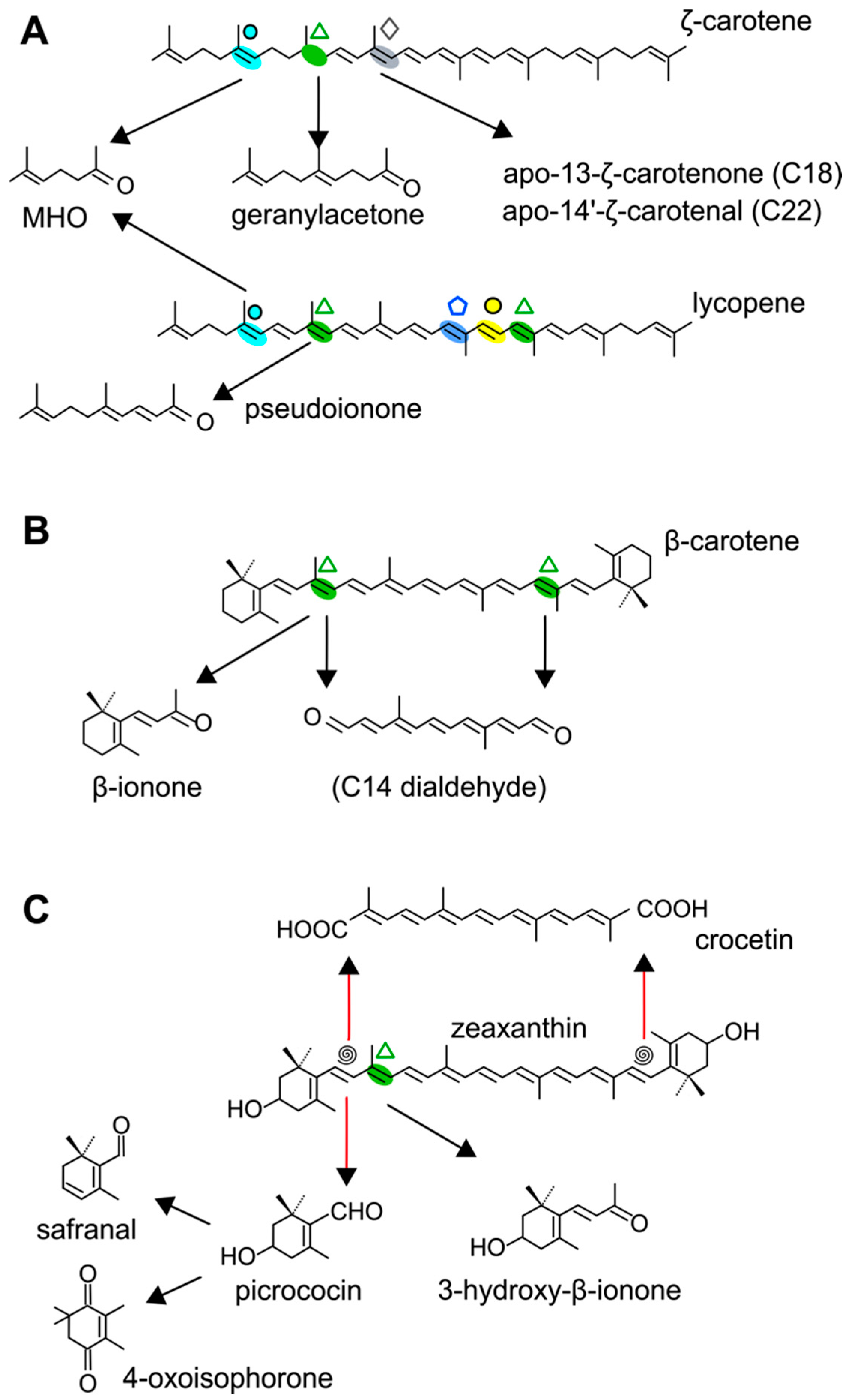
  101. Bouvier, F.; Suire, C.; Mutterer, J.; Camara, B. Oxidative remodeling of chromoplast carotenoids: Identification of the carotenoid dioxygenase CsCCD and CsZCD genes involved in Crocus secondary metabolite biogenesis. Plant Cell 2003, 15, 47–62. [Google Scholar] [CrossRef] [PubMed]
  102. Floss, D.S.; Walter, M.H. Role of carotenoid cleavage dioxygenase 1 (CCD1) in apocarotenoid biogenesis revisited. Plant Signal. Behav. 2009, 4, 172–175. [Google Scholar] [CrossRef] [PubMed]
  103. Fraser, P.D.; Bramley, P.M. The biosynthesis and nutritional uses of carotenoids. Prog. Lipid Res. 2004, 43, 228–265. [Google Scholar] [CrossRef] [PubMed]
  104. Foo, E.; Yoneyama, K.; Hugill, C.; Quittenden, L.J.; Reid, J.B. Strigolactones: Internal and external signals in plant symbioses? Plant Signal. Behav. 2013, 8. [Google Scholar] [CrossRef] [PubMed]
  105. Walter, M.H. Role of carotenoid metabolism in the arbuscular mycorrhizal symbiosis. In Molecular Microbial Ecology of the Rhizosphere; Bruijn, F.J.D., Ed.; Wiley-Blackwell: Hoboken, NJ, USA, 2013. [Google Scholar]
  106. López-Raez, J.A.; Fernández, I.; García, J.M.; Berrio, E.; Bonfante, P.; Walter, M.H.; Pozo, M.J. Differential spatio-temporal expression of carotenoid cleavage dioxygenases regulates apocarotenoid fluxes during AM symbiosis. Plant Sci. 2015, 230, 59–69. [Google Scholar] [CrossRef] [PubMed]
  107. González-Jorge, S.; Ha, S.H.; Magallanes-Lundback, M.; Gilliland, L.U.; Zhou, A.; Lipka, A.E.; Nguyen, Y.N.; Angelovici, R.; Lin, H.; Cepela, J.; et al. CAROTENOID CLEAVAGE DIOXYGENASE4 is a negative regulator of β-carotene content in Arabidopsis seeds. Plant Cell 2013, 25, 4812–4826. [Google Scholar] [CrossRef] [PubMed]
  108. Bouvier, F.; Isner, J.C.; Dogbo, O.; Camara, B. Oxidative tailoring of carotenoids: A prospect towards novel functions in plants. Trends Plant Sci. 2005, 10, 187–194. [Google Scholar] [CrossRef] [PubMed]
  109. Wei, S.; Hannoufa, A.; Soroka, J.; Xu, N.; Li, X.; Zebarjadi, A.; Gruber, M. Enhanced β-ionone emission in Arabidopsis over-expressing AtCCD1 reduces feeding damage in vivo by the crucifer flea beetle. Environ. Entomol. 2011, 40, 1622–1630. [Google Scholar] [CrossRef] [PubMed]
  110. Rodriguez-Avila, N.L.; Narvaez-Zapata, J.A.; Ramírez-Benítez, J.E.; Aguilar-Espinosa, M.L.; Rivera-Madrid, R. Identification and expression pattern of a new carotenoid cleavage dioxygenase gene member from Bixa orellana. J. Exp. Bot. 2011, 62, 5385–5395. [Google Scholar] [CrossRef] [PubMed]
  111. González-Verdejo, C.I.; Obrero, A.; Roman, B.; Gómez, P. Expression profile of carotenoid cleavage dioxygenase genes in summer squash (Cucurbita pepo l.). Plant Foods Hum. Nutr. 2015, 70, 200–206. [Google Scholar] [CrossRef] [PubMed]
  112. Imai, A.; Takahashi, S.; Nakayama, K.; Satoh, H. The promoter of the carotenoid cleavage dioxygenase 4a-5 gene of Chrysanthemum morifolium (CmCCD4a-5) drives petal-specific transcription of a conjugated gene in the developing flower. J. Plant Physiol. 2013, 170, 1295–1299. [Google Scholar] [CrossRef] [PubMed]
  113. Kim, S.H.; Jeong, J.C.; Park, S.; Bae, J.Y.; Ahn, M.J.; Lee, H.S.; Kwak, S.S. Down-regulation of sweetpotato lycopene β-cyclase gene enhances tolerance to abiotic stress in transgenic calli. Mol. Biol. Rep. 2014, 41, 8137–8148. [Google Scholar] [CrossRef] [PubMed]
  114. Sun, Z.; Hans, J.; Walter, M.H.; Matusova, R.; Beekwilder, J.; Verstappen, F.W.; Ming, Z.; van Echtelt, E.; Strack, D.; Bisseling, T.; et al. Cloning and characterisation of a maize carotenoid cleavage dioxygenase (ZmCCD1) and its involvement in the biosynthesis of apocarotenoids with various roles in mutualistic and parasitic interactions. Planta 2008, 228, 789–801. [Google Scholar] [CrossRef] [PubMed]
  115. Ahrazem, O.; Trapero, A.; Gómez, M.D.; Rubio-Moraga, A.; Gómez-Gómez, L. Genomic analysis and gene structure of the plant carotenoid dioxygenase 4 family: A deeper study in Crocus sativus and its allies. Genomics 2010, 96, 239–250. [Google Scholar] [CrossRef] [PubMed]
  116. Reddy, A.S.; Márquez, Y.; Kalyna, M.; Barta, A. Complexity of the alternative splicing landscape in plants. Plant Cell 2013, 25, 3657–3683. [Google Scholar] [CrossRef] [PubMed]
  117. Frusciante, S.; Diretto, G.; Bruno, M.; Ferrante, P.; Pietrella, M.; Prado-Cabrero, A.; Rubio-Moraga, A.; Beyer, P.; Gómez-Gómez, L.; Al-Babili, S.; et al. Novel carotenoid cleavage dioxygenase catalyzes the first dedicated step in saffron crocin biosynthesis. Proc. Natl. Acad. Sci. USA 2014, 111, 12246–12251. [Google Scholar] [CrossRef] [PubMed]
  118. Ahrazem, O.; Rubio-Moraga, A.; Berman, J.; Capell, T.; Christou, P.; Zhu, C.; Gómez-Gómez, L. The carotenoid cleavage dioxygenase CCD2 catalysing the synthesis of crocetin in spring crocuses and saffron is a plastidial enzyme. New Phytol. 2016, 209, 650–663. [Google Scholar] [CrossRef] [PubMed]
  119. Ahrazem, O.; Rubio-Moraga, A.; Nebauer, S.G.; Molina, R.V.; Gómez-Gómez, L. Saffron: Its phytochemistry, developmental processes, and biotechnological prospects. J. Agric. Food Chem. 2015, 63, 8751–8764. [Google Scholar] [CrossRef] [PubMed]
  120. Rubio-Moraga, A.; Ahrazem, O.; Rambla, J.L.; Granell, A.; Gómez-Gómez, L. Crocins with high levels of sugar conjugation contribute to the yellow colors of early-spring flowering crocus tepals. PLoS ONE 2013, 8, e71946. [Google Scholar] [CrossRef] [PubMed]
  121. Jüttner, F.; Watson, S.B.; von Elert, E.; Koster, O. β-cyclocitral, a grazer defence signal unique to the cyanobacterium Microcystis. J. Chem. Ecol. 2010, 36, 1387–1397. [Google Scholar] [CrossRef] [PubMed]
  122. Messing, S.A.; Gabelli, S.B.; Echeverria, I.; Vogel, J.T.; Guan, J.C.; Tan, B.C.; Klee, H.J.; McCarty, D.R.; Amzel, L.M. Structural insights into maize viviparous14, a key enzyme in the biosynthesis of the phytohormone abscisic acid. Plant Cell 2010, 22, 2970–2980. [Google Scholar] [CrossRef] [PubMed]
  123. Ahrazem, O.; Rubio-Moraga, A.; Argandoña, J.; Castillo-López, R.; Gómez-Gómez, L. Intron retention and rhythmic diel pattern regulation of carotenoid cleavage dioxygenase 2 during crocetin biosynthesis in saffron. Plant Mol. Biol. 2016, 91, 355–374. [Google Scholar] [CrossRef] [PubMed]
  124. Moraga, A.R.; Nohales, P.F.; Pérez, J.A.F.; Gómez-Gómez, L. Glucosylation of the saffron apocarotenoid crocetin by a glucosyltransferase isolated from Crocus sativus stigmas. Planta 2004, 219, 955–966. [Google Scholar] [CrossRef] [PubMed]
  125. Simkin, A.J.; Zhu, C.; Kuntz, M.; Sandmann, G. Light-dark regulation of carotenoid biosynthesis in pepper (Capsicum annuum) leaves. J. Plant Physiol. 2003, 160, 439–443. [Google Scholar] [CrossRef] [PubMed]
  126. Simkin, A.J.; Labouré, A.-M.; Kuntz, M.; Sandmann, G. Comparison of carotenoid content, gene expression and enzyme levels in tomato (Lycopersicon esculentum) leaves. Z. Naturforschung C 2003, 58, 371–380. [Google Scholar] [CrossRef]
  127. Kato, M.; Ikoma, Y.; Matsumoto, H.; Sugiura, M.; Hyodo, H.; Yano, M. Accumulation of carotenoids and expression of carotenoid biosynthetic genes during maturation in citrus fruit. Plant Physiol. 2004, 134, 824–837. [Google Scholar] [CrossRef] [PubMed]
  128. Zhang, L.; Ma, G.; Kato, M.; Yamawaki, K.; Takagi, T.; Kiriiwa, Y.; Ikoma, Y.; Matsumoto, H.; Yoshioka, T.; Nesumi, H. Regulation of carotenoid accumulation and the expression of carotenoid metabolic genes in citrus juice sacs in vitro. J. Exp. Bot. 2011, 63, 871–886. [Google Scholar] [CrossRef] [PubMed]
  129. Filichkin, S.A.; Mockler, T.C. Unproductive alternative splicing and nonsense mRNAs: A widespread phenomenon among plant circadian clock genes. Biol. Direct 2012, 7. [Google Scholar] [CrossRef] [PubMed]
  130. Rubio-Moraga, A.; Rambla, J.L.; Fernández-de-Carmen, A.; Trapero-Mozos, A.; Ahrazem, O.; Orzaez, D.; Granell, A.; Gómez-Gómez, L. New target carotenoids for CCD4 enzymes are revealed with the characterization of a novel stress-induced carotenoid cleavage dioxygenase gene from Crocus sativus. Plant Mol. Biol. 2014, 85, 555–569. [Google Scholar] [CrossRef] [PubMed]
  131. Ma, G.; Zhang, L.; Matsuta, A.; Matsutani, K.; Yamawaki, K.; Yahata, M.; Wahyudi, A.; Motohashi, R.; Kato, M. Enzymatic formation of β-citraurin from β-cryptoxanthin and zeaxanthin by carotenoid cleavage dioxygenase4 in the flavedo of citrus fruit. Plant Physiol. 2013, 163, 682–695. [Google Scholar] [CrossRef] [PubMed]
  132. Ohmiya, A. Carotenoid cleavage dioxygenases and their apocarotenoid products in plants. Plant Biotechnol. 2009, 26, 351–358. [Google Scholar] [CrossRef]
  133. Rodrigo, M.J.; Alquézar, B.; Alós, E.; Medina, V.; Carmona, L.; Bruno, M.; Al-Babili, S.; Zacarías, L. A novel carotenoid cleavage activity involved in the biosynthesis of citrus fruit-specific apocarotenoid pigments. J. Exp. Bot. 2013, 64, 4461–4478. [Google Scholar] [CrossRef] [PubMed]
  134. Lätari, K.; Wüst, F.; Hübner, M.; Schaub, P.; Kim, G.B.; Matsubara, S.; Beyer, P.; Welsch, R. Tissue-specific apocarotenoid glycosylation contributes to carotenoid homeostasis in Arabidopsis leaves. Plant Physiol. 2015, 168, 1550–1562. [Google Scholar] [CrossRef] [PubMed]
  135. Avendaño-Vázquez, A.-O.; Cordoba, E.; Llamas, E.; San Román, C.; Nisar, N.; De la Torre, S.; Ramos-Vega, M.; de la Luz Gutiérrez-Nava, M.; Cazzonelli, C.I.; Pogson, B.J. An uncharacterized apocarotenoid-derived signal generated in ζ-carotene desaturase mutants regulates leaf development and the expression of chloroplast and nuclear genes in Arabidopsis. Plant Cell 2014, 26, 2524–2537. [Google Scholar] [CrossRef] [PubMed]
  136. Ohmiya, A.; Kishimoto, S.; Aida, R.; Yoshioka, S.; Sumitomo, K. Carotenoid cleavage dioxygenase (CmCCD4a) contributes to white color formation in chrysanthemum petals. Plant Physiol. 2006, 142, 1193–1201. [Google Scholar] [CrossRef] [PubMed]
  137. Ytterberg, A.J.; Peltier, J.-B.; van Wijk, K.J. Protein profiling of plastoglobules in chloroplasts and chromoplasts. A surprising site for differential accumulation of metabolic enzymes. Plant Physiol. 2006, 140, 984–997. [Google Scholar] [CrossRef] [PubMed]
  138. Brehelin, C.; Kessler, F.; van Wijk, K.J. Plastoglobules: Versatile lipoprotein particles in plastids. Trends Plant Sci. 2007, 12, 260–266. [Google Scholar] [CrossRef] [PubMed]
  139. Vidi, P.A.; Kessler, F.; Brehelin, C. Plastoglobules: A new address for targeting recombinant proteins in the chloroplast. BMC Biotechnol. 2007, 7. [Google Scholar] [CrossRef] [PubMed]
  140. Lundquist, P.K.; Poliakov, A.; Bhuiyan, N.H.; Zybailov, B.; Sun, Q.; van Wijk, K.J. The functional network of the Arabidopsis plastoglobule proteome based on quantitative proteomics and genome-wide coexpression analysis. Plant Physiol. 2012, 158, 1172–1192. [Google Scholar] [CrossRef] [PubMed]
  141. Besagni, C.; Kessler, F. A mechanism implicating plastoglobules in thylakoid disassembly during senescence and nitrogen starvation. Planta 2013, 237, 463–470. [Google Scholar] [CrossRef] [PubMed]
  142. Rottet, S.; Besagni, C.; Kessler, F. The role of plastoglobules in thylakoid lipid remodeling during plant development. Biochim. Biophys. Acta 2015, 1847, 889–899. [Google Scholar] [CrossRef] [PubMed]
  143. Lundquist, P.K.; Poliakov, A.; Giacomelli, L.; Friso, G.; Appel, M.; McQuinn, R.P.; Krasnoff, S.B.; Rowland, E.; Ponnala, L.; Sun, Q.; et al. Loss of plastoglobule kinases ABC1K1 and ABD1K3 causes conditional degreening, modified prenyl-lipids, and recruitment of the jasmonic acid pathway. Plant Cell 2013, 25, 1818–1839. [Google Scholar] [CrossRef] [PubMed]
  144. Vallabhaneni, R.; Bradbury, L.M.; Wurtzel, E.T. The carotenoid dioxygenase gene family in maize, sorghum, and rice. Arch. Biochem. Biophys. 2010, 504, 104–111. [Google Scholar] [CrossRef] [PubMed]
  145. Yoshioka, Y.; Ono, M.; Osaki, M.; Konishi, I.; Sakaguchi, S. Differential effects of inhibition of bone morphogenic protein (BMP) signalling on T-cell activation and differentiation. Eur. J. Immunol. 2012, 42, 749–759. [Google Scholar] [CrossRef] [PubMed]
  146. Zheng, X.; Xie, Z.; Zhu, K.; Xu, Q.; Deng, X.; Pan, Z. Isolation and characterization of carotenoid cleavage dioxygenase 4 genes from different citrus species. Mol. Genet. Genom. 2015, 290, 1589–1603. [Google Scholar] [CrossRef] [PubMed]
  147. Moraga, A.R.; Rambla, J.L.; Ahrazem, O.; Granell, A.; Gómez-Gómez, L. Metabolite and target transcript analyses during Crocus sativus stigma development. Phytochemistry 2009, 70, 1009–1016. [Google Scholar] [CrossRef] [PubMed]
  148. Wei, Y.; Wan, H.; Wu, Z.; Wang, R.; Ruan, M.; Ye, Q.; Li, Z.; Zhou, G.; Yao, Z.; Yang, Y. A comprehensive analysis of carotenoid cleavage dioxygenases genes in Solanum lycopersicum. Plant Mol. Biol. Rep. 2015, 34, 512–543. [Google Scholar] [CrossRef]
  149. Priya, R.; Siva, R. Phylogenetic analysis and evolutionary studies of plant carotenoid cleavage dioxygenase gene. Gene 2014, 548, 223–233. [Google Scholar] [CrossRef] [PubMed]
  150. Campbell, R.; Ducreux, L.J.; Morris, W.L.; Morris, J.A.; Suttle, J.C.; Ramsay, G.; Bryan, G.J.; Hedley, P.E.; Taylor, M.A. The metabolic and developmental roles of carotenoid cleavage dioxygenase 4 from potato. Plant Physiol. 2010, 154, 656–664. [Google Scholar] [CrossRef] [PubMed]
  151. Butchko, R.A.E.; Brown, D.W.; Busman, M.; Tudzynski, B.; Wiemann, P. Lae1 regulates expression of multiple secondary metabolite gene clusters in Fusarium verticillioides. Fungal Genet. Biol. 2012, 49, 602–612. [Google Scholar] [CrossRef] [PubMed]
  152. Zhang, B.; Liu, C.; Wang, Y.; Yao, X.; Wang, F.; Wu, J.; King, G.J.; Liu, K. Disruption of a carotenoid cleavage dioxygenase 4 gene converts flower color from white to yellow in Brassica species. New Phytol. 2015, 206, 1513–1526. [Google Scholar] [CrossRef] [PubMed]
  153. Bruno, M.; Beyer, P.; Al-Babili, S. The potato carotenoid cleavage dioxygenase 4 catalyzes a single cleavage of β-ionone ring-containing carotenes and non-epoxidated xanthophylls. Arch. Biochem. Biophys. 2015, 572, 126–133. [Google Scholar] [CrossRef] [PubMed]
  154. Soares, V.L.; Rodrigues, S.M.; de Oliveira, T.M.; de Queiroz, T.O.; Lima, L.S.; Hora-Júnior, B.T.; Gramacho, K.P.; Micheli, F.; Cascardo, J.C.; Otoni, W.C. Unraveling new genes associated with seed development and metabolism in Bixa orellana l. By expressed sequence tag (EST) analysis. Mol. Biol. Rep. 2011, 38, 1329–1340. [Google Scholar] [CrossRef] [PubMed]
  155. Tuan, P.A.; Park, S.U. Molecular cloning and characterization of cDNAs encoding carotenoid cleavage dioxygenase in bitter melon (Momordica charantia). J. Plant Physiol. 2013, 170, 115–120. [Google Scholar] [CrossRef] [PubMed]
  156. Hai, N.T.L.; Masuda, J.-I.; Miyajima, I.; Thien, N.Q.; Mojtahedi, N.; Hiramatsu, M.; Kim, J.-H.; Okubo, H. Involvement of carotenoid cleavage dioxygenase 4 gene in tepal color change in Lilium brownii var. colchesteri. J. Jpn. Soc. Hort. Sci. 2012, 81, 366–373. [Google Scholar] [CrossRef]
  157. Obrero, A.; González-Verdejo, C.I.; Die, J.V.; Gómez, P.; Del Río-Celestino, M.; Román, B.N. Carotenogenic gene expression and carotenoid accumulation in three varieties of Cucurbita pepo during fruit development. J. Agric. Food Chem. 2013, 61, 6393–6403. [Google Scholar] [CrossRef] [PubMed]
  158. Brandi, F.; Bar, E.; Mourgues, F.; Horváth, G.; Turcsi, E.; Giuliano, G.; Liverani, A.; Tartarini, S.; Lewinsohn, E.; Rosati, C. Study of ‘Redhaven’ peach and its white-fleshed mutant suggests a key role of CCD4 carotenoid dioxygenase in carotenoid and norisoprenoid volatile metabolism. BMC Plant Biol. 2011, 11, 24. [Google Scholar] [CrossRef] [PubMed]
  159. Adami, M.; De Franceschi, P.; Brandi, F.; Liverani, A.; Giovannini, D.; Rosati, C.; Dondini, L.; Tartarini, S. Identifying a carotenoid cleavage dioxygenase (ccd4) gene controlling yellow/white fruit flesh color of peach. Plant Mol. Biol. Rep. 2013, 31, 1166–1175. [Google Scholar] [CrossRef]
  160. Falchi, R.; Vendramin, E.; Zanon, L.; Scalabrin, S.; Cipriani, G.; Verde, I.; Vizzotto, G.; Morgante, M. Three distinct mutational mechanisms acting on a single gene underpin the origin of yellow flesh in peach. Plant J. 2013, 76, 175–187. [Google Scholar] [CrossRef] [PubMed]
  161. Fukamatsu, Y.; Tamura, T.; Hihara, S.; Oda, K. Mutations in the CCD4 carotenoid cleavage dioxygenase gene of yellow-flesh peaches. Biosci. Biotechnol. Biochem. 2013, 77, 2514–1526. [Google Scholar] [CrossRef] [PubMed]
  162. Ma, J.; Li, J.; Zhao, J.; Zhou, H.; Ren, F.; Wang, L.; Gu, C.; Liao, L.; Han, Y. Inactivation of a gene encoding carotenoid cleavage dioxygenase (ccd4) leads to carotenoid-based yellow coloration of fruit flesh and leaf midvein in peach. Plant Mol. Biol. Rep. 2014, 32, 246–257. [Google Scholar] [CrossRef]
  163. Liu, Y.; Zeng, S.; Sun, W.; Wu, M.; Hu, W.; Shen, X.; Wang, Y. Comparative analysis of carotenoid accumulation in two goji (Lycium barbarum L. and L. ruthenicum murr.) fruits. BMC Plant Biol. 2014, 14, 269. [Google Scholar] [CrossRef] [PubMed]
  164. Ramel, F.; Mialoundama, A.S.; Havaux, M. Nonenzymic carotenoid oxidation and photooxidative stress signalling in plants. J. Exp. Bot. 2013, 64, 799–805. [Google Scholar] [CrossRef] [PubMed]
  165. Sorefan, K.; Booker, J.; Haurogne, K.; Goussot, M.; Bainbridge, K.; Foo, E.; Chatfield, S.; Ward, S.; Beveridge, C.; Rameau, C.; et al. MAX4 and RMS1 are orthologous dioxygenase-like genes that regulate shoot branching in Arabidopsis and pea. Genes Dev. 2003, 17, 1469–1474. [Google Scholar] [CrossRef] [PubMed]
  166. Booker, J.; Auldridge, M.; Wills, S.; McCarty, D.; Klee, H.; Leyser, O. MAX3/CCD7 is a carotenoid cleavage dioxygenase required for the synthesis of a novel plant signaling molecule. Curr. Biol. 2004, 14, 1232–1238. [Google Scholar] [CrossRef] [PubMed]
  167. Schwartz, S.H.; Qin, X.; Loewen, M.C. The biochemical characterization of two carotenoid cleavage enzymes from Arabidopsis indicates that a carotenoid-derived compound inhibits lateral branching. J. Biol. Chem. 2004, 279, 46940–46945. [Google Scholar] [CrossRef] [PubMed]
  168. Gómez-Roldán, V.; Fermas, S.; Brewer, P.B.; Puech-Pages, V.; Dun, E.A.; Pillot, J.P.; Letisse, F.; Matusova, R.; Danoun, S.; Portais, J.C.; et al. Strigolactone inhibition of shoot branching. Nature 2008, 455, 189–194. [Google Scholar] [CrossRef] [PubMed]
  169. Umehara, M.; Hanada, A.; Yoshida, S.; Akiyama, K.; Arite, T.; Takeda-Kamiya, N.; Magome, H.; Kamiya, Y.; Shirasu, K.; Yoneyama, K.; et al. Inhibition of shoot branching by new terpenoid plant hormones. Nature 2008, 455, 195–200. [Google Scholar] [CrossRef] [PubMed]
  170. Vogel, J.T.; Walter, M.H.; Giavalisco, P.; Lytovchenko, A.; Kohlen, W.; Charnikhova, T.; Simkin, A.J.; Goulet, C.; Strack, D.; Bouwmeester, H.J.; et al. Slccd7 controls strigolactone biosynthesis, shoot branching and mycorrhiza-induced apocarotenoid formation in tomato. Plant J. 2010, 61, 300–311. [Google Scholar] [CrossRef] [PubMed]
  171. Koltai, H.; LekKala, S.P.; Bhattacharya, C.; Mayzlish-Gati, E.; Resnick, N.; Wininger, S.; Dor, E.; Yoneyama, K.; Hershenhorn, J.; Joel, D.M.; et al. A tomato strigolactone-impaired mutant displays aberrant shoot morphology and plant interactions. J. Exp. Bot. 2010, 61, 1739–1749. [Google Scholar] [CrossRef] [PubMed]
  172. Bouwmeester, H.J.; Roux, C.; López-Raez, J.A.; Becard, G. Rhizosphere communication of plants, parasitic plants and am fungi. Trends Plant Sci. 2007, 12, 224–230. [Google Scholar] [CrossRef] [PubMed]
  173. Akiyama, K.; Matsuzaki, K.; Hayashi, H. Plant sesquiterpenes induce hyphal branching in arbuscular mycorrhizal fungi. Nature 2005, 435, 824–827. [Google Scholar] [CrossRef] [PubMed]
  174. Zhou, F.; Lin, Q.; Zhu, L.; Ren, Y.; Zhou, K.; Shabek, N.; Wu, F.; Mao, H.; Dong, W.; Gan, L.; et al. D14-SCFD3-dependent degradation of D53 regulates strigolactone signalling. Nature 2013, 504, 406–410. [Google Scholar] [CrossRef] [PubMed]
  175. Waldie, T.; McCulloch, H.; Leyser, O. Strigolactones and the control of plant development: Lessons from shoot branching. Plant J. 2014, 79, 607–622. [Google Scholar] [CrossRef] [PubMed]
  176. Cardoso, C.; Ruyter-Spira, C.; Bouwmeester, H.J. Strigolactones and root infestation by plant-parasitic Striga, Orobanche and Phelipanche spp. Plant Sci. 2011, 180, 414–420. [Google Scholar] [CrossRef] [PubMed]
  177. Cook, C.E.; Whichard, L.P.; Turner, B.; Wall, M.E.; Egley, G.H. Germination of witchweed (Striga lutea lour.): Isolation and properties of a potent stimulant. Science 1966, 154, 1189–1190. [Google Scholar] [CrossRef] [PubMed]
  178. Xie, X.; Yoneyama, K. The strigolactone story. Annu. Rev. Phytopathol. 2010, 48, 93–117. [Google Scholar] [CrossRef] [PubMed]
  179. Yoshida, S.; Shirasu, K. Plants that attack plants: Molecular elucidation of plant parasitism. Curr. Opin. Plant Biol. 2012, 15, 708–713. [Google Scholar] [CrossRef] [PubMed]
  180. Jamil, M.; Charnikhova, T.; Verstappen, F.; Bouwmeester, H. Carotenoid inhibitors reduce strigolactone production and Striga hermonthica infection in rice. Arch. Biochem. Biophys. 2010, 504, 123–131. [Google Scholar] [CrossRef] [PubMed]
  181. Arite, T.; Umehara, M.; Ishikawa, S.; Hanada, A.; Maekawa, M.; Yamaguchi, S.; Kyozuka, J. D14, a strigolactone-insensitive mutant of rice, shows an accelerated outgrowth of tillers. Plant Cell Physiol. 2009, 50, 1416–1424. [Google Scholar] [CrossRef] [PubMed]
  182. Bainbridge, K.; Sorefan, K.; Ward, S.; Leyser, O. Hormonally controlled expression of the Arabidopsis MAX4 shoot branching regulatory gene. Plant J. 2005, 44, 569–580. [Google Scholar] [CrossRef] [PubMed]
  183. Ruyter-Spira, C.; Kohlen, W.; Charnikhova, T.; van Zeijl, A.; van Bezouwen, L.; de Ruijter, N.; Cardoso, C.; Lopez-Raez, J.A.; Matusova, R.; Bours, R.; et al. Physiological effects of the synthetic strigolactone analog GR24 on root system architecture in Arabidopsis: Another belowground role for strigolactones? Plant Physiol. 2011, 155, 721–734. [Google Scholar] [CrossRef] [PubMed]
  184. Snowden, K.C.; Simkin, A.J.; Janssen, B.J.; Templeton, K.R.; Loucas, H.M.; Simons, J.L.; Karunairetnam, S.; Gleave, A.P.; Clark, D.G.; Klee, H.J. The decreased apical dominance1/petunia hybrida carotenoid cleavage dioxygenase8 gene affects branch production and plays a role in leaf senescence, root growth, and flower development. Plant Cell 2005, 17, 746–759. [Google Scholar] [CrossRef] [PubMed]
  185. Foo, E.; Bullier, E.; Goussot, M.; Foucher, F.; Rameau, C.; Beveridge, C.A. The branching gene ramosus1 mediates interactions among two novel signals and auxin in pea. Plant Cell 2005, 17, 464–474. [Google Scholar] [CrossRef] [PubMed]
  186. Ledger, S.E.; Janssen, B.J.; Karunairetnam, S.; Wang, T.; Snowden, K.C. Modified carotenoid cleavage dioxygenase8 expression correlates with altered branching in kiwifruit (Actinidia chinensis). New Phytol. 2010, 188, 803–813. [Google Scholar] [CrossRef] [PubMed]
  187. Kohlen, W.; Charnikhova, T.; Lammers, M.; Pollina, T.; Toth, P.; Haider, I.; Pozo, M.J.; de Maagd, R.A.; Ruyter-Spira, C.; Bouwmeester, H.J.; et al. The tomato CAROTENOID CLEAVAGE DIOXYGENASE8 (SlCCD8) regulates rhizosphere signaling, plant architecture and affects reproductive development through strigolactone biosynthesis. New Phytol. 2012, 196, 535–547. [Google Scholar] [CrossRef] [PubMed]
  188. Liang, J.; Zhao, L.; Challis, R.; Leyser, O. Strigolactone regulation of shoot branching in chrysanthemum (Dendranthema grandiflorum). J. Exp. Bot. 2010, 61, 3069–3078. [Google Scholar] [CrossRef] [PubMed]
  189. Djennane, S.; Hibrand-Saint Oyant, L.; Kawamura, K.; Lalanne, D.; Laffaire, M.; Thouroude, T.; Chalain, S.; Sakr, S.; Boumaza, R.; Foucher, F.; et al. Impacts of light and temperature on shoot branching gradient and expression of strigolactone synthesis and signalling genes in rose. Plant Cell Environ. 2013, 37, 742–757. [Google Scholar] [CrossRef] [PubMed] [Green Version]
  190. Liang, Y.S.; Jeon, Y.A.; Lim, S.H.; Kim, J.K.; Lee, J.Y.; Kim, Y.M.; Lee, Y.H.; Ha, S.H. Vascular-specific activity of the Arabidopsis carotenoid cleavage dioxygenase 7 gene promoter. Plant Cell Rep. 2011, 30, 973–980. [Google Scholar] [CrossRef] [PubMed]
  191. Zou, J.; Zhang, S.; Zhang, W.; Li, G.; Chen, Z.; Zhai, W.; Zhao, X.; Pan, X.; Xie, Q.; Zhu, L. The rice high-tillering DWARF1 encoding an ortholog of Arabidopsis MAX3 is required for negative regulation of the outgrowth of axillary buds. Plant J. 2006, 48, 687–698. [Google Scholar] [CrossRef] [PubMed]
  192. Drummond, R.S.; Martinez-Sanchez, N.M.; Janssen, B.J.; Templeton, K.R.; Simons, J.L.; Quinn, B.D.; Karunairetnam, S.; Snowden, K.C. Petunia hybrida carotenoid cleavage dioxygenase7 is involved in the production of negative and positive branching signals in petunia. Plant Physiol. 2009, 151, 1867–1877. [Google Scholar] [CrossRef] [PubMed]
  193. Cheng, X.; Ruyter-Spira, C.; Bouwmeester, H. The interaction between strigolactones and other plant hormones in the regulation of plant development. Front. Plant Sci. 2013, 4. [Google Scholar] [CrossRef] [PubMed]
  194. Kapulnik, Y.; Delaux, P.M.; Resnick, N.; Mayzlish-Gati, E.; Wininger, S.; Bhattacharya, C.; Sejalon-Delmas, N.; Combier, J.P.; Becard, G.; Belausov, E.; et al. Strigolactones affect lateral root formation and root-hair elongation in Arabidopsis. Planta 2011, 233, 209–216. [Google Scholar] [CrossRef] [PubMed]
  195. Scaffidi, A.; Waters, M.T.; Ghisalberti, E.L.; Dixon, K.W.; Flematti, G.R.; Smith, S.M. Carlactone-independent seedling morphogenesis in Arabidopsis. Plant J. 2013, 76, 1–9. [Google Scholar] [CrossRef] [PubMed]
  196. Waters, M.T.; Nelson, D.C.; Scaffidi, A.; Flematti, G.R.; Sun, Y.K.; Dixon, K.W.; Smith, S.M. Specialisation within the DWARF14 protein family confers distinct responses to karrikins and strigolactones in Arabidopsis. Development 2012, 139, 1285–1295. [Google Scholar] [CrossRef] [PubMed]
  197. Liu, J.; Novero, M.; Charnikhova, T.; Ferrandino, A.; Schubert, A.; Ruyter-Spira, C.; Bonfante, P.; Lovisolo, C.; Bouwmeester, H.J.; Cardinale, F. CAROTENOID CLEAVAGE DIOXYGENASE 7 modulates plant growth, reproduction, senescence, and determinate nodulation in the model legume Lotus japonicus. J. Exp. Bot. 2013, 64, 1967–1981. [Google Scholar] [CrossRef] [PubMed]
  198. Yan, H.; Saika, H.; Maekawa, M.; Takamure, I.; Tsutsumi, N.; Kyozuka, J.; Nakazono, M. Rice tillering dwarf mutant dwarf3 has increased leaf longevity during darkness-induced senescence or hydrogen peroxide-induced cell death. Genes Genet. Syst. 2007, 82, 361–366. [Google Scholar] [CrossRef] [PubMed]
  199. Johnson, X.; Brcich, T.; Dun, E.A.; Goussot, M.; Haurogne, K.; Beveridge, C.A.; Rameau, C. Branching genes are conserved across species. Genes controlling a novel signal in pea are coregulated by other long-distance signals. Plant Physiol. 2006, 142, 1014–1026. [Google Scholar] [CrossRef] [PubMed]
  200. Foo, E. Auxin influences strigolactones in pea mycorrhizal symbiosis. J. Plant Physiol. 2013, 170, 523–528. [Google Scholar] [CrossRef] [PubMed]
  201. Rubio-Moraga, A.; Ahrazem, O.; Pérez-Clemente, R.M.; Gómez-Cadenas, A.; Yoneyama, K.; López-Raez, J.A.; Molina, R.V.; Gómez-Gómez, L. Apical dominance in saffron and the involvement of the branching enzymes CDD7 and CCD8 in the control of bud sprouting. BMC Plant Biol. 2014, 14, 171. [Google Scholar] [CrossRef] [PubMed]
  202. Liaaen-Jensen, S. Carotenoids in chemosystematics. In Biosynthesis and Metabolism; Britton, G., Liaaen-Jensen, S., Pfander, H., Eds.; Birkhäuser: Basel, Switzerland, 1998. [Google Scholar]
  203. Takaichi, S. Carotenoids in algae: Distributions, biosyntheses and functions. Mar. Drugs 2011, 9, 1101–1118. [Google Scholar] [CrossRef] [PubMed]
  204. Kreimer, G.; Marner, F.J.; Brohsonn, U.; Melkonian, M. Identification of 11-cis and all-trans-retinal in the photoreceptive organelle of a flagellate green alga. FEBS Lett. 1991, 293, 49–52. [Google Scholar] [CrossRef]
  205. Derguini, F.; Mazur, P.; Nakanishi, K.; Starace, D.M.; Saranak, J.; Foster, K.W. All-trans-retinal is the chromophore bound to the photoreceptor of the alga Chlamydomonas reinhardtii. Photochem. Photobiol. 1991, 54, 1017–1021. [Google Scholar] [CrossRef] [PubMed]
  206. Sineshchekov, O.A.; Govorunova, E.G.; Jung, K.H.; Zauner, S.; Maier, U.G.; Spudich, J.L. Rhodopsin-mediated photoreception in cryptophyte flagellates. Biophys. J. 2005, 89, 4310–4319. [Google Scholar] [CrossRef] [PubMed]
  207. Sineshchekov, O.A.; Jung, K.H.; Spudich, J.L. Two rhodopsins mediate phototaxis to low- and high-intensity light in Chlamydomonas reinhardtii. Proc. Natl. Acad. Sci. USA 2002, 99, 8689–8694. [Google Scholar] [CrossRef] [PubMed]
  208. Schmidt, M.; Gessner, G.; Luff, M.; Heiland, I.; Wagner, V.; Kaminski, M.; Geimer, S.; Eitzinger, N.; Reissenweber, T.; Voytsekh, O.; et al. Proteomic analysis of the eyespot of Chlamydomonas reinhardtii provides novel insights into its components and tactic movements. Plant Cell 2006, 18, 1908–1930. [Google Scholar] [CrossRef] [PubMed]
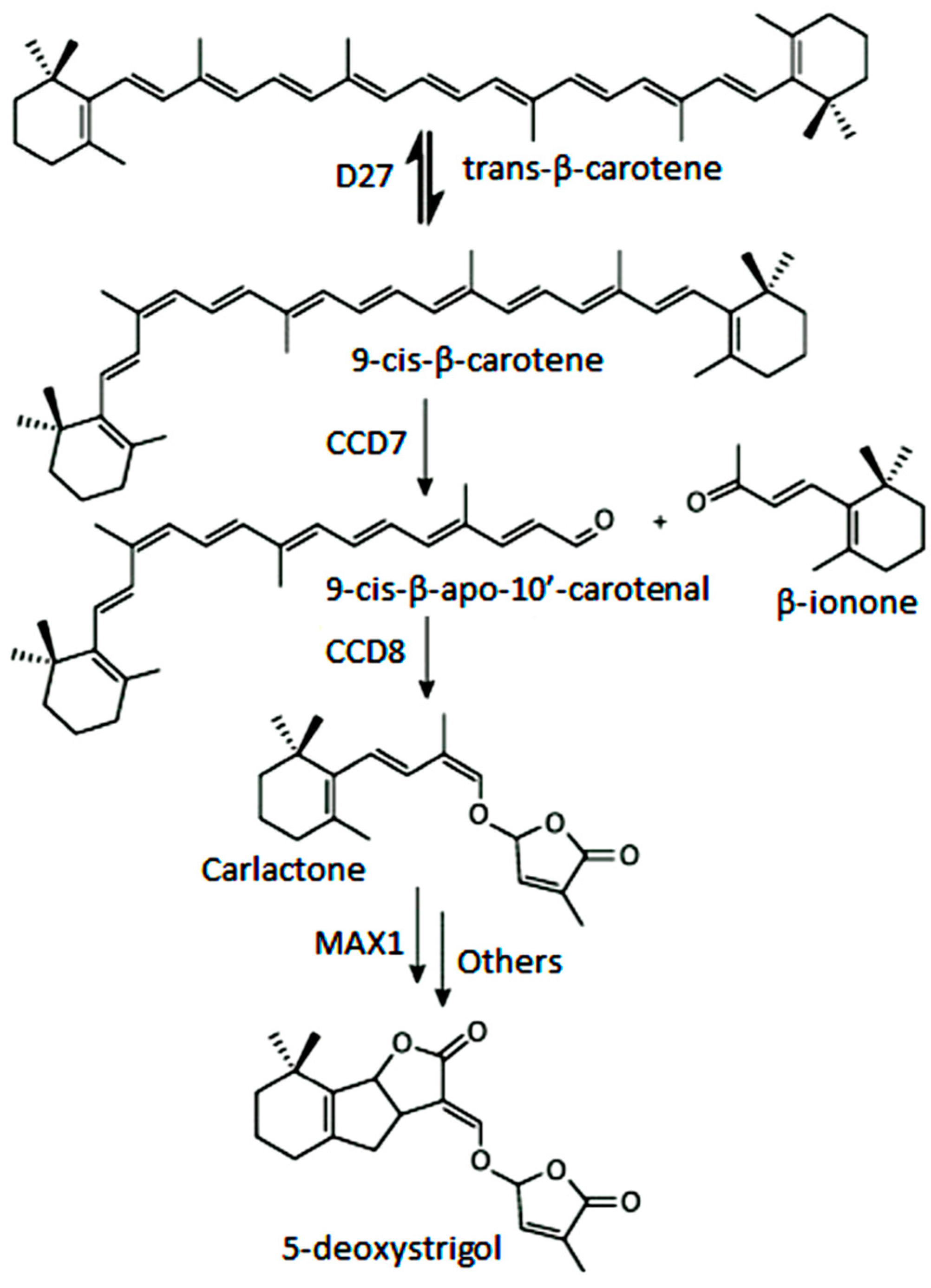
  209. Al-Babili, S.; Bouwmeester, H.J. Strigolactones, a novel carotenoid-derived plant hormone. Annu. Rev. Plant Biol. 2015, 66, 161–186. [Google Scholar] [CrossRef] [PubMed]
  210. Hu, G.P.; Yuan, J.; Sun, L.; She, Z.G.; Wu, J.H.; Lan, X.J.; Zhu, X.; Lin, Y.C.; Chen, S.P. Statistical research on marine natural products based on data obtained between 1985 and 2008. Mar. Drugs 2011, 9, 514–525. [Google Scholar] [CrossRef] [PubMed]
  211. Delaux, P.M.; Xie, X.; Timme, R.E.; Puech-Pages, V.; Dunand, C.; Lecompte, E.; Delwiche, C.F.; Yoneyama, K.; Becard, G.; Sejalon-Delmas, N. Origin of strigolactones in the green lineage. New Phytol. 2012, 195, 857–871. [Google Scholar] [CrossRef] [PubMed]
  212. Yamamoto, M.; Baldermann, S.; Yoshikawa, K.; Fujita, A.; Mase, N.; Watanabe, N. Determination of volatile compounds in four commercial samples of Japanese green algae using solid phase microextraction gas chromatography mass spectrometry. Sci. World J. 2014, 2014, 289780. [Google Scholar] [CrossRef] [PubMed]
  213. Baldermann, S.; Yamamoto, M.; Yang, Z.; Kawahashi, T.; Kuwano, K.; Watanabe, N. C13-apocarotenoids: More than flavor compounds? In Carotenoid Cleavage Products; Winterhalter, P., Ebeler, S.E., Eds.; ACS Symposium Series: Washington, DC, USA, 2013. [Google Scholar]
  214. Ikawa, M.; Sasner, J.J.; Haney, J.F. Activity of cyanobacterial and algal odor compounds found in lake waters on green alga Chlorella pyrenoidosa growth. Hydrobiologia 2001, 443, 19–22. [Google Scholar] [CrossRef]
  215. Baldermann, S.; Mulyadi, A.N.; Yang, Z.; Murata, A.; Fleischmann, P.; Winterhalter, P.; Knight, M.; Finn, T.M.; Watanabe, N. Application of centrifugal precipitation chromatography and high-speed counter-current chromatography equipped with a spiral tubing support rotor for the isolation and partial characterization of carotenoid cleavage-like enzymes in Enteromorpha compressa (L.) nees. J. Sep. Sci. 2011, 34, 2759–2764. [Google Scholar] [PubMed]
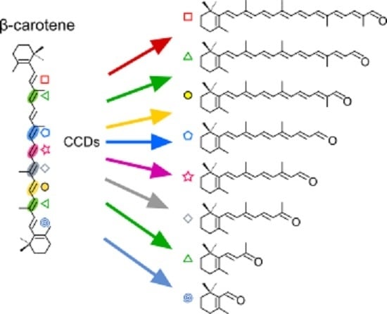
Figure 1. Tridimensional models of 12 carotenoid-cleavage dioxygenases from all the subfamilies included in this review. The VP14 (PBD: 2biwA) structure from maize has been used as a template. (A) Side view of CCDs with β-strands shown in yellow, α-helices in magenta, and loops in grey; (B) Top view rotated 90° towards the viewer from (A); (C) Lateral and top views of CCD2, CCD8, and ACO showing Fe2+ ion in green and histidines in blue. Accession numbers are: VP14: O24592.2, ACOX, P74334; AtCCD1, O65572; AtCCD7, AEC10494.1; AtCCD8, Q8VY26; AtCCD4: O49675; Cao-2, XP001727958.1; CarS, ADU04395.1; CarX, CAH70723.1; CsCCD2L, ALM23547.1; CcCCD4b1, XP006424046; AcaA, 77754.
Ijms 17 01781 g001aIjms 17 01781 g001b
Figure 2. Enzymatic reactions achieved by fungal CCDs. (A) Retinal production from β-carotene by the Cco1 (Ustilago maydis) and CarX (F. fujikuroi) CCDs; (B) β-apo-4′-carotenal production from torulene by the CarT (F. fujikuroi) and Cao-2 (N. crassa) CCDs; (C) β-apo-13-carotenone production from β-carotene by the sequential cleavage by the CarS and AcaA CCDs (P. blakesleeanus). Cleavage sites are shaded and indicated by an arrow.
Ijms 17 01781 g002
Figure 3. Activity of plant CCD1 and CCD2 subfamilies, showing their divergence in substrate specificity and cleavage sites on the following substrates: (A) ζ-carotene and lycopene; (B) β-carotene; and (C) zeaxantine. Symbols indicate double bonds where oxygenases cleave. Ijms 17 01781 i048: C5–C6; Ijms 17 01781 i042: C7–C8; Ijms 17 01781 i043: C9–C10 and C9′–C10′; Ijms 17 01781 i044: C13–C14; Ijms 17 01781 i045: C13′–C14′; Ijms 17 01781 i046: C11′–C12′.
Ijms 17 01781 g003
Figure 4. Biosynthetic pathway of strigolactones (SLs) from β-carotene.
Ijms 17 01781 g004
Table 1. Enzymatic activity of bacterial apo-carotenoid cleavage oxygenases Diox1 and NosAco showing products to different substrates.
SubstratesDiox1 Synechocystis sp. PCC 6803NosAco Nostoc sp. PCC 7120
β-apo-4′-carotenal
Ijms 17 01781 i001
RetinalNot detected
β-apo-8′-carotenal
Ijms 17 01781 i002
RetinalRetinal
β-apo-10′-carotenal
Ijms 17 01781 i003
RetinalRetinal
β-apo-12′-carotenal
Ijms 17 01781 i004
RetinalNot detected
(3R)-3-OH-β-apo-8′-carotenal
Ijms 17 01781 i005
3-OH-retinal3R-3-OH-retinal
(3R)-3-OH-β-apo-12′-carotenal
Ijms 17 01781 i006
3-OH-retinal3R-3-OH-retinal
Apo-8′-lycopenal
Ijms 17 01781 i007
Acycloretinal (slow)Acycloretinal
Apo-10′-lycopenal
Ijms 17 01781 i008
No informationAcycloretinal
β-apo-8′-carotenol
Ijms 17 01781 i009
RetinalRetinal
(3R)-3-OH-β-apo-8′-carotenol
Ijms 17 01781 i010
3-OH-retinal3-OH-retinal
Apo-8′-lycopenol
Ijms 17 01781 i011
No informationAcycloretinal
4-oxo-β-apo-8′-carotenal
Ijms 17 01781 i012
4-oxo-retinal (slow)Not tested
References[29][32]
Ijms 17 01781 i041: Enzyme cleaves at the C15–C15′ double bond, or equivalent positions.
Table 2. Enzimatic activity and substrate specificity of different bacterial carotenoid oxygenases.
Assayed SubstratesEnzymes and Products
β-carotene-oxygenase Microcystis PCC 7806NosCCD (NSC1) Nostoc sp. PCC 7120MtCCO Mycobacterium tuberculosisNSC3 Nostoc sp. PCC 7120NACOX1 NovosphingobiumSaCCO Sphingopyxis alaskensisPpCCO Plesiocystis pacifica
β-apo-8′-carotenal
Ijms 17 01781 i013
β-ionone ( Ijms 17 01781 i043) + Apo-8,10′-apocarotene-dial ( Ijms 17 01781 i043)
Other minority products ( Ijms 17 01781 i042, Ijms 17 01781 i043)
β-apo-13-carotenone ( Ijms 17 01781 i044)
Retinal ( Ijms 17 01781 i041)
β-apo-13-carotenone ( Ijms 17 01781 i045)
β-apo-14′-carotenal Transretinal ( Ijms 17 01781 i041)
β-apo-13-carotenone ( Ijms 17 01781 i044)Apo-12′-carotenal ( Ijms 17 01781 i046)
Apo-10′-carotenal ( Ijms 17 01781 i043)
β-apo-10′-carotenal
Ijms 17 01781 i014
β-ionone ( Ijms 17 01781 i043) + Apo-10,10′-apocarotene-dial ( Ijms 17 01781 i043)β-apo-13-carotenone ( Ijms 17 01781 i044)
Retinal ( Ijms 17 01781 i041)
3-OH-β-apo-8′-carotenal
Ijms 17 01781 i015
3-OH-β-ionone ( Ijms 17 01781 i043) + Apo-8,10′-apocarotene-dial ( Ijms 17 01781 i043)3-OH-β-apo-13-carotenone ( Ijms 17 01781 i044)
3-OH-retinal ( Ijms 17 01781 i041)
3-OH-β-apo-10′-carotenal
Ijms 17 01781 i016
3-OH-β-ionone ( Ijms 17 01781 i043) + Apo-10,10′-apocarotene-dial ( Ijms 17 01781 i043)3-OH-β-apo-13-carotenone ( Ijms 17 01781 i044)
3-OH-retinal ( Ijms 17 01781 i041)
γ-carotene
Ijms 17 01781 i017
β-ionone ( Ijms 17 01781 i043) + Apo-8,10′-apocarotene-dial ( Ijms 17 01781 i047) β-apo-13-carotenone ( Ijms 17 01781 i044)
β-carotene
Ijms 17 01781 i018
2 × β-cyclocitral ( Ijms 17 01781 i042, Ijms 17 01781 i047)
Crocetindial ( Ijms 17 01781 i042, Ijms 17 01781 i047)
Apo-10,10′-apocarotene-dial ( Ijms 17 01781 i043)β-apo-13-carotenone ( Ijms 17 01781 i044)
Retinal ( Ijms 17 01781 i041)
β-apo-14′-carotenal ( Ijms 17 01781 i045)
NoNoNo (in vitro)No
Zeaxanthin
Ijms 17 01781 i019
2 × hydroxi-β-cyclocitral ( Ijms 17 01781 i042, Ijms 17 01781 i047)
Crocetindial ( Ijms 17 01781 i042, Ijms 17 01781 i047)
3-OH-β-ionone ( Ijms 17 01781 i043)
Apo-10,10′-apocarotene-dial ( Ijms 17 01781 i043)
3-OH-β-apo-13-carotenone ( Ijms 17 01781 i044)
3-OH-β-apo-14′-carotenal ( Ijms 17 01781 i045)
3-OH-retinal ( Ijms 17 01781 i041)
3-OH-β-apo-11-carotenal?
NoNoNo (in vitro)Apo-13′-zeaxanthinone ( Ijms 17 01781 i045)
Apo-14′-zeaxanthinal ( Ijms 17 01781 i045)
Apo-12′-zeaxanthinal ( Ijms 17 01781 i046)
Lycopene
Ijms 17 01781 i020
NoNopoorlyYes (in vivo)
Unknown product
Lutein
Ijms 17 01781 i021
3-OH-β-apo-13-carotenone ( Ijms 17 01781 i044)
3-OH-β-apo-14′-carotenal ( Ijms 17 01781 i045)
3-OH-retinal ( Ijms 17 01781 i041)
3-OH-α-apo-15′-carotenal( Ijms 17 01781 i041)
No
Echinenone
Ijms 17 01781 i022
No4-oxo-β-ionone ( Ijms 17 01781 i043)
Apo-10,10′-apocarotene-dial ( Ijms 17 01781 i043)
β-ionone ( Ijms 17 01781 i043)
Canthaxantin
Ijms 17 01781 i023
2 x 4-oxo-β-ionone ( Ijms 17 01781 i043)
Apo-10,10′-apocarotene-dial ( Ijms 17 01781 i043)
Apo-13′-canta-xanthinone ( Ijms 17 01781 i045)
Apo-14′-cantaxanthinal ( Ijms 17 01781 i045)
Apo-12′-cantaxanthinal ( Ijms 17 01781 i046)
Astaxanthin
Ijms 17 01781 i024
3-OH-β-ionone ( Ijms 17 01781 i043)
3-OH, 4-oxo-β-inone ( Ijms 17 01781 i043)
Apo-10,10′-apocarotene-dial ( Ijms 17 01781 i043)
Apo-13′-astaxanthinone ( Ijms 17 01781 i045)
Apo-14′-astaxanthinal ( Ijms 17 01781 i045)
References[19][30,33][36][31][37][38][38]
Symbols indicate double bonds where oxygenases cleave. Ijms 17 01781 i042: C7–C8; Ijms 17 01781 i043: C9–C10; Ijms 17 01781 i044: C13–C14; Ijms 17 01781 i041: C15–C15′; Ijms 17 01781 i045: C13′–C14′; Ijms 17 01781 i046: C11′-C12′; Ijms 17 01781 i043: C9′–C10′; Ijms 17 01781 i047: C7′–C8′. When an enzyme cleaves at different positions, the predominant product is indicated in bold. Lack of bold means no product preference or information not available.
Table 3. Substrates tested and cleaved with a unique oxygenase.
SubstratesOxygenaseProducts
β-apo-4′-carotenal
Ijms 17 01781 i025
NACOX1β-apo-13-carotenone ( Ijms 17 01781 i044)
3,3′-dihydroxy-isorenieratene
Ijms 17 01781 i026
MtCCO3-OH-β-apo-13-carotenone ( Ijms 17 01781 i044)
3-OH-β-apo-15′-carotenal ( Ijms 17 01781 i041)
3-OH-β-apo-14′-carotenal ( Ijms 17 01781 i045)
Echinenone
Ijms 17 01781 i027
NosCCDApo-10,10′-apocarotenoid ( Ijms 17 01781 i043)
2 × C13 ( Ijms 17 01781 i043)
Nostoxanthin
Ijms 17 01781 i028
PpCCOApo-14′-nostoxanthinal ( Ijms 17 01781 i045)
Apo-12′-nostoxanthinal ( Ijms 17 01781 i046)
Hydroxylycopene
Ijms 17 01781 i029
PpCCOUnknown
Dihydroxylycopene
Ijms 17 01781 i030
PpCCOUnknown
Torulene
Ijms 17 01781 i031
NSC3Transretinal ( Ijms 17 01781 i041)
4,4′-diapotorulene
Ijms 17 01781 i032
NSC3Apo-14′-diapotorulenal ( Ijms 17 01781 i044)
4,4′-diapotorulene-4-al
Ijms 17 01781 i033
NSC3Apo-10′-diapotorulenal ( Ijms 17 01781 i045)
4,4′-diaponeurosporene
Ijms 17 01781 i034
NSC3Apo-14′-diaponeurosporenal ( Ijms 17 01781 i044)
4,4′-diaponeurosporen-4′-al
Ijms 17 01781 i035
NSC3Apo-14′-diaponeurosporenal ( Ijms 17 01781 i044)
Apo-10′-diaponeurosporenal ( Ijms 17 01781 i046)
4,4′-diaponeurosporen-4′-oic acid
Ijms 17 01781 i036
NSC3Apo-10′-diaponeurosporenal ( Ijms 17 01781 i046)
Myxol
Ijms 17 01781 i037
NosCCD3-OH-β-ionone ( Ijms 17 01781 i043)
Apo-8′-10-apocarotenedial ( Ijms 17 01781 i043)
Myxol-2′-fucoside
Ijms 17 01781 i038
NosCCD3-OH-β-ionone ( Ijms 17 01781 i043)
Apo-8′-10-apocarotenedial ( Ijms 17 01781 i043)
acetate adduct C10
4-ketomyxol-2′-fucoside
Ijms 17 01781 i039
NosCCD3-OH-4-oxo-β-ionone ( Ijms 17 01781 i043)
Apo-8′-10-apocarotenedial ( Ijms 17 01781 i043)
4-hydroxymyxol-2′-fucoside
Ijms 17 01781 i040
NosCCD3, 4-OH-β-ionone ( Ijms 17 01781 i043)
Apo-8′-10-apocarotenedial ( Ijms 17 01781 i043)
Symbols indicate double bonds where oxygenases cleave. Ijms 17 01781 i043: C9=C10; Ijms 17 01781 i044: C13=C14; Ijms 17 01781 i041: C15=C15′; Ijms 17 01781 i045: C13′=C14′; Ijms 17 01781 i046: C11′=C12′; Ijms 17 01781 i043: C9′=C10′; Ijms 17 01781 i047: C7′=C8′.
Table 4. Functional characterization of CCD4 enzymes.
SpeciesProteinAssay aParent CarotenoidCleavage PositionProduct DetectedReferences
Arabidopsis thalianaCCD4in plantaβ-carotene, Violaxanthinn.d.n.d.[107]
in plantaPhytofluene, ζ-carotenen.d.n.d.[135]
in planta (leaf)Epoxy-β-xanthophyllsC9–C10 or C9′–C10′C13-glycosids[134]
in planta (root)β-carotenen.d.long-chain free apocarotenals (C15 to C30)[134]
in vitroApo-β-caroten-8′-alC9–C10β-ionone[96]
Brassica napusCCD4_C3in plantaα-carotene, δ-caroteneC9–C10α-ionone[152]
Chrysantemum morifoliumCCD4ain vivo and in vitroβ-caroteneC9–C10 (C9′–C10′)β-ionone[96]
Citrus clementinaCCD4b1in vitroβ-carotene, β-cryptoxanthin, Zeaxanthin, Lutein, α-caroteneC7–C8 or C7′–C8′3-OH-apo-β-8-carotenal, apo-β-8-carotenal (C30);
β-cyclocitral and 3-OH-β-cyclocitral (C10)
[133]
Citrus unshiuCCD4in vitro and in vivoβ-cryptoxanthin, ZeaxanthinC7–C8 or C7′–C8′3-OH-apo-β-8-carotenal[131]
Crocus sativusCCD4a/bin vivoβ-carotene, ZeaxanthinC9–C10 (C9′–C10′)β-ionone, β-OH-ionone[89,147]
CCD4cin vivoβ-caroteneC9–C10 or C9′–C10′β-ionone, β-cyclocitral[130]
in plantaC7–C8 or C7′–C8′
in plantaLuteinC9–C10Megastigma-4,6,8-triene (derived from 3-OH-α-ionone)[130]
Malus domesticaCCD4in vivoβ-caroteneC9–C10 (C9′–C10′)β-ionone[96]
Osmanthus fragansCCD4in vivoβ-caroteneC9–C10 (C9′–C10′)β-ionone[96]
Rosa damascenaCCD4in vivoβ-caroteneC9–C10 (C9′–C10′)β-ionone[96]
in vitroapo-β-8-carotenalC9–C10β-ionone[96]
Solanum tuberosumCCD4in plantaViolaxanthinn.d.n.d.[150]
in vitro and in vivoβ-caroteneC9–C10 or C9′–C10′Apo-β-caroten-10′-al; β-ionone[153]
in vitroα-carotene, Lutein, Zeaxanthin, β-cryptoxanthinC9–C10 or C9′–C10′3-OH-β-apo-10′-carotenal, β-apo-10′-carotenal3-OH-ε-apo-10´-carotenal[153]
Vitis viniferaCCD4a/bin vivoε-carotene,C9–C10 (C9′–C10′)α-ionone,[98]
CCD4a/bin vivoNeurosporeneC9′–C10′Geranylacetone[98]
CCD4a/bin vivoLycopeneC5–C6 (C5′–C6′)6-methyl-5-hepten-2-one[98]
CCD4bin vivoζ-caroteneC9–C10 (C9′–C10′)α-ionone, Geranylacetone[98]
a In planta refers to data inferred from changes in carotenoid and/or apocarotenoid profiles in overexpressing or knockout CCD4 mutants; In vivo assay indicates data obtained from bacteria over-accumulating carotenoids and expressing CCD4; in vitro assay refers to enzymatic assays performed with recombinant CCD4 enzyme. n.d. not determined.

