Article

Flavonoids from Chionanthus retusus (Oleaceae) Flowers and Their Protective Effects against Glutamate-Induced Cell Toxicity in HT22 Cells

1
Graduate School of Biotechnology and Department of Oriental Medicinal Biotechnology, Kyung Hee University, Yongin 17104, Korea
2
College of Pharmacy, Chosun University, Gwangju 61452, Korea
3
Strategic Planning Division, National Institute of Biological Resources, Incheon 22689, Korea
4
Department of Herbal Crop Research, National Institute of Horticultural and Herbal Science, RDA, Eumseong 27709, Korea
*
Author to whom correspondence should be addressed.
Int. J. Mol. Sci. 2019, 20(14), 3517; https://0-doi-org.brum.beds.ac.uk/10.3390/ijms20143517
Submission received: 31 May 2019 / Revised: 13 July 2019 / Accepted: 15 July 2019 / Published: 18 July 2019
(This article belongs to the Special Issue Natural Products and Neuroprotection)

Abstract

:
The dried flowers of Chionanthus retusus were extracted with 80% MeOH, and the concentrate was divided into EtOAc, n-BuOH, and H2O fractions. Repeated SiO2, octadecyl SiO2 (ODS), and Sephadex LH-20 column chromatography of the EtOAc fraction led to the isolation of four flavonols (14), three flavones (57), four flavanonols (811), and one flavanone (12), which were identified based on extensive analysis of various spectroscopic data. Flavonoids 46 and 811 were isolated from the flowers of C. retusus for the first time in this study. Flavonoids 1, 2, 5, 6, 8, and 1012 significantly inhibited NO production in RAW 264.7 cells stimulated by lipopolysaccharide (LPS) and glutamate-induced cell toxicity and effectively increased HO-1 protein expression in mouse hippocampal HT22 cells. Flavonoids with significant neuroprotective activity were also found to recover oxidative-stress-induced cell damage by increasing HO-1 protein expression. This article demonstrates that flavonoids from C. retusus flowers have significant potential as therapeutic materials in inflammation and neurodisease.

1. Introduction

With the rapid growth of the aging population, the treatment of age-related diseases has become an important global issue, including in Korea [1]. Neurodisease is among the various illnesses induced by aging [2]. Previous studies have revealed the neuroprotective activities of bioactive compounds such as alkaloids, sterols, and flavonoids [3,4]. Flavonoids perform various neuroprotective actions, such as suppressing neuroinflammation; protecting neurons; and promoting memory, cognitive function, and learning [5,6]. Given the many experiments demonstrating their neuroprotective effects, these compounds may have therapeutic potential in neurodisease [3,6,7,8,9].
Flavonoids have a phenylchromane (C6-C3-C6) structure and are synthesized from l-phenylalanine and l-tyrosine via the shikimic acid pathway [10]. They comprise one of the most widespread and diverse groups of compounds in nature [11,12,13]. Among various natural resources, flowers (the reproductive organs of plants) contain diverse secondary metabolites, including volatiles, pigments, and flavonoids, which lure pollinating insects and facilitate pollination [14,15,16]. Sun et al. previously determined the total flavonoid content of Chionanthus retusus flowers to be 10.7% [17]. Thus, in this study, we focused on the isolation, identification, and investigation of the potential therapeutic effects of flavonoids from C. retusus flowers.
C. retusus (Oleaceae), a deciduous tree with oval leaves, is widely cultivated and distributed in Korea, China, Taiwan, and Japan, growing to 20–25 m high [18]. This plant has been used as an antipyretic, treatment for palsy and diarrhea in Oriental medicine and is known to contain many kinds of secondary metabolites, including flavonoids, lignans, sterols, and terpenoids [18,19,20]. These compounds have been reported to exert antioxidant, anti-inflammatory, and neuroprotective effects [6,7,18]. Although numerous active components have been isolated from C. retusus leaves and stems, the flowers of C. retusus have rarely been studied. This paper describes the isolation of 12 flavonoids from C. retusus flowers, determination of their chemical structures through extensive analysis of various spectroscopic data, evaluation of their anti-inflammatory and neuroprotective effects, and the relationship of their structure to their activity.

2. Results and Discussion

2.1. Contents of Total Phenols and Total Flavonoids in C. retusus Flowers

The contents of total phenols and flavonoids in the extract and fractions were determined as gallic acid and catechin equivalent values, respectively. As shown in Table 1, MeOH extract and EtOAc fraction (fr.) showed the highest contents compared to other fr.s. MeOH extract and EtOAc fr. showed a yellowish color on a thin-layer-chromatography (TLC) plate by spraying 10% H2SO4 and baking (data not shown), suggesting the extract and EtOAc fr. to include high amounts of flavonoids.

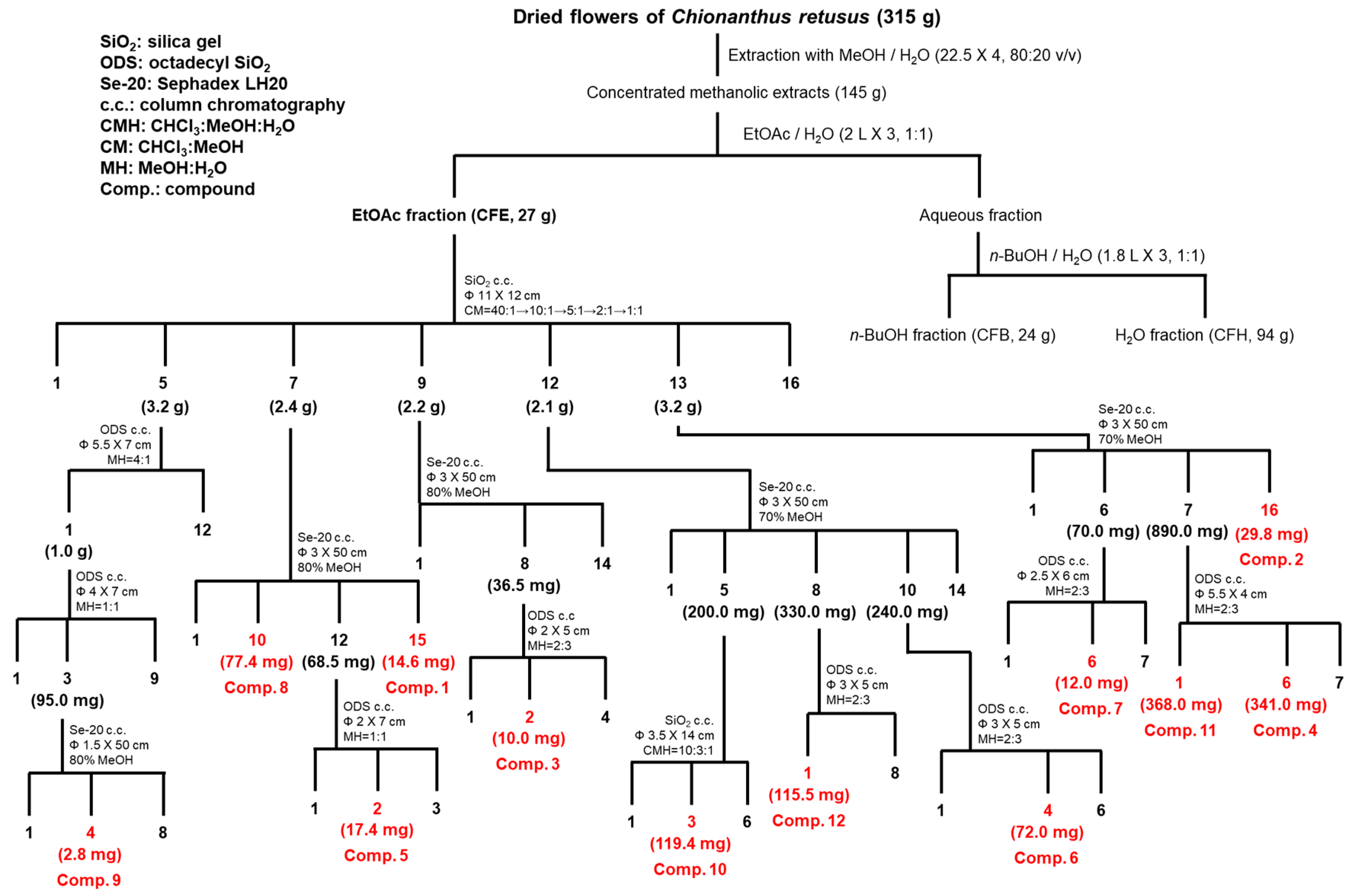
2.2. Isolation and Identification of Flavonoids from C. retusus Flowers
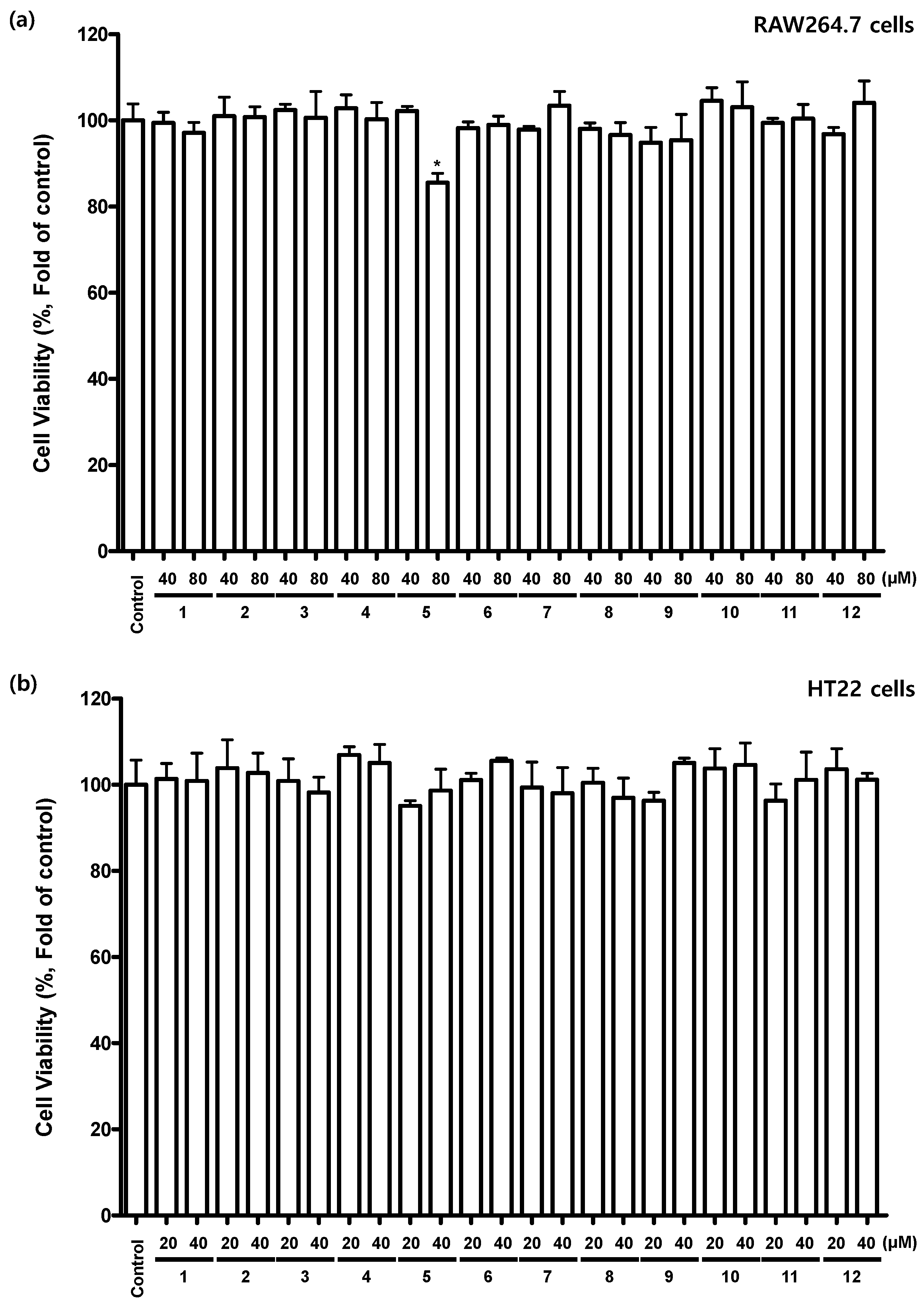
The dried flowers of C. retusus were extracted with MeOH, and the concentrate was divided into EtOAc, n-BuOH, and H2O fr.s. Repeated SiO2, octadecyl SiO2 (ODS), and Sephadex LH-20 column chromatography (c.c.). on the EtOAc Fr enabled the isolation of four flavonols (14), three flavones (57), four flavanonols (811), and one flavanone (12). These compounds were identified to be quercetin (1) [20], kaempferol (2) [20], astragalin (3) [21], nicotiflorin (4) [22], luteolin (5) [20], luteolin 4′-O-β-d-glucopyranoside (6) [23], isorhoifolin (7) [24], taxifolin (8) [25], aromadendrin (9) [20], aromadendrin 7-O-β-d-glucopyranoside (10) [26], taxifolin 7-O-β-d-glucopyranoside (11) [27], and eriodictyol 7-O-β-d-glucopyranoside (12) [24] based on extensive analysis of data from various spectroscopic methods, including IR, FAB/MS, 1d-NMR (1H, 13C, DEPT), and 2d-NMR (COSY, HSQC, HMBC) (Figure 1). The identities of the compounds were confirmed by comparing their NMR and MS values with those reported in the literature. We determined the stereochemistry of the chiral centers (C-2 and C-3) in flavonoids 812 by examining the coupling constants between H-2 and H-3 in the 1H-NMR spectra. They were mostly observed to be 12 Hz, which suggested that the two protons were in a 2,3-trans configuration.

2.3. Inhibition Effects of Flavonoids 112 on NO Production in Lipopolysaccharide (LPS)-Induced RAW 264.7 Cells
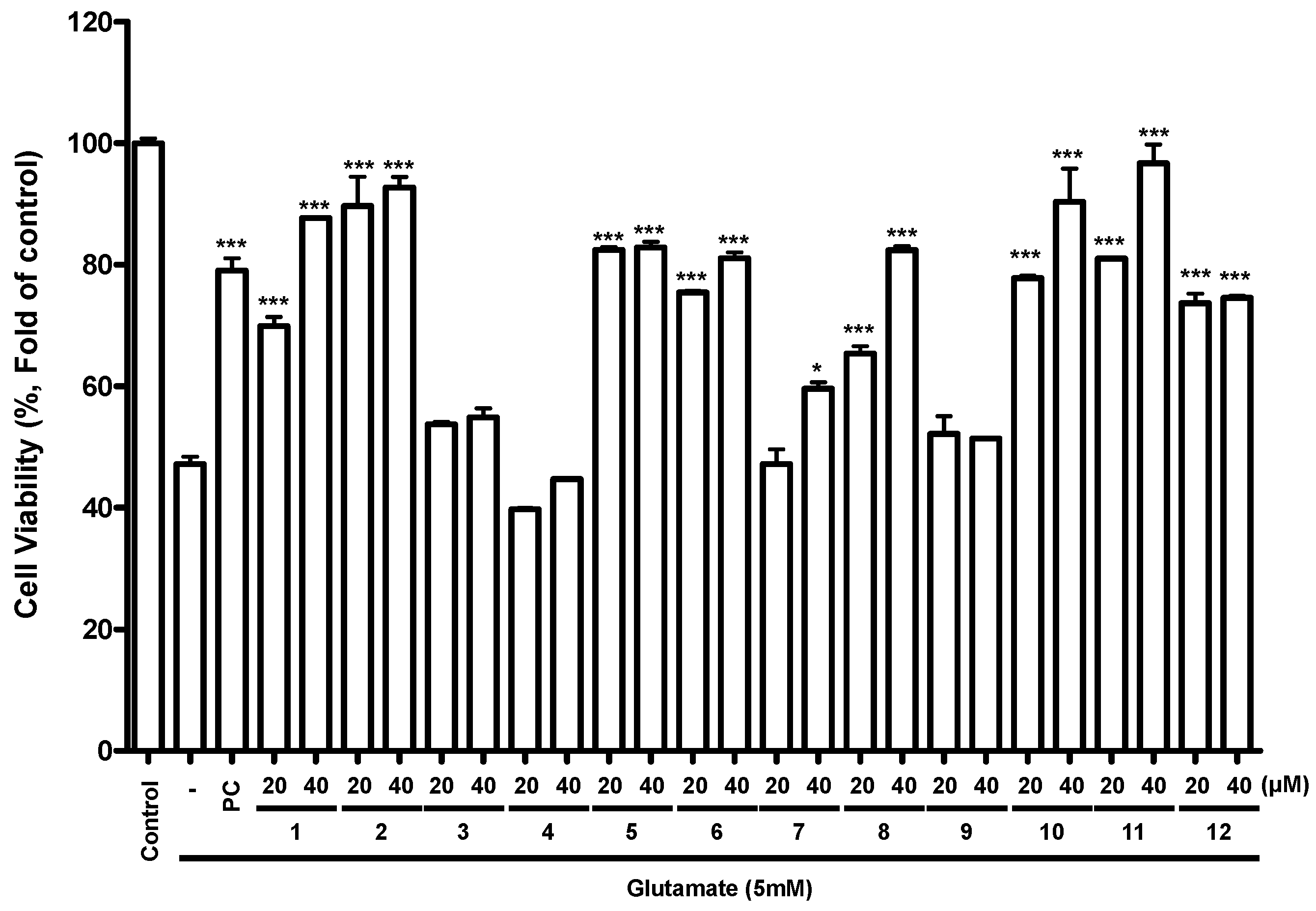
Oxidative stress is not only an important feature of several neurodegenerative processes, but also actively triggers intracellular signaling pathways that lead to cell death [28]. We first examined the viability of RAW264.7 cells treated with compounds 112 using a 3-(4,5-dimethylthiazol-2-yl)-2,5-diphenyltetrazolium bromide (MTT) assay. It did not show cytotoxicity or cellular proliferation when treated with compounds 14 and 612 at concentrations of 40 or 80 µM in RAW264.7 cells. However, compound 5 exhibited cytotoxic effects at 80 µM (Figure 2a). To investigate the anti-inflammatory effects of compounds 112, we appreciated their inhibitory effects on NO production in LPS-induced RAW 264.7 cells. These cells were pretreated with flavonoids 112 and butein, a positive control, before one day LPS treatment. As shown in Table 2, compounds 1, 2, 5, and 6 highly inhibited NO production, while compounds 8 and 1012 showed moderate inhibition effect. Flavonoids with a catechol structure in the B ring (1, 5, 6, 8, 11, and 12) exerted stronger anti-inflammatory effects than those with a phenol structure (3, 4, 7, 9, and 10). In addition, as the number of glucose moieties increased in compounds 16, the NO inhibitory effects of these compounds in RAW 264.7 cells decreased. However, compounds with glucopyranosyl moieties at C-7 (10 and 11) exhibited higher activity than aglycones (8 and 9). These results indicate that the presence of a catechol structure in the B ring and a glucopyranosyl moiety in the flavonoid structure were key factors of the anti-inflammatory effects of these flavonoids.

2.4. Effects of Flavonoids 112 on Glutamate-Induced Cell Toxicity in Mouse Hippocampal HT22 Cells

To investigate the protective effects of compounds 112 against glutamate-induced oxidative neuronal cell death, we also examined their effects on the viability of mouse hippocampal HT22 cells. To investigate the potential for cellular proliferation or cytotoxic effects of compounds 112, we first examined the viability of mouse hippocampal HT22 cells treated with compounds 112 using an MTT assay. No cytotoxic effects or cellular proliferation by compounds 112 were observed at concentrations <40 µM (Figure 2b). These cells were pretreated with compounds 112 at concentrations of 20 or 40 μM for 3 h and then were treated with glutamate and reacted for 12 h. Thereafter, cell viability was assessed with an MTT assay. None of the compounds exhibited toxicity at the highest concentration (40 μM). Compounds 1, 2, 5, 6, 8, 10, 11, and 12 significantly increased cell viability following glutamate treatment (Figure 3). Butein derived from Rhus verniciflua, which is known to protect mouse hippocampal HT22 cells from glutamate-induced death [29], was used as a positive control and indeed exhibited cytoprotective effects (Figure 3). Flavonoids with a catechol structure in the B ring (1, 5, 6, 8, 11, and 12) exerted stronger cytoprotective effects than those with a phenol structure (3, 4, 7, 9, and 10). In addition, as the number of glucose moieties increased in compounds 16, the cytoprotective effects of these compounds in HT22 cells decreased. However, compounds with glucopyranosyl moieties at C-7 (10 and 11) exhibited higher activity than aglycones (8 and 9). These results indicate that the presence of a catechol structure in the B ring and a glucopyranosyl moiety in the flavonoid structure were key determinants of the effects of these flavonoids on mouse hippocampal HT22 cells.

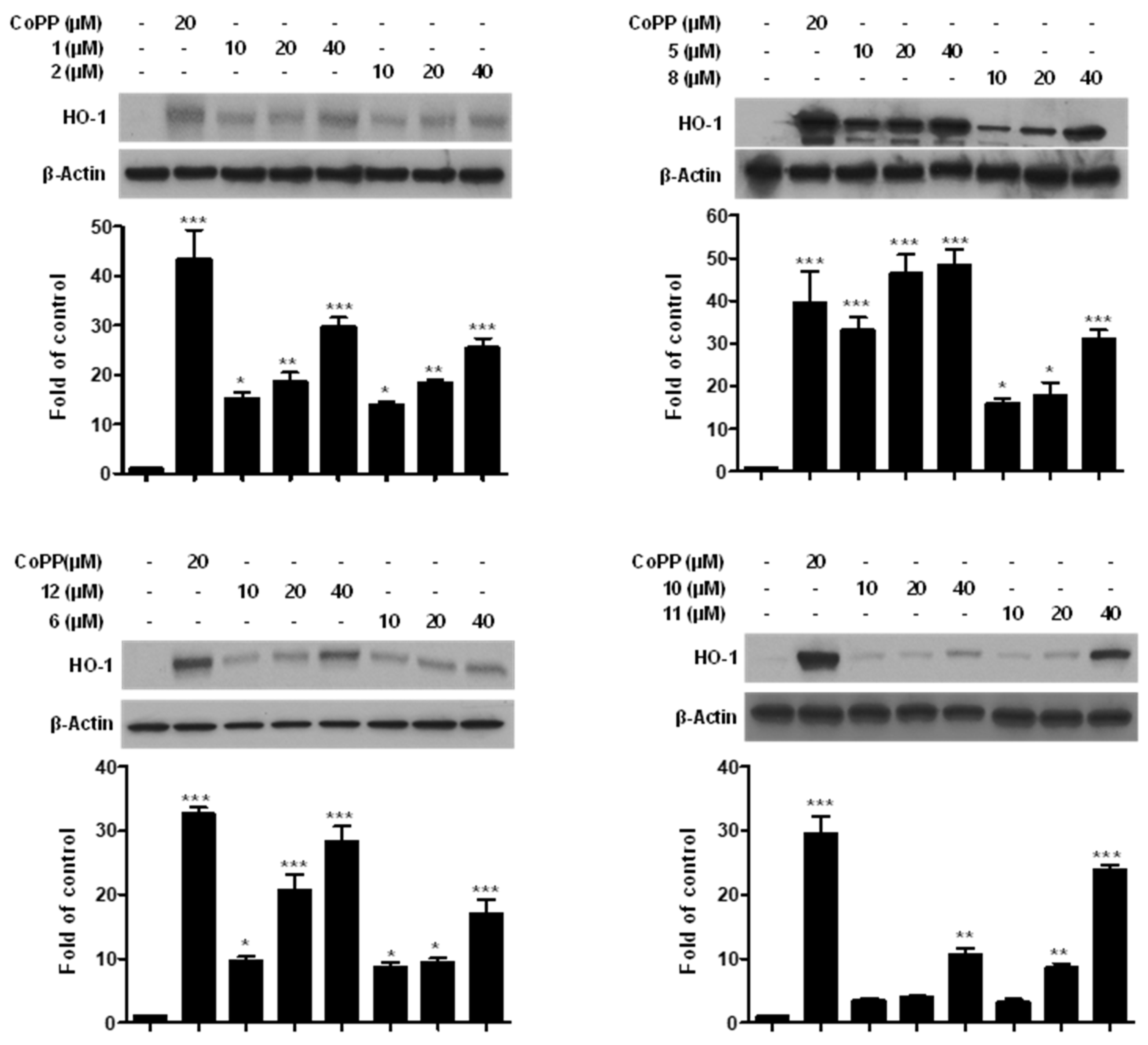
2.5. Effects of Compounds 1, 2, 5, 6, 8, and 1012 on HO-1 Expression in Mouse Hippocampal HT22 Cells

Heme oxygenase (HO) is an important enzyme in the antioxidant cell system. HO-1, one of the HO derivatives, decomposes heme in the cell to produce carbon monoxide, iron, and biliverdin [30]. HO-1 expression has been reported to inhibit brain cell damage resulting from oxidative stress [31]. We examined whether compounds 1, 2, 5, 6, 8, and 1012 affected the protein expression of HO-1, given their protection against glutamate-induced toxicity in mouse hippocampal HT22 cells. Mouse hippocampal HT22 cells were treated with compounds 1, 2, 5, 6, 8, and 1012 at three concentrations (10, 20, and 40 μM) and then cultured for 12 h. Cobalt protoporphyrin (CoPP), a well-known HO-1 inducer, was used as a positive control. As shown in Figure 4, compounds 1, 2, 5, 6, 8, and 1012 all increased HO-1 protein expression in a dose-dependent manner in mouse hippocampal HT22 cells. Flavonoid aglycones (1, 2, 5, and 8) exhibited higher activity than the glycosides (1012). The flavonol and flavanonol with a catechol structure in the B ring (1 and 11) displayed stronger HO-1 expression than those with a phenol structure (2 and 10). Flavonoids with a hydroxy group at C-3 (8 and 11) exhibited weaker HO-1 expression than those without (5 and 12). In addition, a flavonoid with a double bond between C2 and C3 (1) was a weaker inhibitor of oxidative-stress-induced brain-cell damage than one with a single bond (8). These results indicate that the presence of a hydroxy group at C-3, the structure of the B ring and the type of C2‒C3 bond are key determinants of the extent to which these flavonoids protect brain cells from damage due to oxidative stress.

2.6. Effects of Compounds 1, 2, 5, 6, 8, and 1012 on Cell Viability through HO Signaling Pathway

Compounds 1, 2, 5, 6, 8, and 1012, which exhibited cytoprotective effects, also increased HO-1 expression (Figure 3 and Figure 4). To investigate whether HO-1 expression regulates cell viability, we assessed the protective effects of compounds 1, 2, 5, 6, 8, and 1012 when tin protoporphyrin IX (SnPP) was used as a HO-1 activity inhibitor. Cells were treated with compounds 1, 2, 5, 6, 8, and 1012 (40 μM) in the presence or absence of SnPP (50 μM) and then exposed to glutamate (5 mM) for 12 h. When cells were pre-treated with SnPP, the protective effects of the compounds decreased (Figure 5); that is, cell viability was significantly lower in SnPP-pretreated cells than in the cells not treated with SnPP. These results indicate that compounds 1, 2, 5, 6, 8, and 1012 inhibited oxidative-stress-induced cell damage by increasing HO-1 protein expression.

3. Materials and Methods

3.1. Plant Materials

The flowers of C. retusus Lindl. And Paxton were gathered near Kyung Hee University, Yong-In, South Korea, in August 2014, and were identified by Prof. Dae-Keun Kim, College of Pharmacy, Woosuk University, Jeonju, South Korea. A voucher specimen (KHU-NPCl-201408) has been deposited at the Natural Products Chemistry Laboratory, Kyung Hee University.

3.2. General Experimental Procedures

The equipment and chemicals used to isolate and identify flavonoids from C. retusus flowers and evaluate their neuroprotective activity were obtained from the literature [32,33,34,35].

3.3. Isolation Procedure of Flavonoids (112) from C. retusus Flowers

Dried C. retusus flowers (315 g) were extracted in 80% aqueous MeOH (22.5 L × 4) at room temperature for 24 h, and then filtered and concentrated in vacuo. The concentrated MeOH extracts (145 g) were poured into H2O (2.0 L) and successively extracted with EtOAc (2.0 L × 3) and n-BuOH (1.8 L × 3). Each layer was concentrated under reduced pressure to obtain EtOAc (CFE, 27 g), n-BuOH (CFB, 24 g), and H2O (CFH, 94 g). Frs. CFE (27 g) was subjected to SiO2 c.c. (Φ 11 × 12 cm) and eluted with CHCl3‒MeOH (CM; 40:1 → 10:1 → 5:1 → 2:1 → 1:1, 600 mL of each), with monitoring by TLC, yielding 15 frs (CFE-1 to CFE-15).
CFE-5 (3.2 g, Ve/Vt 0.360–0.415) was subjected to ODS c.c. (Φ 5.5 × 7 cm, MeOH-H2O [MH] = 4:1, 1.7 L) to yield 12 Frs (CFE-5-1 to CFE-5-12). CFE-5-1 (1.0 g, Ve/Vt 0.000–0.110) was subjected to ODS c.c. (Φ 4.0 × 7 cm, MH = 1:1, 1.5 L) to yield 9 Frs (CFE-5-1-1 to CFE-5-1-9). CFE-5-1-3 (95.0 mg, Ve/Vt 0.150–0.260) was subjected to Sephadex LH-20 c.c. (Φ 1.5 × 60 cm, 80% MeOH, 560 mL) to yield 8 Frs (CFE-5-1-3-1 to CFE-5-1-3-8), along with purified compound 9 (CFE-5-1-3-4, 2.8 mg, Ve/Vt 0.550–0.560, TLC [SiO2] Rf 0.37, CM = 10:1, TLC [ODS] Rf 0.58, MH = 2:1).
CFE-7 (2.4 g, Ve/Vt 0.430–0.480) was subjected to Sephadex LH-20 c.c. (Φ 3 × 50 cm, 80% MeOH, 1.3 L) to yield 15 Frs (CFE-7-1 to CFE-7-15), along with purified compound 8 (CFE-7-10, 77.4 mg, Ve/Vt 0.488-0.542, TLC [SiO2] Rf 0.45, CHCl3-MeOH-H2O [CMH] = 10:3:1, TLC [ODS] Rf 0.60, MH = 3:2) and purified compound 1 (CFE-7-15, 14.6 mg, Ve/Vt 0.885-1.000, TLC [SiO2] Rf 0.47, CMH = 10:3:1, TLC [ODS] Rf 0.74, MH = 4:1). CFE-7-12 (68.5 mg, Ve/Vt 0.650–0.720) was subjected to ODS c.c. (Φ 2.0 × 7 cm, MH = 1:1, 620 mL) to yield 3 Frs (CFE-7-12-1 to CFE-7-12-3), along with purified compound 5 (CFE-7-12-2, 17.4 mg, Ve/Vt 0.194–0.677, TLC [SiO2] Rf 0.50, CMH = 10:3:1, TLC [ODS] Rf 0.50, MH = 4:1).
CFE-9 (2.2 g, Ve/Vt 0.580–0.610) was subjected to Sephadex LH-20 c.c. (Φ 3 × 50 cm, 80% MeOH, 2.2 L) to yield 14 Frs (CFE-9-1 to CFE-9-14). CFE-9-8 (36.5 mg, Ve/Vt 0.480-0.510) was subjected to ODS c.c. (Φ 2.0 × 5 cm, MH = 2:3, 200 mL) to yield 4 Frs (CFE-9-8-1 to CFE-9-8-4), along with purified compound 3 (CFE-9-8-2, 10.0 mg, Ve/Vt 0.125–0.425, TLC [SiO2] Rf 0.50, CM = 4:1, TLC [ODS] Rf 0.65, MH = 3:1).
CFE-12 (2.1 g, Ve/Vt 0.710-0.790) was subjected to Sephadex LH-20 c.c. (Φ 3.0 × 50 cm, 70% MeOH, 2.3 L) to yield 14 Frs (CFE-12-1 to CFE-12-14). CFE-12-5 (200.0 mg) was subjected to SiO2 c.c. (Φ 3.5 × 14 cm) and eluted with CMH = 10:3:1 (560 mL), with monitoring by TLC, yielding 6 Frs (CFE-12-5-1 to CFE-12-5-6), along with purified compound 10 (CFE-12-5-3, 119.4 mg, Ve/Vt 0.102–0.250, TLC [SiO2] Rf 0.50, CMH = 65:35:10, TLC [ODS] Rf 0.50, MH = 2:3). CFE-12-8 (330.0 mg, Ve/Vt 0.370–0.410) was subjected to ODS c.c. (Φ 3.0 × 5 cm, MH = 2:3, 1.2 L) to yield 8 Frs (CFE-12-8-1 to CFE-12-8-8), along with purified compound 12 (CFE-12-8-1, 115.5 mg, Ve/Vt 0.000–0.058, TLC [SiO2] Rf 0.50, CMH = 65:35:10, TLC [ODS] Rf 0.70, MH = 3:2). CFE-12-10 (240.0 mg, Ve/Vt 0.460-0.550) was subjected to ODS c.c. (Φ 3.0 × 5 cm, MH = 2:3, 840 mL) to yield 6 Frs (CFE-12-10-1 to CFE-12-10-6), along with purified compound 6 (CFE-12-10-4, 72.0 mg, Ve/Vt 0.286–0.414, TLC [SiO2] Rf 0.55, CMH = 65:35:10, TLC [ODS] Rf 0.45, MH = 3:2).
CFE-13 (3.2 g, Ve/Vt 0.710-0.790) was subjected to Sephadex LH-20 c.c. (Φ 3.0 × 50 cm, 70% MeOH, 2.3 L) to yield 16 Frs (CFE-13-1 to CFE-13-16), along with purified compound 2 (CFE-13-16, 29.0 mg, Ve/Vt 0.846–0.912, TLC [SiO2] Rf 0.50, CM = 5:1, TLC [ODS] Rf 0.40, MH = 3:1). CFE-13-6 (70.0 mg, Ve/Vt 0.270–0.320) was subjected to ODS c.c. (Φ 2.5 × 6 cm, MH = 2:3, 740 mL) to yield six Frs (CFE-13-6-1 to CFE-13-6-6), along with purified compound 7 (CFE-13-6-4, 12.0 mg, Ve/Vt 0.657–0.730, TLC [SiO2] Rf 0.50, CM = 2:1, TLC [ODS] Rf 0.55, MH = 3:2). CFE-13-7 (890.0 mg, Ve/Vt 0.330-0.480) was subjected to ODS c.c. (Φ 5.5 × 4 cm, MH = 2:3, 2.6 L) to yield 7 Frs (CFE-13-7-1 to CFE-13-7-7), along with purified compound 11 (CFE-13-7-1, 368.0 mg, Ve/Vt 0.000–0.102, TLC [SiO2] Rf 0.50, CM = 2:1, TLC [ODS] Rf 0.55, MH = 1:3) and compound 4 (CFE-13-7-6, 341.0 mg, Ve/Vt 0.512–0.923, TLC [SiO2] Rf 0.50, CM = 2:1, TLC [ODS] Rf 0.65, MH = 3:2) (Scheme 1).
quercetin (1): Yellowish powder (MeOH); m.p. 276–277 °C; ultraviolet (UV) (MeOH) λmax (nm) 370, 305, 267, 255; infrared (IR) (KBr) νmax 3350, 1680, 1615 cm−1; positive FAB/MS m/z 303 [M + H]+.
kaempferol (2): Yellowish powder (MeOH); m.p. 278–279 °C; UV (MeOH) λmax (nm) 364, 320, 294, 265, 254; IR (KBr) νmax 3345, 1658, 1605 cm−1; positive FAB/MS m/z 309 [M + Na]+.
astragalin (3): Yellowish powder (MeOH); m.p. 230–231 °C; [ α ] D 21 +16.0 (c 0.1, MeOH); UV (MeOH) λmax (nm) 348, 259; IR (KBr) νmax 3350, 2930, 2365, 1655, 1610 cm−1; positive FAB/MS m/z 471 [M + Na]+.
nicotiflorin (4): Yellowish powder (MeOH); m.p. 268–269°C; [ α ] D 21 −15.0 (c 1.0, MeOH); UV (MeOH) λmax (nm) 365, 267, 254; IR (KBr) νmax 3365, 2940, 2360, 1655, 1600, 1515 cm−1; positive FAB/MS m/z 639 [M + Na]+.
luteolin (5): Yellowish powder (MeOH); m.p. 329–330 °C; UV (MeOH) λmax (nm) 349, 269, 254; IR (KBr) νmax 3320, 2930, 1600, 1520 cm−1; positive FAB/MS m/z 271 [M + H]+.
luteolin 4′-O-β-d-glucopyranoside (6): Yellowish powder (MeOH); m.p. 178–179 °C; UV (MeOH) λmax (nm) 341, 272; IR (KBr) νmax 3320, 2930, 1600, 1520, 1510, 1480 cm−1; positive FAB/MS m/z 449 [M + H]+.
isorhoifolin (7): Yellowish needles; m.p. 269–270 °C; [ α ] D 21 −96.7 (c 1.0, MeOH); UV (MeOH) λmax (nm) 331, 266; IR (KBr) νmax 3365, 2360, 1635, 1600, 1515 cm−1; positive FAB/MS m/z 579 [M + H]+.
taxifolin (8): Yellowish powder (MeOH); m.p. 236–237 °C; [ α ] D 21 +23.1 (c 0.1, MeOH); UV (MeOH) λmax (nm) 330, 280; IR (KBr) νmax 3415, 1625, 1515, 1472 cm−1; positive FAB/MS m/z 327 [M + Na]+.
aromadendrin (9): White powder; m.p. 216–217 °C; [ α ] D 21 +58.5 (c 0.3, MeOH); UV (MeOH) λmax (nm) 329, 292, 228; IR (KBr) νmax 3420, 1655, 1518 cm−1; positive FAB/MS m/z 311 [M + Na]+.
aromadendrin 7-O-β-d-glucopyranoside (10): Yellowish powder (MeOH); m.p. 172–173 °C; [ α ] D 21 −18.7 (c 0.2, MeOH); UV (MeOH) λmax (nm) 321, 285; IR (KBr) νmax 3435, 1645, 1520, 1365 cm1; positive FAB/MS m/z 473 [M + Na]+.
taxifolin 7-O-β-d-glucopyranoside (11): Yellowish powder (MeOH); m.p. 169–170 °C; [ α ] D 21 −48.2 (c 0.2, MeOH); UV (MeOH) λmax (nm) 331, 283; IR (KBr) νmax 3420, 1635, 1450, 1510, 1390 cm−1; positive FAB/MS m/z 467 [M + H]+.
eriodictyol 7-O-β-d-glucopyranoside (12): Yellowish powder (MeOH); m.p. 173–174 °C; [ α ] D 21 −35.5 (c 0.2, MeOH); UV (MeOH) λmax (nm) 283, 233; IR (KBr) νmax 3455, 1690, 1595, 1510 cm−1; positive FAB/MS m/z 451 [M + H]+.
1H-NMR (400 MHz, δH) and 13C-NMR (100 MHz, δC) spectroscopic data of flavonoids 112, see Table 3 and Table 4.

3.4. Cell Culture and MTT Assay

Mouse hippocampal HT22 cells were donated by Wonkwang University, Iksan, Korea (Prof. Youn-Chul Kim). Cytoprotective activity assay was performed, as per the previously described method [35]. Cell viability was evaluated using the MTT assay reported in the literature [36].

3.5. Macrophage RAW 264.7 Culture, Viability Assay, and NO Measurement

Macrophage RAW 264.7 culture, viability assay, and NO measurement were carried out as per the previously described method [35].

3.6. Determination of Total Phenols and Flavonoids Contents in C. retusus Flower

Determination of the total phenolic and flavonoid contents of C. retusus flower was carried out as per the previously described method [37].

3.7. Western Blot Analysis

Pelleted HT22 cells were washed with PBS and lysed with an RIPA buffer from Sigma Chemical Co. The same amount of protein from each sample was mixed into a sample loading buffer, subjected to SDS-PAGE, and transferred to a membrane.

3.8. Statistical Analysis

Statistical analysis was performed with GraphPad Prism 5 software (ver. 3.03, San Diego, CA, USA). Data are presented as the mean ± standard deviation of 3 independent experiments. The mean differences were derived using one-way ANOVA and Tukey’s multiple comparison test, and statistical significance was defined as p < 0.05, p < 0.01, and p < 0.001.

4. Conclusions

In conclusion, four flavonols (14), three flavones (57), four flavanonols (811), and one flavanone (12) were isolated from C. retusus flowers. Flavonoids 46 and 811 were isolated from the flowers of C. retusus for the first time in this study. Flavonoids 1, 2, 5, 6, 8, and 1012 exhibited significant anti-inflammatory and neurocytoprotective activity, and effectively increased HO-1 protein expression. The flavonoids that displayed significant neuroprotective activity were found to recover oxidative stress-induced cell damage by increasing HO-1 protein expression. The relationships between the structural characteristics of these flavonoids and their anti-inflammatory and neuroprotective activity were revealed. Further studies are needed to investigate the potential therapeutic effects of flavonoids in innovative anti-inflammatory and neuroprotective strategies.

Author Contributions

Conceptualization, Y.-G.L. and N.-I.B.; methodology, Y.-G.L., D.-S.L., and N.-I.B.; formal analysis, Y.-G.L. and N.-I.B.; investigation, Y.-G.L., H.L., D.-S.L., and N.-I.B.; resources, Y.-G.L., J.-W.J., K.-H.S., D.Y.L., H.-G.K., J.-H.K., and N.-I.B.; data curation, Y.-G.L., J.-W.J., K.-H.S., D.Y.L., H.-G.K., J.-H.K., and N.-I.B.; writing-original draft preparation, Y.-G.L.; writing-review and editing, supervision, project administration, and funding acquisition, N.-I.B.

Funding

This work was carried out with the support of the “Cooperative Research Program for Agriculture Science & Technology Development (Project No. PJ01420403)”, Rural Development Administration, Republic of Korea.

Conflicts of Interest

The authors declare no conflict of interest.

Abbreviations

c.c.column chromatography
CoPPcobalt protoporphyrin
Frfraction
HOheme oxygenase
IRinfrared
SnPPtin protoporphyrin IX
SiO2silica gel
ODSoctadecyl SiO2
PCpositive control
TLCthin layer chromatography
UVultraviolet
Ve/Vtelution volume/total volume

References

  1. Han, A.R.; Park, S.A.; Ahn, B.E. Reduced stress and improved physical functional ability in elderly with mental health problems following a horticultural therapy program. Complement. Ther. Med. 2018, 38, 19–23. [Google Scholar] [CrossRef] [PubMed]
  2. Nabavi, S.F.; Braidy, N.; Habtemariam, S.; Orhan, I.E.; Daglia, M.; Manayi, A.; Gortzi, O.; Nabavi, S.M. Neuroprotective effects of chrysin: From chemistry to medicine. Neurochem. Int. 2015, 90, 224–231. [Google Scholar] [CrossRef] [PubMed]
  3. Levi, M.S.; Brimble, M.A. A review of neuroprotective agents. Curr. Med. Chem. 2004, 11, 2383–2397. [Google Scholar] [CrossRef] [PubMed]
  4. Lee, A.Y.; Lee, M.H.; Lee, S.H.; Cho, E.J. Alpha-linolenic acid regulates amyloid precursor protein processing by mitogen-activated protein kinase pathway and neuronal apoptosis in amyloid beta-induced SH-SY5Y neuronal cells. Appl. Biol. Chem. 2018, 61, 61–71. [Google Scholar] [CrossRef]
  5. Vauzour, D.; Vafeiadou, K.; Rodriguez-Mateos, A.; Rendeiro, C.; Spencer, J.P.E. The neuroprotective potential of flavonoids: A multiplicity of effects. Genes Nutr. 2008, 3, 91. [Google Scholar] [CrossRef] [PubMed]
  6. Dajas, F.; Rivera-Megret, F.; Blasina, F.; Arredondo, F.; Abin-Carriquiry, J.A.; Costa, G.; Echeverry, C.; Lafon, L.; Heizen, H.; Ferreira, M.; et al. Neuroprotection by flavonoids. Braz. J. Med. Biol. Res. 2003, 36, 1613–1620. [Google Scholar] [CrossRef] [PubMed] [Green Version]
  7. Spagnuolo, C.; Moccia, S.; Russo, G.L. Anti-inflammatory effects of flavonoids in neurodegenerative disorders. Eur. J. Med. Chem. 2018, 153, 105–115. [Google Scholar] [CrossRef] [PubMed]
  8. Dok-Go, H.; Lee, K.H.; Kim, H.J.; Lee, E.H.; Lee, J.Y.; Song, Y.S.; Lee, Y.H.; Jin, C.B.; Lee, Y.S.; Cho, J.S. Neuroprotective effects of antioxidative flavonoids, quercetin, (+)-dihydroquercetin and quercetin 3-methyl ether, isolated from Opuntia ficus-indica var. saboten. Brain Res. 2003, 965, 130–136. [Google Scholar] [CrossRef]
  9. Hwang, S.L.; Shih, P.H.; Yen, G.C. Neuroprotective effects of citrus flavonoids. J. Agric. Food Chem. 2012, 60, 877–885. [Google Scholar] [CrossRef]
  10. Kang, H.M.; Kim, J.H.; Lee, M.Y.; Son, K.H.; Yang, D.C.; Baek, N.I.; Kwon, B.M. Relationship between flavonoid structure and inhibition of farnesyl protein transferase. Nat. Prod. Res. 2004, 18, 349–356. [Google Scholar] [CrossRef]
  11. Yang, H.J.; Shin, Y.J. Antioxidant compounds and activities of edible roses (Rosa hybrida spp.) from different cultivars grown in Korea. Appl. Biol. Chem. 2017, 60, 129–136. [Google Scholar] [CrossRef]
  12. Lee, J.M.; Rodriguez, J.P.; Quilantang, N.G.; Lee, M.H.; Cho, E.J.; Jacinto, S.D.; Lee, S.H. Determination of flavonoids from Perilla frutescens var. japonica seeds and their inhibitory effect on aldose reductase. Appl. Biol. Chem. 2017, 60, 155–162. [Google Scholar] [CrossRef]
  13. Rodriguez, J.P.; Lee, J.M.; Park, J.Y.; Kang, K.S.; Hahm, D.H.; Lee, S.C.; Lee, S.H. HPLC-UV analysis of sample preparation influence on flavonoid yield from Cirsium japonicum var. maackii. Appl. Biol. Chem. 2017, 60, 519–525. [Google Scholar] [CrossRef]
  14. Dötterl, S.; Vereecken, N.J. The chemical ecology and evolution of bee-flower interactions: A review and perspectives. Can. J. Zool. 2010, 88, 668–697. [Google Scholar] [CrossRef]
  15. Raguso, R.A. Wake up and smell the roses: The ecology and evolution of floral scent. Annu. Rev. Ecol. Evol. Syst. 2008, 39, 549–569. [Google Scholar] [CrossRef]
  16. Wright, G.A.; Schiestl, F.P. The evolution of floral scent: The influence of olfactory learning by insect pollinators on the honest signalling of floral rewards. Funct. Ecol. 2009, 23, 841–851. [Google Scholar] [CrossRef]
  17. Sun, X.M.; Li, X.F.; Deng, R.X.; Liu, Y.Q.; Hou, X.W.; Xing, Y.P.; Liu, P. Extraction technology and antioxidant activity of total flavonoids from the flower of Chionanthus retusa. Food Sci. 2015, 36, 266–271, 278. [Google Scholar]
  18. Lee, Y.N.; Jeong, C.H.; Shim, K.H. Isolation of antioxidant and antibrowning substance from Chionanthus retusa leaves. J. Korean Soc. Food Sci. Nutr. 2004, 33, 1419–1425. [Google Scholar]
  19. Deng, R.X.; Zhang, C.F.; Liu, P.; Duan, W.L.; Yin, W.P. Separation and identification of flavonoids from Chinese Fringetree Flowers (Chionanthus retusa Lindl et Paxt). Food Sci. 2014, 35, 74–78. [Google Scholar]
  20. Kwak, J.H.; Kang, M.W.; Roh, J.H.; Choi, S.U.; Zee, O.P. Cytotoxic phenolic compounds from Chionanthus retusus. Arch. Pharm. Res. 2009, 32, 1681–1687. [Google Scholar] [CrossRef]
  21. Baek, Y.S.; Song, N.Y.; Nam, T.G.; Kim, D.O.; Kang, H.C.; Kwon, O.K.; Baek, N.I. Flavonoids from Fragaria ananassa calyx and their antioxidant capacities. Appl. Biol. Chem. 2015, 58, 787–793. [Google Scholar] [CrossRef]
  22. Han, J.T.; Bang, M.H.; Chun, O.K.; Kim, D.O.; Lee, C.Y.; Baek, N.I. Flavonol glycosides from the aerial parts of Aceriphyllum rossii and their antioxidant activities. Arch. Pharm. Res. 2004, 27, 390–395. [Google Scholar] [CrossRef] [PubMed]
  23. Braca, A.; Tommasi, N.D.; Bari, L.D.; Pizza, C.; Politi, M.; Morelli, I. Antioxidant principles from Bauhinia tarapotensis. J. Nat. Prod. 2001, 64, 892–895. [Google Scholar] [CrossRef] [PubMed]
  24. Weber, B.; Herrmann, M.; Hartmann, B.; Joppe, H.; Schmidt, C.O.; Bertram, H.J. HPLC/MS and HPLC/NMR as hyphenated techniques for accelerated characterization of the main constituents in Chamomile (Chamomilla recutita [L.] Rauschert). Eur. Food Res. Technol. 2008, 226, 755–760. [Google Scholar] [CrossRef]
  25. Shrestha, S.; Lee, D.Y.; Park, J.H.; Cho, J.G.; Lee, D.S.; Li, B.; Kim, Y.C.; Jeon, Y.J.; Yeon, S.W.; Baek, N.I. Flavonoids from the fruits of Nepalese sumac (Rhus parviflora) attenuate glutamate-induced neurotoxicity in HT22 cells. Food Sci. Biotechnol. 2013, 22, 895–902. [Google Scholar] [CrossRef]
  26. Hiep, N.T.; Kwon, J.Y.; Kim, D.W.; Hong, S.G.; Guo, Y.Q.; Hwang, B.Y.; Kim, N.H.; Mar, W.C.; Lee, D. Neuroprotective constituents from the fruits of Maclura tricuspidata. Tetrahedron 2017, 73, 2747–2759. [Google Scholar] [CrossRef]
  27. Latté, K.P.; Ferreira, D.; Venkatraman, M.S.; Kolodziej, H. O-Galloyl-C-glycosylflavones from Pelargonium reniforme. Phytochemistry 2002, 59, 419–424. [Google Scholar] [CrossRef]
  28. Simonian, N.A.; Coyle, J.T. Oxidative stress in neurodegenerative diseases. Annu. Rev. Pharmacol. Toxicol. 1996, 36, 83–106. [Google Scholar] [CrossRef] [PubMed]
  29. Lee, D.S.; Jeong, G.S. Butein provides neuroprotective and anti-neuroinflammatory effects through Nrf2/ARE-dependent haem oxygenase 1 expression by activating the PI3K/Akt pathway. Br. J. Pharmacol. 2016, 173, 2894–2909. [Google Scholar] [CrossRef] [Green Version]
  30. Lee, M.S.; Lee, J.N.; Kwon, D.Y.; Kim, M.S. Ondamtanggamibang protects neurons from oxidative stress with induction of heme oxygenase-1. J. Ethnopharmacol. 2006, 108, 294–298. [Google Scholar] [CrossRef]
  31. Choi, B.M.; Kim, H.J.; Oh, G.S.; Pae, H.O.; Oh, H.C.; Jeong, S.J.; Kwon, T.O.; Kim, Y.M.; Chung, H.T. 1,2,3,4,6-Penta-O-galloyl-beta-d-glucose protects rat neuronal cells (Neuro 2A) from hydrogen peroxide-mediated cell death via the induction of heme oxygenase-1. Neurosci. Lett. 2002, 328, 185–189. [Google Scholar] [CrossRef]
  32. Nhan, N.T.; Song, H.S.; Oh, E.J.; Lee, Y.G.; Ko, J.H.; Kwon, J.E.; Kang, S.C.; Lee, D.Y.; Baek, N.I. Phenylpropanoids from Lilium Asiatic hybrid flowers and their anti-inflammatory activities. Appl. Biol. Chem. 2017, 60, 527–533. [Google Scholar]
  33. Lee, Y.G.; Lee, D.G.; Gwag, J.E.; Kim, M.S.; Kim, M.J.; Kim, H.G.; Ko, J.H.; Yeo, H.J.; Kang, S.H.; Baek, N.I. A 1,1′-biuracil from Epidermidibacterium keratini EPI-7 shows anti-aging effects on human dermal fibroblasts. Appl. Biol. Chem. 2019, 62, 14. [Google Scholar] [CrossRef]
  34. Lee, Y.G.; Rodriguez, I.; Nam, Y.H.; Gwag, J.E.; Woo, S.H.; Kim, H.G.; Ko, J.H.; Hong, B.N.; Kang, T.H.; Baek, N.I. Recovery effect of lignans and fermented extracts from Forsythia koreana flowers on pancreatic islets damaged by alloxan in zebrafish (Danio rerio). Appl. Biol. Chem. 2019, 62, 7. [Google Scholar] [CrossRef]
  35. Lee, Y.G.; Seo, K.H.; Lee, D.S.; Gwag, J.E.; Kim, H.G.; Ko, J.H.; Park, S.H.; Lee, D.Y.; Baek, N.I. Phenylethanoid glycoside from Forsythia koreana (Oleaceae) flowers shows a neuroprotective effect. Braz. J. Bot. 2018, 41, 523–528. [Google Scholar] [CrossRef]
  36. Mosmann, T. Rapid colorimetric assay for cellular growth and survival: Application to proliferation and cytotoxicity assays. J. Immunol. Methods 1983, 65, 55–63. [Google Scholar] [CrossRef]
  37. Lee, Y.G.; Lee, J.H.; Lee, N.Y.; Kim, N.K.; Jung, D.W.; Wang, W.; Kim, Y.S.; Kim, H.G.; Nguyen, N.T.; Park, H.S.; et al. Evaluation for the flowers of compositae plants as whitening cosmetics functionality. J. Appl. Biol. Chem. 2017, 60, 5–11. [Google Scholar] [CrossRef] [Green Version]
Figure 1. Chemical structures of flavonoids 1-12 isolated from C. retusus flowers.
Ijms 20 03517 g001
Figure 2. Cytotoxicity of compounds 112 on (a) RAW264.7 cells and (b) mouse hippocampal HT22 cells. (a) RAW264.7 cells were treated with 40 or 80 μM of compounds 112 for 48 h. (b) Mouse hippocampal HT22 cells were treated with 20 or 40 μM of compounds 112 for 24 h. Data are presented as the mean ± standard deviation of three independent experiments. * p < 0.05 vs. non-treated control.
Ijms 20 03517 g002
Figure 3. Effects of compounds 112 on glutamate-induced oxidative neurotoxicity in mouse hippocampal HT22 cells. Mouse hippocampal HT22 cells were pretreated with 20 or 40 μM of compounds 112 and then were treated with glutamate (5 mM) for 12 h. Butein (5 μM) was used as a positive control. Data are presented as the mean ± standard deviation of three independent experiments. * p < 0.05, *** p < 0.001 vs. glutamate.
Ijms 20 03517 g003
Figure 4. Effects of compounds 1, 2, 5, 6, 8, and 1012 on HO-1 expression in mouse hippocampal HT22 cells. Mouse hippocampal HT22 cells were treated with compounds 1, 2, 5, 6, 8, and 1012 at three concentrations (10, 20, and 40 μM) and then cultured for 12 h. Expression of HO-1 was measured by Western blot analysis. Cobalt protoporphyrin (CoPP, 20 μM) was used as a positive control. Representative blots of three independent experiments are shown. * p < 0.05, ** p < 0.01, *** p < 0.001 vs. non-treated control.
Ijms 20 03517 g004
Figure 5. Effects of HO-1 expression induced by compounds 1, 2, 5, 6, 8, and 1012 on glutamate-induced oxidative cell damage in mouse hippocampal HT22 cells. Mouse hippocampal HT22 cells were treated with compounds 1, 2, 5, 6, 8, and 1012 (40 μM) in the presence or absence of tin protoporphyrin IX (SnPP, 50 μM) and then exposed to glutamate (5 mM) for 12 h. Data are presented as the mean ± standard deviation of three independent experiments. ** p < 0.01, *** p < 0.001. # p < 0.05, ## p < 0.01, ### p < 0.001.
Ijms 20 03517 g005
Scheme 1. Isolation procedure of flavonoids from the flowers of Chionanthus retusus. Words in red indicate fraction number, quantity, and compound number of isolated flavonoids.
Ijms 20 03517 sch001
Table 1. Total phenols and flavonoids contents of the extract and fractions from Chionanthus retusus flowers.
SamplesExtractEtOAc fr.n-BuOH fr.H2O fr.
Total phenols (mg GA/g DW)125.4 ± 3.3245.6 ± 5.2130.1 ± 2.553.1 ± 1.8
Total flavonoids (mg CA/g DW)119.1 ± 2.7172.1 ± 2.198.2 ± 0.918.2 ± 1.2
GA: gallic acid; CA: catechin; fr., fraction.
Table 2. IC50 values of flavonoids 112 from C. retusus flowers on NO production in lipopolysaccharide (LPS)-induced RAW264.7 cells. The cells were pre-treated with each compound for 12 h, and then stimulated with LPS (1 μg/mL) for 18 h. The production of NO was determined as described in Section 3. Data shown represent the mean ± SD of three experiments.
No.IC50 (μM)No.IC50 (μM)No.IC50 (μM)
137.93 ± 0.0355.99 ± 0.029>100
221.25 ± 0.03630.60 ± 0.051071.56 ± 0.08
3>1007>1001157.18 ± 0.03
4>100878.53 ± 0.031260.86 ± 0.01
Table 3. 1H-NMR data of flavonoids 112 (δH in ppm, coupling pattern, J in Hz).
No.1 (a)2 (a)3 (a)4 (a)5 (b)6 (b)7 (b)8 (a)9 (a)10 (a)11 (a)12 (a)
2-------5.09, d, 12.05.05, d, 11.65.03, d, 12.05.09, d, 11.65.14, dd, 12.4, 2.8
3----6.91, s6.90, s6.80, s4.71, d, 12.04.58, d, 11.64.59, d, 12.04.71, d, 11.63.00, dd, 16.4, 12.4
2.63, dd, 16.4, 2.8
66.17, brs6.18, brs6.20, brs6.20, brs6.75, d, 1.66.75, brs6.68, brs6.05, d, 1.25.87, d, 2.06.15, d, 1.66.05, d, 1.25.09, brs
86.38, brs6.38, brs6.36, brs6.37, brs6.76, d, 1.66.77, brs6.78, brs6.13, d, 1.25.89, d, 2.06.21, d, 1.66.09, d, 1.26.00, brs
2′7.73, d, 1.88.10, d, 8.48.06, d, 8.08.07, d, 8.07.83, brs7.85, d, 1.27.87, d, 7.67.20, d, 1.27.29, d, 8.47.38, d, 8.07.18, d, 1.26.73, brs
3′-6.91, d, 8.46.90, d, 8.06.90, d, 8.0--7.19, d, 7.6-6.80, d, 8.46.85, d, 8.0--
5′6.88, d, 8.46.91, d, 8.46.90, d, 8.06.90, d, 8.07.39, d, 8.07.36, d, 8.07.19, d, 7.67.01, d, 7.66.80, d, 8.46.85, d, 8.07.01, d, 7.66.61, d, 8.0
6′7.63, dd, 8.4, 1.88.10, d, 8.48.06, d, 8.08.07, d, 8.07.61, brd, 8.47.63, dd, 8.0, 1.27.87, d, 7.67.08, dd, 7.6, 1.27.29, d, 8.47.38, d, 8.07.05, dd, 7.6, 1.26.62, brd, 8.0
glc-1--5.28, d, 7.65.12, d, 7.6-5.24, d, 8.05.09, d, 7.6--5.05, d, 7.65.20, d, 8.05.09, d, 7.6
glc-2--3.51, O3.27–3.79, O-3.59, O3.25–3.81, O--3.57, O3.50, O3.47, dd, 7.6, 7.2
glc-3--3.28, O3.27–3.79, O-3.35, O3.25–3.81, O--3.33, O3.49, O3.39, dd, 7.2, 7.2
glc-4--3.38, dd, 7.6, 8.03.27–3.79, O-3.45, dd, 7.6, 8.03.25–3.81, O--3.63, O3.37, O3.34, O
glc-5--3.50, O3.27–3.79, O-3.48, O3.25–3.81, O--3.55, O3.37, O3.33, O
glc-6--3.74, dd, 12.4, 6.03.27–3.79, O-3.68, dd, 11.6, 5.43.25–3.81, O--3.75, dd, 12.0, 4.83.86, dd, 11.6, 4.43.85, dd, 11.6, 5.2
3.60, dd, 12.4, 2.43.27–3.79, O3.55, dd, 11.6, 2.03.25–3.81, O3.65, dd, 12.0, 1.63.64, dd, 11.6, 1.23.67, dd, 11.6, 1.8
rha-1---4.50, brs--4.49, brs-----
rha-2---3.27–3.79, O--3.25–3.81, O-----
rha-3---3.27–3.79, O--3.25–3.81, O-----
rha-4---3.27–3.79, O--3.25–3.81, O-----
rha-5---3.27–3.79, O--3.25–3.81, O-----
rha-6---1.10, d, 6.0--1.10, d, 6.0-----
(a) CD3OD, 400 MHz; (b) pyridine-d5, 400 MHz; glc: β-d-glucopyranosyl; rha: α-l-rhamnopyranosyl; O: overlapped.
Table 4. 13C-NMR data of flavonoids 112.
No.1 (a)2 (a)3 (a)4 (a)5 (b)6 (b)7 (b)8 (a)9 (a)10 (a)11 (a)12 (a)
2158.7158.0158.1158.1163.3163.2164.283.882.984.983.880.9
3135.8137.2135.2135.3104.5104.1103.872.471.573.972.444.3
4179.4177.3179.3161.1182.0181.6182.4196.9197.9201.1196.9198.7
5163.2162.5162.6162.6162.5162.2163.0164.0166.9165.0164.0165.0
6100.099.399.599.899.799.698.996.296.098.496.298.0
7166.1165.5165.9165.6166.4166.0166.5167.4163.3167.2167.4166.9
894.994.594.894.794.594.494.995.195.096.895.197.0
9159.2158.2159.0159.1157.9157.9158.1163.2162.6166.5163.2164.6
10105.7104.5105.8105.4104.0103.8116.5100.5100.4103.0100.5103.8
1′123.4123.7122.5122.4127.0126.9121.9128.4127.6129.3128.4121.6
2′116.1130.7132.1132.1114.6114.3128.9114.7129.5130.5114.8116.1
3′150.2116.3116.1115.9150.5150.5104.9145.7114.9116.1145.7147.0
4′145.8160.5161.5161.1149.4149.3162.7144.9157.8160.1144.8144.5
5′117.8116.3116.1115.9117.3117.5104.9114.8114.9116.1115.0117.2
6′123.2130.7132.1132.1119.8119.7128.9119.7129.5130.5119.8119.0
glc-1--104.0104.5-104.1104.5--101.2103.9101.1
glc-2--75.575.5-75.775.8--75.975.774.7
glc-3--78.177.9-78.377.8--78.278.177.9
glc-4--71.173.7-71.174.0--71.871.370.9
glc-5--77.876.9-77.876.8--78.078.078.1
glc-6--62.668.4-62.368.7--62.462.462.4
rha-1---102.1--102.1-----
rha-2---71.8--72.1-----
rha-3---72.1--72.3-----
rha-4---71.2--71.5-----
rha-5---69.5--70.1-----
rha-6---17.9--18.0-----
(a) CD3OD, 100 MHz; (b) pyridine-d5, 100 MHz; glc: β-d-glucopyranosyl; rha: α-l-rhamnopyranosyl.

