Review

Cadmium and Plant Development: An Agony from Seed to Seed

Environmental Biology, Centre for Environmental Sciences, Hasselt University, B-3590 Diepenbeek, Belgium
*
Author to whom correspondence should be addressed.
Int. J. Mol. Sci. 2019, 20(16), 3971; https://0-doi-org.brum.beds.ac.uk/10.3390/ijms20163971
Submission received: 5 July 2019 / Revised: 8 August 2019 / Accepted: 9 August 2019 / Published: 15 August 2019
(This article belongs to the Special Issue Heavy Metals Accumulation, Toxicity and Detoxification in Plants)

Abstract

:
Anthropogenic pollution of agricultural soils with cadmium (Cd) should receive adequate attention as Cd accumulation in crops endangers human health. When Cd is present in the soil, plants are exposed to it throughout their entire life cycle. As it is a non-essential element, no specific Cd uptake mechanisms are present. Therefore, Cd enters the plant through transporters for essential elements and consequently disturbs plant growth and development. In this review, we will focus on the effects of Cd on the most important events of a plant’s life cycle covering seed germination, the vegetative phase and the reproduction phase. Within the vegetative phase, the disturbance of the cell cycle by Cd is highlighted with special emphasis on endoreduplication, DNA damage and its relation to cell death. Furthermore, we will discuss the cell wall as an important structure in retaining Cd and the ability of plants to actively modify the cell wall to increase Cd tolerance. As Cd is known to affect concentrations of reactive oxygen species (ROS) and phytohormones, special emphasis is put on the involvement of these compounds in plant developmental processes. Lastly, possible future research areas are put forward and a general conclusion is drawn, revealing that Cd is agonizing for all stages of plant development.

1. Introduction

Cadmium (Cd) pollution, as a consequence of both geological and anthropogenic activities, affects many regions worldwide [1]. Although Cd is non-essential and no specific Cd uptake mechanisms have been identified in plants, it is taken up in root cells through transporters for essential bivalent cations such as calcium (Ca), iron (Fe), manganese (Mn) and zinc (Zn) [2]. Depending on the plant species and growth conditions, plants can endure low Cd concentrations, but in general Cd disturbs photosynthesis, respiration and the uptake of water and nutrients. As a consequence, Cd pollution negatively affects plant growth and development, thereby significantly reducing crop yield [3].
This Cd-induced phytotoxicity is related to its ability to bind to thiol, histidyl and carboxyl groups of structural proteins and enzymes, thereby interfering with their function. Furthermore, Cd can also disturb protein function by replacing essential ions in their active sites due to its strong chemical similarity with other divalent cations. Despite its non-redox-active nature, Cd indirectly induces the production of reactive oxygen species (ROS), resulting in an oxidative challenge, which is defined as an imbalance between cellular pro- and antioxidants in favor of the former [4,5]. This Cd-induced ROS production is achieved through multiple mechanisms. Firstly, Cd is able to replace Fe in various proteins, thereby increasing free cellular Fe levels. As a redox-active metal, Fe can directly induce ROS production through Fenton and Haber-Weiss reactions [6]. Furthermore, Cd indirectly induces ROS production by depleting cellular levels of the non-enzymatic antioxidant glutathione (GSH), as a consequence of increased phytochelatin (PC) synthesis. The latter contributes to Cd chelation but reduces the amount of GSH available for antioxidative defense [7,8]. In addition, indirect Cd-induced ROS production can result from its ability to inhibit enzymes involved in antioxidative defense mechanisms [8]. Cadmium can also contribute to ROS production through its effects on ROS-producing enzymes such as nicotinamide adenine dinucleotide phosphate (NADPH) oxidases and increases ROS production in plant organelles by interfering with metabolic processes such as photosynthesis, respiration and photorespiration [4].
When present in elevated concentrations, ROS can evoke damage to a multitude of cellular macromolecules including lipids, proteins and DNA. However, ROS are not only damaging agents, but are also key players in signal transduction during physiological processes as well as responses to biotic and abiotic stresses [9,10,11]. In order to enable ROS-induced signaling and prevent damage to cellular macromolecules, ROS levels should be tightly controlled. To this end, plants have developed an extensive antioxidative defense system consisting of both enzymatic antioxidants such as superoxide dismutase (SOD), catalase (CAT) and several peroxidases and non-enzymatic antioxidants such as the water-soluble GSH and ascorbate (AsA) and the lipid-soluble carotenes and α-tocopherol [9].
Besides interfering with ROS homeostasis, Cd exposure also affects phytohormone signaling [12,13]. It was shown, for example, that Cd exposure induces a fast and transient increase in ethylene levels in Arabidopsis thaliana through increasing the expression of 1-aminocyclopropane-1-carboxylate (ACC) synthase 2 (ACS2) and ACS6, which are involved in the biosynthesis of the ethylene precursor ACC [14]. Furthermore, Cd was shown to enhance ethylene and jasmonic acid (JA) concentrations in Pisum sativum [15] and abscisic acid (ABA), salicylic acid (SA) and JA levels in Oryza sativa [16]. It also interfered with auxin (AUX) homeostasis in A. thaliana seedlings, where it significantly altered AUX concentrations and distribution in primary root tips and cotyledons. It decreased indole-3-acetic acid (IAA) content, enhanced IAA oxidase activity and affected transcript levels of putative AUX biosynthetic and catabolic genes [17]. Similarly, a negative effect of Cd exposure on AUX biosynthesis, transport and distribution was also reported in O. sativa [18]. Interestingly, extensive cross-talk exists between ROS and phytohormones in plant development and stress tolerance, as was recently reviewed by Xia and colleagues (2015) [19].
In order to improve plant growth on Cd-polluted soils, for example for phytoremediation purposes [20], it is of crucial importance to increase our knowledge on the mechanisms underlying the negative impact of Cd exposure on plant development. Therefore, the main aim of this work is to provide an overview of Cd-induced effects on plant growth and development, specifically addressing (1) seed dormancy and germination, (2) vegetative plant growth and (3) reproductive plant growth. Important underlying mechanisms are discussed, highlighting the involvement of ROS and phytohormones. Furthermore, perspectives for future research are proposed, focusing on the investigation of transgenerational effects of Cd exposure and the involvement of the plant microbiome in response to Cd stress.

2. Seed Germination

Seed germination, accompanied with a release from seed dormancy, is one of the most important events in a plant’s life cycle. It determines whether a plant is willing to take the risk of environmental exposure in order to reach reproductive maturity and produce seeds of its own. In the past, this trait has been exploited in cereals to obtain seeds with a low dormancy level, so that when the seeds were sown onto the field, they would germinate quickly and evenly. This led to the problem known as pre-harvest sprouting, during which seeds already germinate when they are still attached to the mother plant, resulting in a significant reduction of yield and quality of the seeds [21,22,23].
Abscisic acid and gibberellic acid (GA) are the major phytohormones regulating germination. They act antagonistically, with high levels of ABA causing the preservation of a dormant seed and high levels of GA initiating germination [24,25]. The ABA/GA ratio thereby acts as a central hub integrating environmental signals [26]. However, recent studies also indicate a role for other plant hormones. Auxin, previously thought not to have an important role in seed germination, acts alongside ABA in keeping dormancy high [27]. Furthermore, AUX and ABA are dependent on each other in this process, as AUX operates by keeping the expression of a major ABA signaling downstream regulator, ABSCISIC ACID INSENSITIVE 3 (ABI3), high [28]. In addition, ABA represses the elongation of the embryonic axis through AUX-regulated signaling [29]. Another hormone, ethylene, works in the opposite way. Ethylene is able to stop the inhibitory effect of ABA on endosperm weakening, thereby facilitating seed germination [30,31]. Several other phytohormones including brassinosteroids, cytokinins (CK), JA, strigolactones and SA, have been shown to either stimulate or inhibit germination. However, their mutual interactions and importance in this process are still unclear [27].
Next to the hormonal regulation of seed germination, the event is also characterized by an increase in ROS levels, especially hydrogen peroxide (H2O2) [32]. Reactive oxygen species are assumed to function upstream of the hormonal interactions by stimulating GA biosynthesis and metabolism and inducing ABA catabolism [33,34]. However, Bahin and colleagues (2011) stated that ROS mainly function through GA signaling since H2O2 did not influence ABA metabolism and signaling in Hordeum vulgare seeds [35]. On the other hand, ABA was shown to prevent the accumulation of ROS within the embryonic axis of the seeds [33]. Currently, the interactions between ROS and ABA/GA are still under debate [36]. The role of ROS in seed germination is dual and is defined as the oxidative window of germination [37]. Too low concentrations of ROS within the seed will fail to induce seed germination, whereas excessive ROS levels will lead to irreversible seed damage. In dry seeds, ROS levels tend to be rather low [37]. This is accomplished by maintaining a high level of antioxidant capacity scavenging ROS molecules. Depletion of antioxidants leads to a ROS increase, which initiates germination under favorable conditions [38]. Once H2O2 is elevated, it has the capacity to selectively oxidize mRNAs and proteins [39]. In A. thaliana seeds, ROS-induced carboxylation of 12S cruciferins, the major storage proteins, occurs. Upon seed imbibition, these oxidized molecules disappear rapidly, indicating their role in early seedling establishment [40]. A similar mechanism of proteome oxidation was found in sunflower seeds [41]. Furthermore, upon exposure to methylviologen, a ROS-inducing agent, genes involved in Ca and redox signaling were differentially expressed [33]. Hou and colleagues (2019) found that three major events were important in releasing Leymus chinensis seeds from dormancy [42]. A decrease in proteins related to AsA and aldarate metabolism accompanied with an increase of thioredoxins (Trx) changed the antioxidant system and led to an increase in ROS. This was followed by the oxidation of stored mRNAs and proteins. Furthermore, an increase in β-tubulin led to cytoskeleton changes and resulted in physical dormancy release by transporting substances related to germination and cell wall loosening. Thirdly, these cytoskeleton changes in turn affected chromatin remodeling and proteins [42].
Surrounded by a rigid seed coat, the plant embryo is well protected from environmental stresses [43,44]. Germination is initiated with the uptake of water followed by embryonic expansion [45]. Metabolic reactivation accompanied with metabolic respiration is characterized by a steep increase in oxygen consumption just after imbibition of the seed. Furthermore, protein synthesis, DNA repair and remobilization of stored reserves are essential processes in the successful germination of the seeds [46]. Physical constraints are imposed by outer seed tissue, and seed germination involves rupture of the testa and the endosperm [47]. Upon imbibition of the seed, the testa becomes more permeable over time and Cd content begins to increase in inner seed tissues [48]. Thereby, genotypic variations of seed coat permeability might be an important factor contributing to the effects of metals on seed germination [49,50]. Subsequently, Cd-inhibited seed germination occurs in a dose-dependent manner [51,52,53]. Seeds of Trigonella foenum-graecum exposed to solutions of chromium (Cr), lead (Pb) and Cd showed that Cd had the strongest germination inhibition effect at 10 mg L−1, which was the highest concentration tested [54]. Likewise, in Triticum aestivum, less Cd than Pb was needed to inhibit the seed germination process [55]. Nevertheless, large differences can occur between plant species. Ahsan and colleagues (2007) reported that adding 1 mM of CdCl2 to the solution completely inhibited seed germination of the O. sativa cultivar Hwayeong [56]. Seeds of H. vulgare were apparently more tolerant to Cd and their germination rate was only fully inhibited around 9.5 mM CdCl2 [57]. In addition, Cd tolerance of germination might differ largely within one plant species, as was shown within both T. aestivum and O. sativa cultivars [49,51]. Some cultivars still germinate vigorously with high Cd concentrations, while others will fail. Interestingly, under the threshold of 0.5 mM CdCl2, most tested O. sativa cultivars showed an increased germination compared to the control seeds [49]. A similar observation was made by Lefèvre and colleagues (2009) with Dorycnium pentaphyllum Scop. seeds [58]. Here, adding 10 µM CdCl2 also significantly increased germination compared to control seeds, while after exposure to 1 mM CdCl2 for 17 days, no more than 40% germination was obtained. These last findings could be indicative for seed germination stimulation at low Cd concentrations, i.e., hormesis, which are undoubtedly dependent on the plant species examined.
Cadmium is known to inhibit seed germination through different mechanisms (Figure 1). In Vigna unguiculata seeds, the inhibitory effect of Cd was proposed to be due to an impairment of water uptake, thereby limiting the water availability for the developing embryo [59]. A limited water supply is not the only problem for proper germination. An inhibition of starch mobilization from the endosperm accompanied with an impaired translocation of soluble sugars to the embryonic axis can lead to further starvation of this embryonic axis [50]. A reduction of hydrolyzing enzymes, such as α-amylase, proteases and acid phosphatases, in Sorghum bicolor seeds was suggested to be responsible for this reduced storage mobilization. A decrease in α-amylase activity has been reported multiple times in relation to a decrease in starch release from cotyledons [50,60,61]. He and colleagues (2008) pointed out that Ca is a vital element for amylase activity and the replacement of the chemically similar Cd ion could disrupt normal enzyme functioning [62]. Furthermore, in radish seeds, a direct competition for Ca-calmodulin binding sites occurred between Ca and Cd ions [63]. The interaction between Ca and calmodulin is suggested to serve a role in metabolic activation during the early phases of seed germination [64]. An alteration in the remobilization process is also observed in Vicia faba seeds by the leakage of soluble sugars and amino acids into the imbibition medium, which is probably related to the loss of membrane integrity [65]. An increase of malondialdehyde content is observed in Cd-exposed P. sativum embryos, which might indicate membrane lipid peroxidation [66].
Cadmium is a known inducer of oxidative stress resulting in elevated ROS levels [9,15,67]. Cadmium-induced oxidative stress was able to oxidize Trx isoforms in P. sativum seeds [68]. These proteins are potentially involved in monitoring the redox state of storage proteins in both cereals and dicotyledons [69]. Furthermore, the GSH levels were twofold lower, accompanied with a decrease in glutathione reductase (GR) activity, which suggests that intracellular oxidative stress occurred in seeds under Cd exposure [70]. This depletion of the reduced GSH pool might be partially compensated by a higher level of glutaredoxin (Grx) level, which is able to bind Cd at its active site. Peroxiredoxin (Prx) expression was elevated under Cd exposure in both cotyledons and embryonic axes of P. sativum seeds. Cadmium could bind to the cysteine residues of Prx, which could serve as a Cd sink [71]. This mechanism might protect the seed from Cd-induced oxidative stress.
Studies that link the interaction between Cd and phytohormones during seed germination are still scarce. Treatment with ethylene is shown to have an alleviating role on Cd-inhibited germination in Cajanus cajan [72]. In O. sativa, the α-amylase activity is enhanced when Cd-exposed seeds are pre-treated with SA and seedlings show a reduced Cd uptake [73]. In conclusion, seed germination comes forward as a tightly regulated orchestra between hormones and the ROS balance. Within the oxidative window of seed germination hypothesis, one might assume that a small amount of Cd could stimulate the initiation of germination as long as the adverse effects of Cd are not impossible to overcome by the plant’s antioxidant defense mechanisms.

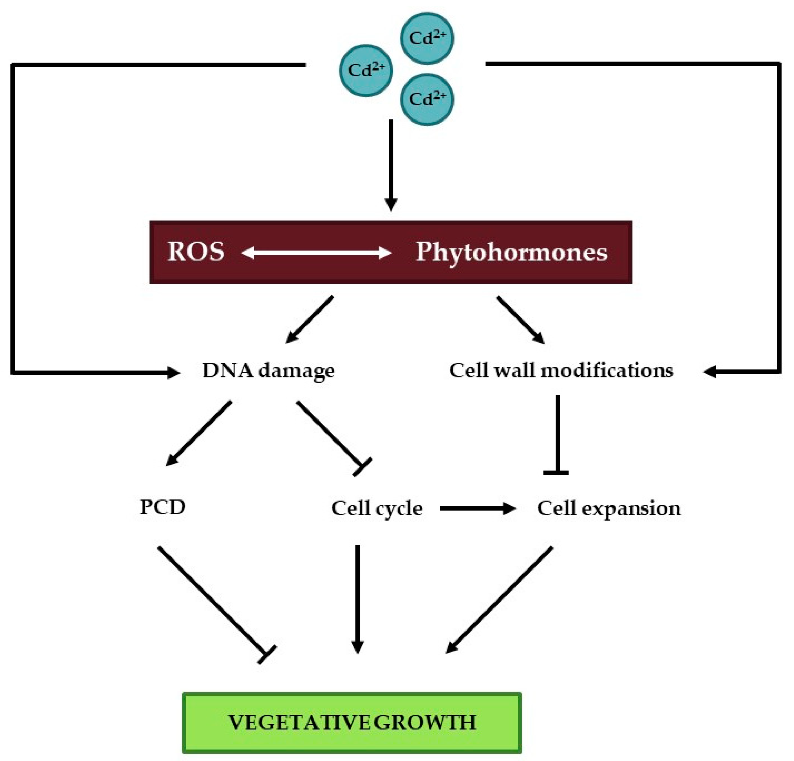
3. Vegetative Plant Growth

Once germination has occurred, two major processes drive plant growth, namely cell division and cell expansion, which is limited by the cell wall during vegetative growth. In the following sections, the effect of Cd on these processes is uncovered in detail (Figure 2).

3.1. The DNA Damage Response

Stress-induced effects on cell cycle progression often result from the activation of the DNA damage response (DDR). Upon perceiving DNA damage, cells trigger this response, which includes the activation of DNA repair pathways. When the extent of DNA damage is low, cell cycle progression is transiently inhibited in order to repair the DNA before DNA replication or cell division take place. When the damaged DNA cannot be repaired, cells undergo terminal differentiation or programmed cell death (PCD) [74].
The induction of cell cycle arrest upon the perception of DNA damage requires the activation of one of two phosphatidylinositol-3-OH-kinase-like kinases: ataxia telangiectasia mutated (ATM) and ATM- and RAD3-related (ATR). Whereas ATM is mainly activated by the presence of DNA double-strand breaks (DSBs), ATR is involved in responses to stalled replication forks. However, both types of DNA damage often occur simultaneously, causing the activation of both kinases [74]. Whereas cell cycle regulation in response to DNA damage depends on p53 in animals, plants lack a p53 orthologue. Instead, SUPPRESSOR OF GAMMA RESPONSE 1 (SOG1) is considered as the plant counterpart of p53. This transcription factor belongs to the NO APICAL MERISTEM/ARABIDOPSIS TRANSCRIPTION ACTIVATION FACTOR/CUP-SHAPED COTYLEDON (NAC) domain family and is activated through phosphorylation by ATM. Once active, SOG1 induces the expression of a multitude of genes involved in DNA repair, cell cycle progression and cell death [74,75]. In addition to SOG1, also WEE1 plays a key role in the plant DDR. This kinase can be activated by both ATM and ATR and mainly controls S phase progression [74].

3.1.1. DNA Damage

Cadmium exposure is well known to induce DNA damage in mammalian cells, as reviewed by Bertin and Averbeck (2006) and Filipic (2012) [76,77]. The mechanisms responsible for this Cd-induced DNA damage include ROS-induced formation of 8-hydroxyguanosine and the inhibition of DNA repair systems. Furthermore, Cd is able to interfere with proteins containing a zinc finger motif, implicated in the maintenance of genome stability, DNA repair and DNA damage signaling [76,77].
At present, in-depth knowledge on Cd-induced DNA damage and its underlying mechanisms in plants is lacking. However, many studies have demonstrated that Cd exposure induces DNA damage in multiple plant species. Table 1 provides an overview of recently published research (since 2014) demonstrating different types of Cd-induced DNA damage in a broad range of plant species. Several authors reported that Cd exposure increases the percentage tail length, tail intensity and tail moment determined through single cell gel electrophoresis, also known as the comet assay (Table 1). The detection of DNA damage using this method relies on the fact that DNA strand breaks facilitate DNA migration from the nucleoids towards the anode during gel electrophoresis, thereby forming a “comet tail”. Whereas the alkaline comet assay (performed at pH > 13) detects both DNA single-stranded breaks (SSBs) and DSBs, only DSBs are detected through the neutral comet assay [78,79]. Taken together, the results obtained through this method suggest that Cd exposure induces DNA strand breaks in different plant species including V. faba [80,81,82], Nicotiana tabacum [83,84], Allium sativum [82], Solanum tuberosum [85], Allium cepa [80,86,87], Lemna minor [88,89], Lactuca sativa [86,90], H. vulgare [91], Brassica oleracea and Trifolium repens [92]. In S. tuberosum, the Cd-induced increase in percentage tail DNA was more pronounced in roots as compared to leaves and appeared later in the latter organ [85]. Similarly, Cd exposure for 24 and 72 h caused significant increases in the percentage tail DNA and the tail moment in N. tabacum roots, whereas no effects were observed in leaves [83]. This is likely due to the fact that roots form the entry route for Cd into the plant and are therefore exposed earlier than leaves. Furthermore, roots of both plant species were shown to accumulate higher Cd levels than leaves [83,85].
In addition to inducing DNA strand breaks, Cd is also shown to induce micronucleus formation and chromosomal aberrations, which are both detected through microscopic analysis (Table 1). Micronuclei arise when chromosome fragments or entire chromosomes fail to be included in the nuclei of the daughter cells at the end of mitosis, as they do not manage to properly attach to the mitotic spindle during anaphase. They eventually become enclosed by a nuclear membrane and appear as small nuclei after conventional nuclear staining. Micronuclei are generally considered as biomarkers for genotoxicity [93,94]. Chromosomal aberrations frequently detected during mitosis in Cd-exposed plants include anaphase and telophase bridges, sticky chromosomes, chromosome breaks, non-oriented chromosomes and laggards chromosomes [80,86,95,96]].
Another approach frequently used to determine Cd-induced genotoxicity in plants is the investigation of random amplified polymorphic DNA (RAPD) profiles. This method consists of a PCR amplification of genomic DNA using multiple short (approximately 10 nucleotides) random primers, followed by gel electrophoresis and visualization of the amplified PCR products. Mutations and other types of DNA damage induced by exposure to stress factors such as Cd can result in the disappearance as well as de novo creation of primer annealing sites, thereby yielding an altered RAPD profile [97]. This subsequently allows for calculation of the genomic template stability (GTS) using the following formula: GTS (%) = (1 – a/n) × 100, where “a” represents the number of polymorphic bands in the treated samples and “n” identifies the total number of bands in the control samples [98,99]. As indicated in Table 1, Cd exposure was shown to alter the RAPD profile in both roots and leaves of a broad range of plant species, resulting in a reduced GTS.
Other markers used to assess Cd-induced DNA damage are the full peak coefficient of variation determined via flow cytometric analysis [113] and DNA polymorphisms determined by PCR-based methods (besides RAPD) such as amplified fragment length polymorphism (AFLP), simple sequence repeat (SSR), inter-simple sequence repeat (ISSR) and sequence-related amplified polymorphism (SRAP) [103,104,108,109]]. However, in comparison to the other DNA damage indicators described, the use of these markers in studies evaluating Cd genotoxicity in plants is currently limited.
Despite the large number of studies indicating Cd-induced genotoxicity in plants, its underlying mechanisms are still largely unknown. However, as Cd exposure is well known to induce ROS production, it is likely that DNA oxidation contributes to the observed damage. Therefore, it would be of interest to study the extent of oxidative DNA damage in Cd-exposed plants. Furthermore, it would be interesting to assess the extent of DNA repair in Cd-exposed plants to gain an insight into plant responses to the observed Cd-induced damage. Although methods for the assessment of oxidative DNA damage and DNA repair are widely adopted in animal samples, these methods have so far received little attention in plant research. However, their optimization might significantly enhance our knowledge on DNA damage and repair induced by Cd and other stress factors in plants.

3.1.2. The Cell Cycle

As a consequence of inducing DNA damage, Cd exposure can affect cell cycle progression. The formation of new cells through cell division is the primary driving force for organ growth in plants. Similar to that in other eukaryotes, the classical plant cell cycle consists of four phases: gap 1 (G1) phase, DNA synthesis (S) phase, gap 2 (G2) phase and mitotic (M) phase [114]. The gap phases enable the control of accurate and full completion of previous phases. As a consequence, many important regulatory mechanisms controlling cell cycle progression operate at the G1/S and G2/M transitions. During the S phase, nuclear DNA is replicated, whereas the replicated sister chromatids are divided over the two daughter cells arising through cytokinesis during the M phase. The cell cycle is regulated by the activity of cyclin-dependent kinases (CDKs), which are serine/threonine protein kinases that phosphorylate target proteins crucial for cell cycle progression. As their name implies, CDKs form heterodimers with regulatory cyclins in order to become activated [115]. During progression throughout the cell cycle, CDK activity shows a typical pattern, reaching two thresholds: one for DNA replication (S phase) and one for cell division (M phase) [116].
In addition to dividing through the classical cell cycle, plant cells can also undergo endoreduplication. During this alternative cell cycle mode, plant cells replicate their nuclear DNA (S phase) without intermittent cell division (M phase), resulting in endopolyploidy (i.e., the existence of different ploidy levels in adjacent cells of a species) [117].
Similar to the classical cell cycle, endoreduplication is also regulated by the action of CDK-cyclin complexes. During an endocycle, CDK activity only reaches the threshold for DNA replication but not for cell division. Endoreduplication is important for normal plant growth and development, as it is tightly related to cell differentiation and expansion [116]. The importance of endoreduplication in plant development and its connection to cell size become apparent during trichome development. Trichomes are large, single epidermal cells that develop on most aerial parts of A. thaliana plants and typically contain three to four branches. These highly specialized structures—involved in plant protection against external stress factors such as herbivory, frost and ultraviolet radiation—require endoreduplication for their development and reach a final DNA content of 32C, with C representing the haploid DNA content [118,119]. Interestingly, trichomes were previously shown to accumulate Cd, possibly to prevent Cd-induced damage at more sensitive sites within the plant [120,121]. In addition to its involvement in plant growth and development, endoreduplication could also serve as a strategy in plant defense against biotic and abiotic stress factors, as reviewed by Scholes and Paige (2015) [122]. Under stress conditions, an increased ploidy level and therefore a higher number of DNA templates could help to sustain genome integrity and stimulate genetic pathways responsible for plant defense [122].
Cadmium-induced disturbances of root and leaf growth were shown to coincide with effects on cell division in a wide range of plant species in a multitude of experimental set-ups. An overview of recently published research (since 2014) demonstrating Cd-induced effects on multiple cell cycle-related parameters in several plant species is provided in Table 2. In general, Cd negatively affects cell cycle progression, as becomes apparent from a decreased mitotic index (i.e., the ratio between the number of cells undergoing mitosis and the total cell number), determined via microscopic analysis. As suggested by Monteiro et al. (2012), Cd might bind to the sulfhydryl groups of cysteine residues present in tubulins, thereby affecting microtubule formation and disturbing cell division [90]. Although many studies have addressed the influence of Cd exposure on the cell cycle in roots, knowledge regarding its effects on this process in leaves is scarce. However, Baryla et al. (2001) demonstrated that leaves of Cd-exposed Brassica napus plants contained a smaller number of stomatal guard cells and were characterized by a larger mesophyll cell size as compared to their control counterparts, suggesting that Cd also affects the cell cycle in leaves [123]. Similarly, Cd exposure inhibited cell division and increased cell size in both the upper and lower cell layers of young Elodea canadensis leaves. Interestingly, this response coincided with a strong Cd-induced disturbance of the typical cell wall structure, which was likely the consequence of the accumulation of large amounts of Cd in the apoplast and the binding of Cd ions to cell wall components [124] (cfr. infra). Furthermore, Cd exposure was shown to reduce both adaxial pavement cell number and surface area in different leaves of A. thaliana in a time-dependent manner. The decreased cell surface area might be related to a lower extent of endoreduplication in leaves of Cd-exposed plants, as indicated by a significantly decreased endoreduplication factor (i.e., the average number of endocycles that has taken place per cell) [105]. In contrast, other studies reported a decreased percentage of cells with a 2C nuclear DNA content and an increased percentage of cells with a higher nuclear DNA content in roots of A. thaliana [101,102] and P. sativum [125,126,127]. Although an increased proportion of 4C cells might indicate a cell cycle arrest in G2 phase, an increased level of 8C cells points towards an elevated extent of endoreduplication. These data suggest that Cd exposure stimulates this alternative cell cycle variant in roots, whereas inhibiting it in leaves. However, in young A. thaliana leaves, an increased extent of endoreduplication was observed shortly after the start of Cd exposure [Hendrix et al., personal communication]. It could be hypothesized that increased ploidy levels confer tolerance to Cd stress, since Talukdar (2014) demonstrated that roots of tetraploid and triploid Lathyrus sativus plants were less sensitive to Cd as compared to their diploid counterparts [95]. Furthermore, it is tempting to speculate that the increased extent of endoreduplication is related to a stimulation of trichome development, as these structures are characterized by a high nuclear DNA content [118,119] and provide sites for Cd sequestration [120,121]. However, this hypothesis requires further investigation.
Cadmium-induced effects on the cell cycle in roots and leaves often coincide with alterations in the expression of cell-cycle related genes, such as those encoding cyclins and CDKs (Table 2). Indeed, CYCB1 expression decreased upon Cd exposure in Glycine max cell suspension cultures, possibly affecting G2/M progression [128,129]. Exposure to 100 µM Cd for 15 days affected the expression of a large number of cell cycle-related genes in roots of O. sativa. Interestingly, Cd-induced effects on a number of these genes were altered by simultaneous treatment with 2,3,5-triiodobenzoic acid (an inhibitor of polar AUX transport), indole-3-butytric acid (an AUX hormone), Tiron (a superoxide (O2•−) scavenger) or sodium diethyldithiocarbamate (an SOD inhibitor) [130]. In another study, the authors demonstrated that treatment with ABA or tungstate (an ABA inhibitor) also influenced Cd-induced effects on transcript levels of certain genes involved in cell cycle regulation [131]. These data emphasize the involvement of plant hormones and ROS in Cd-induced effects on the cell cycle. The significance of various phytohormones and their complex interactions in regulating cell division and endoreduplication was extensively reviewed by Tank et al. (2014) [132]. Although the precise role of ROS in cell cycle regulation is still unclear, their importance in this process is illustrated by the fact that oxidative and reductive signals are required for certain cell cycle transitions. In addition, transcript levels and activities of cyclins and CDKs were demonstrated to be altered upon redox perturbations. The antioxidative metabolite GSH might constitute an important redox-related cell cycle regulator, as it was reported to be translocated into the nucleus during cell division [133] and the severely GSH-deficient root meristemless 1 (rml1) A. thaliana mutant is unable to form a root apical meristem due to a lack of cell division [134]. Furthermore, ROS and redox homeostasis are also crucial mediators of cytokinesis, as indicated by a disturbed cell division in mutants characterized by impaired ROS production or signal transduction [135,136]. The involvement of ROS and redox regulators in the cell cycle is discussed in more detail in a recent review by Mhamdi and Van Breusegem (2018) [137].
Additional evidence for Cd-induced effects on transcript levels of cell cycle-related genes comes from A. thaliana. In roots of this species, Cd exposure caused a downregulation of several G1/S marker genes such as HISTONE H4 and E2Fa and G2/M marker genes, including CYCB1;1 and CYCB1;2. This response coincided with an altered expression of DNA repair genes and was less pronounced in mutants with an impaired DNA mismatch repair pathway, suggesting that DNA damage contributes to the observed cell cycle arrest [101,102]. In agreement with this hypothesis, Hendrix et al. (2018) demonstrated significant increases in the expression of several DNA repair genes and genes encoding CDK inhibitors of the SIAMESE-related (SMR) family in leaves of A. thaliana exposed to 5 µM Cd for 72 h [105]. Interestingly, these SMR genes were previously reported to be transcriptionally upregulated in response to ROS-induced DNA damage in plants exposed to hydroxyurea [138], further pointing towards the involvement of DNA damage in the Cd-induced cell cycle inhibition.

3.1.3. Cell Death

In case DNA damage is severe and cannot be repaired, the DDR is responsible for activating PCD [74]. Plant PCD is defined as a genetically encoded and actively controlled form of cellular suicide and is generally subdivided into developmental PCD and environmentally induced PCD. Developmental PCD plays a crucial role in normal plant growth and development, being involved in seed development and germination, as well as vegetative development. During the latter, PCD is for example involved in the differentiation of xylem tracheary elements, the emergence of lateral and adventitious roots and in senescence [140]. It is well-known that ROS and NO are important regulators of both developmental and environmentally induced plant PCD, as was recently reviewed by Locato et al. (2016) [140].
Interestingly, Cd-induced DNA damage and cell cycle effects observed in different plant species often coincide with a decrease in cell viability. For example, Kuthanova et al. (2008) demonstrated that exposure to 50 µM Cd affected cell cycle progression in synchronized tobacco BY-2 cell cultures and significantly reduced cell viability [141]. The type of cell death induced upon Cd exposure strongly depends on the cell cycle phase at which Cd exposure started. Whereas Cd application during S and G2 phase resulted in an apoptosis-like PCD type, characterized by DNA fragmentation, no such effect was observed in cells exposed to Cd during the other cell cycle phases. Instead, cells exposed during M phase displayed a rapid cell death, coinciding with fragmented late telophasic nuclei, while the decrease in viability of cells treated with Cd at G1 phase took place at a slower rate [141]. Similarly, the decreased mitotic index and chromosomal aberrations observed in A. cepa root tips upon exposure to a range of Cd concentrations was accompanied by a strong increase in Evans blue staining, indicative of cell death [80]. However, no effect on cell viability was observed in root tips of T. aestivum exposed to a range of Cd concentrations for 48 h, although these conditions induced oxidative modifications of cell cycle-related proteins and cell cycle arrest [142]. These data emphasize that the occurrence of Cd-induced cell death depends on many factors including the plant species, Cd concentration and exposure duration.
The Cd-induced PCD response in tobacco BY-2 was shown to depend on three ROS waves: (1) an initial transient NADPH oxidase-dependent increase in H2O2 levels, (2) the accumulation of mitochondrial O2•− and (3) fatty acid hydroperoxide accumulation, which coincides with the occurrence of cell death [143]. The importance of ROS in Cd-induced PCD is also supported by the work from Tamás et al. (2017) [144], who showed that cell death in roots of Cd-exposed H. vulgare is mostly pronounced at locations where O2•− generation was observed (i.e., the transition and distal elongation zones). However, Cd-induced cell death was only observed in roots of plants exposed to 60 µM Cd, whereas no effect was observed upon exposure to lower concentrations, suggesting that cell death only occurs when ROS concentrations exceed a certain threshold [144]. In an Arabidopsis cell suspension culture, the H2O2 production required for PCD was shown to depend on NO production, as inhibition of NO synthesis partially prevented H2O2 accumulation and cell death [145]. As summarized by Locato et al. (2016) [140], different underlying mechanisms have been proposed for the observed NO-dependency of Cd-induced PCD. First, NO-dependent nitrosylation of PCs could reduce their ability to chelate Cd, thereby increasing free Cd levels and enhancing toxicity symptoms [145,146]. Furthermore, NO could increase Cd accumulation by affecting transcript levels of genes involved in Cd uptake and detoxification [147,148]. A final mechanism proposed is the Cd-induced activation of MPK6, which subsequently activates caspase-3-like, a PCD executor [149].
It is likely that phytohormones are also involved in regulating Cd-induced PCD, possibly through interactions with ROS. Simultaneous exposure of a tomato cell suspension culture to Cd and ethylene caused a stronger Cd-induced PCD response as compared to treatment with Cd alone. Furthermore, Cd-induced PCD was mitigated by application of the ethylene biosynthesis inhibitor 2-aminoethoxyvinyl glycine and the ethylene receptor blocker silver thiosulfate [150], clearly indicating the key role of ethylene signaling in Cd-induced PCD. Furthermore, a Cd-induced increase in cellular SA concentrations of tobacco cells was shown to induce a MAPK signaling pathway involved in mediating PCD [151]. Similarly, exogenously applied SA was able to alleviate Cd-induced ROS production, photosynthetic damage and cell death in Cd-exposed A. thaliana [152]. The cell death observed in this study was likely a consequence of a strong Cd-induced activation of autophagy. This process involves the vacuolar degradation and recycling of cellular macromolecules or entire organelles and was previously reported to be induced upon Cd exposure in various plant species including A. thaliana [152], G. max [153], T. aestivum [154] and Theobroma cacao [155]. Although their interplay has not yet been studied in Cd-exposed plants, crosstalk between autophagy and several phytohormones exists in plants exposed to other environmental stimuli or during normal plant development [156]. Furthermore, an interplay between autophagy and ROS was also shown, with ROS contributing to the establishment of autophagy and autophagy contributing to ROS scavenging. A comprehensive overview of the involvement of ROS and phytohormones and their interplay in autophagy in stress-exposed plants was recently published by Signorelli et al. (2019) [157]. However, the involvement and interplay of ROS and phytohormones in Cd-induced autophagy remains largely unknown. Interestingly, autophagy contributes to nutrient recycling and remobilization during senescence [157], a process which was shown to be prematurely induced upon Cd exposure in different plant species, as indicated by increases in senescence-related parameters such as protease activity, lipid peroxidation and the expression of senescence-associated genes (SAGs) [9].
Taken together, these data suggest that Cd-induced DNA damage is an important trigger for effects on plant growth either through the induction of cell death or through influences on the cell cycle. In order to prevent Cd entry into cells and subsequent DNA damage, cell wall structure can be altered to increase the number of Cd binding sites. This often coincides with an increased cell wall rigidity, which can in turn limit cell expansion and hence plant growth.

3.2. The Cell Wall

The primary cell wall consists of structural proteins that are embedded in a matrix of polysaccharides which includes cellulose, hemicellulose and pectin [158,159,160]. These components ensure that inner structures have sufficient support, but at the same time remain adjustable for the expanding cell. Furthermore, these polysaccharides contain functional groups such as hydroxyl, thiol and carboxyl groups that enable the cell wall to bind large amounts of divalent and trivalent metals including Cd [161]. The homogalacturonan (HG) domain of pectin is created by the golgi apparatus and secreted into the cell wall in a highly methylesterified form. Using a Ca ion, two HG domains can be linked together by their carboxylic groups [162]. Calcium can be replaced within this structure by other metals such as Pb, Cu, Cd and Zn that show greater affinity [161].

3.2.1. The Cell Wall as Major Storage Compartment for Cadmium

The cell wall is the primary defense structure of a plant’s cell against pathogen attacks or unfavorable environmental conditions such as drought and metals [163,164,165]. By keeping excess Cd out of the cytoplasm, it prevents damage to macromolecules, proteins and DNA caused directly or upon Cd-induced oxidative stress. Since roots are the organs in direct contact with Cd from the soil, their cell walls play an important role in this process. Many studies show that most Cd is stored within the cell walls of roots by various plant species [166,167]. For example, in the plant Coptis chinensis, the roots and rhizomes retained between 62 and 77% of all Cd [168]. When the capacity of the cell wall is exceeded, Cd might additionally form complexes with PCs and is subsequently sequestered within the vacuole [167,169,170]. However, Dong and colleagues (2016) reported that upon exposure to 200 µM CdCl2, most Cd in the leaves was present in the soluble fraction in Arachis hypogaea and compartmentalization in vacuoles might be of greater importance here [171].

3.2.2. Cadmium-Induced Cell Wall Modifications

In addition to the formation of a rigid barrier, the cell wall might also be actively modified under Cd exposure [172,173,174]. By increasing the pectin methylesterase (PME) activity, the amount of de-esterified pectin increases, thereby creating more negative charges to bind Cd [175,176]. Moreover, an increase in pectin and hemicellulose content can also contribute to the latter [177,178]. Recently, in O. sativa seedlings it was shown that root aeration increases pectin content and PME activity resulting in Cd toxicity alleviation [179]. Since pectin is especially synthesized in young, expanding cells, a delay in root maturation due to aeration might enhance pectin deposit. Next to cell wall modifications related to primary components, another effect that occurs under Cd exposure, is the activation of lignin biosynthesis [180,181]. Lignin is incorporated in the secondary cell walls of specialized cells such as tracheids and vessel elements of the xylem and is created by the oxidative polymerization of monolignol subunits by class III peroxidases (PODs) and laccases [182]. Adding lignin to the cells’ walls decreases the permeability to Cd, but at the same time restricts cell elongation, thereby inhibiting growth. An increase of POD activity in response to Cd has been observed in various plant species [180,181,183]. It is of particular interest that POD uses H2O2 as a substrate, but at the same time H2O2 comes forward as a major signaling molecule within the redox network, which is strongly disturbed by Cd exposure. These close interactions between lignification and redox regulation have been extensively reviewed by Loix and colleagues (2017) [173]. Cell wall expansion is largely regulated by ROS homeostasis in the apoplast [184]. Whereas H2O2 contributes to cell wall stiffening, hydroxyl radicals (OH) lead to cell wall loosening due to pectin and xyloglucan cleavage [185,186].
The role of cellulose under Cd exposure is still a matter of debate. The cell wall cellulose content was shown to decrease in O. sativa and Zea mays under Cd exposure, but an increase was observed in Linum usitatissimum. Therefore, it is proposed that different defense strategies are present between monocots and dicots [175,177,187]. Recently, it was shown that Cd accumulates preferably within cellulose in the Cd-sensitive G. max BX10 variety, but contrarily, the Cd-tolerant G. max HX3 accumulated more Cd in cell wall-related pectin [188]. This might even suggest intraspecies differences in sequestration of Cd in different cell wall components. Cellulose microfibrils are cable-like structures composed of many β-1,4-linked glucose molecules, which are added together by cellulose synthase using uridine diphosphate glucose as a donor molecule [189]. Cadmium is known to reduce the amount of cytosolic sucrose, which could have detrimental effects on cellulose biosynthesis, as UDP-glucose is thought to be mainly produced by sucrose synthase (SUSY) from sucrose [174,190]. In a recent proteomic study by Gutsch and colleagues (2018), SUSY was shown to be upregulated in response to long-term Cd exposure in Medicago sativa [191]. The photosynthetic capacity of these plants was inhibited, as indicated by a lower abundance of photosynthetic proteins, which cuts off glucose supply for cell wall biosynthesis. However, a higher SUSY activity might suggest that the plants still invested in cellulose synthesis. In Miscanthus sacchariflorus, several genes involved in cellulose biosynthesis were upregulated under Cd exposure as well. These included cellulose synthase A, cellulose synthase-like protein D4 and cellulose synthase-like protein H1 [192].
In addition to these polysaccharides and lignin, two protein families, i.e., xyloglucan endotransglucosylases/hydrolases (XTH) and expansins (EXP), were demonstrated to affect cell wall expansion as well [193]. Expansins have a function in breaking the non-covalent binding of polysaccharides resulting in cell wall loosening [158,194]. In N. tabacum, NtEXP1, NtEXP4 and NtEXP5 transcripts were abundant in the shoot apices and young leaves, but not in roots and mature leaves, supporting their function in cell wall extension [195]. Furthermore, these genes were induced by growth-stimulating hormones such as AUX, GA and CK, but Cd exposure inhibited their transcription. This was supported by a study in Brassica juncea by Sun and colleagues (2011) who found that overexpression of BjEXPA1 led to a higher Cd sensitivity [196]. However, the opposite was found for the TaEXP2 gene in T. aestivum [197]. This gene was upregulated under Cd exposure and its overexpression led to an increase in biomass and root elongation. This improved plant performance was stated to be due to an enhanced translocation of Cd to the vacuoles, a higher antioxidant capacity and a higher water retention. Secondly, XTHs have the ability to cut and rejoin xyloglucan, which locks cellulose microfibrils and thereby contributes to cell wall loosening [198]. In A. thaliana, XTH33 was required for Cd accumulation within the roots [199]. In addition, XTH33 was shown to be a direct target of EIN3 which acts as a master transcription factor in ethylene-mediated Cd-induced root growth inhibition. When PeXTH from Populus euphratica was introduced in N. tabacum, a decrease of xyloglucan was observed in the cell wall of the roots, leading to a reduced number of Cd binding sites, thereby reducing Cd influx into the roots and limiting Cd toxicity [200]. Lastly, in a proteomic study with two cultivars of A. hypogaea, it was hypothesized that XTHs and α-expansins might be important in keeping cell wall extensibility under Cd exposure in the Cd-sensitive cultivar, which showed a higher capacity of cell wall modification [201]. Both XTH and EXP proteins are activated by AUX, inducing acidic growth of the cell wall [202].

3.2.3. The Role of The Cell Wall in Cadmium Hyperaccumulators

Plants that are able to withstand very high metal concentrations in the soil and show an enhanced metal accumulation within the aboveground organs are defined as hyperaccumulators [169]. These remarkable plant species account for less than 0.2% of all angiosperms, although additional heavy metal-accumulating plants are likely to be identified [203]. The trait is expected to have evolved multiple times but is of very high occurrence within the Brassicaceae family [169]. It is a nice coincidence that frequently studied hyperaccumulators such as Arabidopsis halleri and Noccea caerulescens, previously known as Thlaspi caerulescens, are phylogenetic closely related to the model plant A. thaliana, which does not accumulate metals. As shown by Benzarti et al. (2008), the Cd hyperacummulator N. caerulescens has a more than tenfold higher EC50 value for Cd-induced root growth inhibition in comparison to several non-accumulator plants [204]. However, large differences in the ability to accumulate Cd also exist between populations of hyperaccumulating species [203]. In A. halleri, some individuals of metallicolous populations were able to survive in the presence of 450 µM CdSO4, whereas concentrations as low as 100 µM CdSO4 caused mortality within non-metallicolous populations [205]. Tolerance mechanisms to cope with high soil metal concentrations include enhanced metal uptake and xylem loading, followed by detoxification in the shoot, as reviewed by Verbruggen and colleagues (2009) [206].
The role of Cd storage in the cell wall of hyperaccumulators might not be that straightforward. In a comparative study between a non-hyperaccumulating and a hyperaccumulating ecotype of Sedum alfredii, it was shown that for the latter more cell wall-bound Cd was available for xylem loading [207]. Furthermore, no increase of pectin or hemicellulose 2 was detected in the hyperaccumulating ecotype, which was accompanied with a lower PME activity. This led the authors to the conclusion that Cd translocation could at least partly be due to a difference in cell wall modification regulation [207]. In addition, cell wall modifications in the shoot cell walls of A. halleri under Cd exposure were more pronounced in less tolerant populations [205]. A Cd tolerance mechanism might already be present under controlled conditions in A. halleri in contrast to A. thaliana [208]. Gene expression related to redox balance, Ca signaling, and cell wall remodeling was more affected in A. thaliana, yet PME and CESA transcripts (encoding for cellulose synthases) were more upregulated in A. halleri. This could result in a larger extent of Cd binding to the cell wall and cell wall stiffening, which in turn leads to a greater barrier for cytosolic entering [208]. Moreover, Peng and colleagues (2017) showed that the cell wall of a newly discovered hyperaccumulator Sedum plumbizincicola plays a crucial role in Cd tolerance [209]. In contrast to a non-hyperaccumulating ecotype of S. alfredii, Fourier transform infrared (FT-IR) spectroscopy displayed a higher absorbance ratio of –COO against –COOR, indicating a lower esterification of pectin and a more efficient binding of metal ions for S. plumbizincicola.

4. Reproductive Growth

In order for a plant to complete its life cycle, it must commence the reproductive phase, which in higher plants involves the setting of flowers, pollination and fertilization, followed by the production of seeds. In Brassica campestris, the GSH and AsA content dropped the most when plants were exposed to Cd at their flowering stage, indicating that the reproductive phase is highly susceptible [210]. In L. usitatissimum plants grown in Cd-contaminated soils, a decrease in fitness was noted due to 31.8% fewer seeds and 25.6% fewer fruits [211].
Although A. thaliana plants showed a reduction in silique counts under long-term Cd exposure, they were still able to complete their life cycle, thereby producing seeds with equal germination capacity as control plants [212]. The transition from the vegetative to the reproductive phase under 10 µM Cd was unaffected, which was in agreement with the results from Maistri and colleagues (2011), however an accelerated emergence of inflorescence was observed under 5 µM Cd [213]. Limited research has been done on the later stages of plant development in relation to Cd exposure. Nevertheless, accumulation of large amounts of Cd in the seeds, even without visible toxic effects to the plant, can be a great threat to human health [214]. Cadmium uptake by roots and translocation to aboveground parts is a process that continues throughout the plant’s life cycle. In O. sativa, most of the Cd accumulated in the grains during the early phases of grain development either directly via the xylem or through remobilization through the phloem [215]. However, in Solanum lycopersicum the majority of Cd was taken up in the final stage of fruit development [216]. This co-occurred with a disturbance in nutritional status, as potassium (K), Fe and Zn contents in fruits decreased, while Ca and magnesium (Mg) increased. Exposure to 100 µM Cd resulted in an absence of fruit setting. Furthermore, an interesting finding was reported in the monoecious plant Crocus sativus, where a shift to more male flowers was apparent upon Cd exposure [217]. This was also observed in a study with Cannabis sativa, where treatment with Pb resulted likewise in male flowers and was demonstrated to be due to a hormonal shift with increasing GA levels in these plants, while zeatine (a specific form of CK) decreased [218].
Cadmium has been shown to negatively affect pollen germination accompanied with a disruption of pollen tube morphology in multiple plant species [219,220]. Even very low Cd concentrations of 0.01 µg mL−1 were able to inhibit either pollen germination or tube growth in Vicia angustifolia and Vicia tetrasperma, indicating that this process is very sensitive to Cd [221]. The impaired cell elongation of the pollen tube by Cd is a consequence of its interference with the anionic content of secretory vesicles and its interaction with the cell wall, which contains large quantities of pectin and callose [222]. Cell wall thickening, an increase in cell diameter and abnormal pollen tube growth were observed in all Prunus avium cultivars tested in vitro under Cd exposure [223]. Pollen tubes of Picea wilsonii showed swelling of the tips accompanied by cytoplasmic vacuolization [224]. Furthermore, the importance of ROS/Ca signaling in pollen tube formation has been well documented [137,225]. Exogenously applied OH to pollen of N. tabacum caused loosening of the intine (i.e., the inner layer of the pollen tube cell wall), leading to a disrupted polar growth, while H2O2 stiffened the cell wall [226]. Both treatments resulted in a reduced pollen germination, since only the region of the germinating pore should be weakened through OH-dependent reactions. In conclusion, Cd negatively affects plant fitness by interfering with various processes including pollen tube formation and pollen germination resulting in smaller numbers of seeds that show a reduced germination as was described in the section on seed germination. In conclusion, knowledge on the effects of Cd on the reproductive phase of plant development is still relatively scarce in contrast to information on its effects on vegetative growth and deserves more attention in the future.

5. Conclusion and Future Perspectives

Cadmium exposure interferes with all stages of plant development, inhibiting seed germination, vegetative growth and reproductive growth. Important players in the Cd-induced disturbance of root and leaf growth are DNA damage, which subsequently affects cell cycle progression or even causes cell death, and structural alterations of the cell wall. Furthermore, Cd alters pollen tube morphology, inhibits pollen germination and can be transported into seeds and fruits. The type and extent of Cd-induced effects strongly depends on many factors, including the plant species, organ, cell type, Cd concentration and exposure duration.
Additional research on the mechanisms underlying these Cd-induced disturbances of plant growth and development is required to develop and further improve strategies to enhance plant growth on Cd-polluted soils. In this context, it would be interesting to also focus on transgenerational effects involved in plant adaptation to Cd exposure. Although research in this field is currently limited, a recent study showed that Cd-induced alterations of RAPD profiles in Urtica pilulifera parent plants are transmitted to the next generation [112]. Similarly, Carvalho et al. (2018) demonstrated that exposure of S. lycopersicum to Cd resulted in Cd accumulation in seeds, which altered their nutrient profile and caused a decreased mitotic index in root tips of the offspring [227]. Although the mechanisms underlying these transgenerational effects are currently unknown, epigenetic changes might be involved, as Cd exposure was previously shown to alter DNA methylation patterns in A. thaliana [104]. In addition, the possible involvement of the microbiome in plant adaptation to Cd exposure should also be taken into account, as transgenerational exposure of A. thaliana to Cd significantly altered the seed endophytic community [228,229]. Furthermore, inoculation with specific plant-associated bacteria improved root growth of Cd-exposed A. thaliana plants [230], emphasizing the importance of the plant microbiome in coping with environmental stress factors and optimizing plant growth and development under suboptimal conditions. Finally, the use of soil amendments such as biochar and silicon-based fertilizers is promising for future applications to alleviate Cd toxicity, thereby improving crop growth and quality [231,232].

Author Contributions

All authors participated in the conception of the topic. M.H., A.C. and S.H. wrote the manuscript. M.H. and S.H. made the figures and tables. All authors read and approved the final manuscript after critically revising it for important intellectual content.

Funding

This work was supported by the Research Foundation Flanders (FWO) by project funding for M.H. [G0B6716], S.H. [G0C7518CUY], M.J. [SBOS000119N], J.D. [FWO PhD grant], and bijzonder onderzoeksfonds (BOF) from Hasselt University to V.I. [BOF-18D02CUYA] and S.V. [PhD grant].

Conflicts of Interest

The authors declare no conflict of interest.

Abbreviations

ABAAbscisic acid
ABI3Abscisic acid insensitive 3
ACC1-aminocyclopropane-1-carboxylate
ACSACC synthase
AFLPAmplified fragment length polymorphism
AsAAscorbate
AUXAuxin
ATMAtaxia telangiectasia mutated
ATRRAD3-related
CaCalcium
CATCatalase
CdCadmium
CDKsCyclin-dependent kinases
CrChromium
CKCytokinin
DDRDNA damage response
DSBsDouble-stranded breaks
EdU5-ethynyl-2′-deoxyuridine
ETEthylene
EXPExpansin
FCMFlow cytometry
FeIron
FT-IRFourier transform infrared
GAGibberellic acid
GRGlutathione reductase
GrxGlutaredoxin
GSHGlutathione
GTSGenomic template stability
H2O2Hydrogen peroxide
HGHomogalacturonan
IAAIndole-3-acetic acid
ISSRInter-simple sequence repeat
JAJasmonic acid
MnManganese
NACNO APICAL MERISTEM/ARABIDOPSIS TRANSCRIPTION ACTIVATION FACTOR/CUP-SHAPED COTYLEDON
NADPHNicotinamide adenine dinucleotide phosphate
OHHydroxyl radical
O2Superoxide
PbLead
PCPhytochelatin
PCDProgrammed cell death
PMEPectin methylesterase
PODClass III peroxidase
PrxPeroxiredoxin
qRT-PCRQuantitative reverse transcription polymerase chain reaction
RAPDRandom amplified polymorphic DNA
ROSReactive oxygen species
RT-PCRReverse transcription polymerase chain reaction
SASalicylic acid
SAGsSenescence-associated genes
SMRSIAMESE-related
SODSuperoxide dismutase
SOG1SUPPRESSOR OF GAMMA RESPONSE 1
SRAPSequence-related amplified polymorphism
SSBsSingle-stranded breaks
SSRSimple sequence repeat
SUSYSucrose synthase
TFsTranscription factors
TrxThioredoxin
XTHEndotransglucosylases/hydrolases
ZnZinc

References

  1. Khan, M.A.; Khan, S.; Khan, A.; Alam, M. Soil contamination with cadmium, Consequences and remediation using organic amendments. Sci. Total Environ. 2017, 601, 1591–1605. [Google Scholar] [CrossRef] [PubMed]
  2. Verbruggen, N.; Hermans, C.; Schat, H. Mechanisms to cope with arsenic or cadmium excess in plants. Curr. Opin. Plant Biol. 2009, 12, 364–372. [Google Scholar] [CrossRef] [PubMed]
  3. DalCorso, G.; Farinati, S.; Maistri, S.; Furini, A. How plants cope with cadmium: Staking all on metabolism and gene expression. J. Integr. Plant Biol. 2008, 50, 1268–1280. [Google Scholar] [CrossRef] [PubMed]
  4. Cuypers, A.; Keunen, E.; Bohler, S.; Jozefczak, M.; Opdenakker, K.; Gielen, H.; Vercampt, H.; Bielen, A.; Schellingen, K.; Vangronsveld, J.; et al. Cadmium and copper stress induce a cellular oxidative challenge leading to damage versus signalling. In Metal Toxicity in Plants: Perception, Signaling and Remediation; Gupta, D.K., Sandalio, L.M., Eds.; Springer: Berlin/Heidelberg, Germany, 2012; pp. 65–90. [Google Scholar] [CrossRef]
  5. Sharma, S.S.; Dietz, K.J. The relationship between metal toxicity and cellular redox imbalance. Trends Plant Sci. 2009, 14, 43–50. [Google Scholar] [CrossRef] [PubMed]
  6. Cuypers, A.; Plusquin, M.; Remans, T.; Jozefczak, M.; Keunen, E.; Gielen, H.; Opdenakker, K.; Nair, A.R.; Munters, E.; Artois, T.J.; et al. Cadmium stress: An oxidative challenge. Biometals 2010, 23, 927–940. [Google Scholar] [CrossRef] [PubMed]
  7. Jozefczak, M.; Keunen, E.; Schat, H.; Bliek, M.; Hernandez, L.E.; Carleer, R.; Remans, T.; Bohler, S.; Vangronsveld, J.; Cuypers, A. Differential response of Arabidopsis leaves and roots to cadmium: Glutathione-related chelating capacity vs antioxidant capacity. Plant Physiol. Biochem. 2014, 83, 1–9. [Google Scholar] [CrossRef] [PubMed]
  8. Schutzendubel, A.; Polle, A. Plant responses to abiotic stresses: Heavy metal-induced oxidative stress and protection by mycorrhization. J. Exp. Bot. 2002, 53, 1351–1365. [Google Scholar] [CrossRef] [PubMed]
  9. Cuypers, A.; Hendrix, S.; dos Reis, R.A.; De Smet, S.; Deckers, J.; Gielen, H.; Jozefczak, M.; Loix, C.; Vercampt, H.; Vangronsveld, J.; et al. Hydrogen peroxide, Signaling in disguise during metal phytotoxicity. Front. Plant Sci. 2016, 7, 25. [Google Scholar] [CrossRef] [PubMed]
  10. Moller, I.M.; Jensen, P.E.; Hansson, A. Oxidative modifications to cellular components in plants. Annu. Rev. Plant Biol. 2007, 58, 459–481. [Google Scholar] [CrossRef] [PubMed]
  11. Petrov, V.D.; Van Breusegem, F. Hydrogen peroxide-A central hub for information flow in plant cells. AoB Plants 2012. [Google Scholar] [CrossRef] [PubMed]
  12. Bucker-Neto, L.; Paiva, A.L.S.; Machado, R.D.; Arenhart, R.A.; Margis-Pinheiro, M. Interactions between plant hormones and heavy metals responses. Genet. Mol. Biol. 2017, 40, 373–386. [Google Scholar] [CrossRef] [PubMed]
  13. De Smet, S.; Cuypers, A.; Vangronsveld, J.; Remans, T. Gene networks involved in hormonal control of root development in Arabidopsis thaliana: A framework for studying its disturbance by metal stress. Int. J. Mol. Sci. 2015, 16, 19195–19224. [Google Scholar] [CrossRef] [PubMed]
  14. Schellingen, K.; Van Der Straeten, D.; Vandenbussche, F.; Prinsen, E.; Remans, T.; Vangronsveld, J.; Cuypers, A. Cadmium-induced ethylene production and responses in Arabidopsis thaliana rely on ACS2 and ACS6 gene expression. BMC Plant Biol. 2014, 14, 14. [Google Scholar] [CrossRef] [PubMed]
  15. Rodriguez-Serrano, M.; Romero-Puertas, M.C.; Pazmino, D.M.; Testillano, P.S.; Risueno, M.C.; del Rio, L.A.; Sandalio, L.M. Cellular response of pea Plants to cadmium toxicity: Cross talk between reactive oxygen species, nitric oxide, and calcium. Plant Physiol. 2009, 150, 229–243. [Google Scholar] [CrossRef] [PubMed]
  16. Kim, Y.H.; Khan, A.L.; Kim, D.H.; Lee, S.Y.; Kim, K.M.; Waqas, M.; Jung, H.Y.; Shin, J.H.; Kim, J.G.; Lee, I.J. Silicon mitigates heavy metal stress by regulating P-type heavy metal ATPases, Oryza sativa low silicon genes, and endogenous phytohormones. BMC Plant Biol. 2014, 14, 13. [Google Scholar] [CrossRef] [PubMed]
  17. Hu, Y.F.; Zhou, G.Y.; Na, X.F.; Yang, L.J.; Nan, W.B.; Liu, X.; Zhang, Y.Q.; Li, J.L.; Bi, Y.R. Cadmium interferes with maintenance of auxin homeostasis in Arabidopsis seedlings. J. Plant Physiol. 2013, 170, 965–975. [Google Scholar] [CrossRef]
  18. Ronzan, M.; Piacentini, D.; Fattorini, L.; Della Rovere, F.; Eiche, E.; Rieman, M.; Altamura, M.M.; Falasca, G. Cadmium and arsenic affect root development in Oryza sativa L. negatively interacting with auxin. Environ. Exp. Bot. 2018, 151, 64–75. [Google Scholar] [CrossRef]
  19. Xia, X.J.; Zhou, Y.H.; Shi, K.; Zhou, J.; Foyer, C.H.; Yu, J.Q. Interplay between reactive oxygen species and hormones in the control of plant development and stress tolerance. J. Exp. Bot. 2015, 66, 2839–2856. [Google Scholar] [CrossRef] [Green Version]
  20. Vangronsveld, J.; Herzig, R.; Weyens, N.; Boulet, J.; Adriaensen, K.; Ruttens, A.; Thewys, T.; Vassilev, A.; Meers, E.; Nehnevajova, E.; et al. Phytoremediation of contaminated soils and groundwater: Lessons from the field. Environ. Sci. Pollut. Res. 2009, 16, 765–794. [Google Scholar] [CrossRef]
  21. Gubler, F.; Millar, A.A.; Jacobsen, J.V. Dormancy release, ABA and pre-harvest sprouting. Curr. Opin. Plant Biol. 2005, 8, 183–187. [Google Scholar] [CrossRef]
  22. Nee, G.; Xiang, Y.; Soppe, W.J.J. The release of dormancy, A wake-up call for seeds to germinate. Curr. Opin. Plant Biol. 2017, 35, 8–14. [Google Scholar] [CrossRef] [PubMed]
  23. Rodriguez, M.V.; Barrero, J.M.; Corbineau, F.; Gubler, F.; Benech-Arnold, R.L. Dormancy in cereals (not too much, not so little): About the mechanisms behind this trait. Seed Sci. Res. 2015, 25, 99–119. [Google Scholar] [CrossRef]
  24. Finkelstein, R.; Reeves, W.; Ariizumi, T.; Steber, C. Molecular aspects of seed dormancy. Annu. Rev. Plant Biol. 2008, 59, 387–415. [Google Scholar] [CrossRef] [PubMed]
  25. Graeber, K.; Nakabayashi, K.; Miatton, E.; Leubner-Metzger, G.; Soppe, W.J.J. Molecular mechanisms of seed dormancy. Plant Cell Environ. 2012, 35, 1769–1786. [Google Scholar] [CrossRef] [PubMed]
  26. Bassel, G.W. To grow or not to grow? Trends Plant Sci. 2016, 21, 498–505. [Google Scholar] [CrossRef] [PubMed]
  27. Shu, K.; Liu, X.D.; Xie, Q.; He, Z.H. Two faces of one seed: Hormonal regulation of dormancy and germination. Mol. Plant. 2016, 9, 34–45. [Google Scholar] [CrossRef] [PubMed]
  28. Liu, X.D.; Zhang, H.; Zhao, Y.; Feng, Z.Y.; Li, Q.; Yang, H.Q.; Luan, S.; Li, J.M.; He, Z.H. Auxin controls seed dormancy through stimulation of abscisic acid signaling by inducing ARF-mediated ABI3 activation in Arabidopsis. Proc. Natl. Acad. Sci. USA 2013, 110, 15485–15490. [Google Scholar] [CrossRef] [PubMed]
  29. Belin, C.; Megies, C.; Hauserova, E.; Lopez-Molina, L. Abscisic acid represses growth of the Arabidopsis embryonic axis after germination by enhancing auxin signaling. Plant Cell 2009, 21, 2253–2268. [Google Scholar] [CrossRef]
  30. Arc, E.; Sechet, J.; Corbineau, F.; Rajjou, L.; Marion-Poll, A. ABA crosstalk with ethylene and nitric oxide in seed dormancy and germination. Front. Plant Sci. 2013, 4, 19. [Google Scholar] [CrossRef]
  31. Linkies, A.; Muller, K.; Morris, K.; Tureckova, V.; Wenk, M.; Cadman, C.S.C.; Corbineau, F.; Strnad, M.; Lynn, J.R.; Finch-Savage, W.E.; et al. Ethylene interacts with abscisic acid to regulate endosperm rupture during germination: A comparative approach using Lepidium sativum and Arabidopsis thaliana. Plant Cell 2009, 21, 3803–3822. [Google Scholar] [CrossRef]
  32. Wojtyla, L.; Lechowska, K.; Kubala, S.; Garnczarska, M. Different modes of hydrogen peroxide action during seed germination. Front. Plant Sci. 2016, 7, 16. [Google Scholar] [CrossRef] [PubMed]
  33. El-Maarouf-Bouteau, H.; Sajjad, Y.; Bazin, J.; Langlade, N.; Cristescu, S.M.; Balzergue, S.; Baudouin, E.; Bailly, C. Reactive oxygen species, abscisic acid and ethylene interact to regulate sunflower seed germination. Plant Cell Environ. 2015, 38, 364–374. [Google Scholar] [CrossRef] [PubMed]
  34. Huang, Y.T.; Lin, C.; He, F.; Li, Z.; Guan, Y.J.; Hu, Q.J.; Hu, J. Exogenous spermidine improves seed germination of sweet corn via involvement in phytohormone interactions, H2O2 and relevant gene expression. BMC Plant Biol. 2017, 17, 1. [Google Scholar] [CrossRef]
  35. Bahin, E.; Bailly, C.; Sotta, B.; Kranner, I.; Corbineau, F.; Leymarie, J. Crosstalk between reactive oxygen species and hormonal signalling pathways regulates grain dormancy in barley. Plant Cell Environ. 2011, 34, 980–993. [Google Scholar] [CrossRef] [PubMed]
  36. Li, Z.; Gao, Y.; Zhang, Y.C.; Lin, C.; Gong, D.T.; Guan, Y.J.; Hu, J. Reactive oxygen species and gibberellin acid mutual induction to regulate tobacco seed germination. Front. Plant Sci. 2018, 9, 1279. [Google Scholar] [CrossRef] [PubMed]
  37. Bailly, C.; El-Maarouf-Bouteau, H.; Corbineau, F. From intracellular signaling networks to cell death: The dual role of reactive oxygen species in seed physiology. C. R. Biol. 2008, 331, 806–814. [Google Scholar] [CrossRef] [PubMed]
  38. Bykova, N.V.; Hoehn, B.; Rampitsch, C.; Banks, T.; Stebbing, J.A.; Fan, T.; Knox, R. Redox-sensitive proteome and antioxidant strategies in wheat seed dormancy control. Proteomics 2011, 11, 865–882. [Google Scholar] [CrossRef] [PubMed]
  39. El-Maarouf-Bouteau, H.; Meimoun, P.; Job, C.; Job, D.; Bailly, C. Role of protein and mRNA oxidation in seed dormancy and germination. Front. Plant Sci. 2013, 4, 77. [Google Scholar] [CrossRef] [Green Version]
  40. Job, C.; Rajjou, L.; Lovigny, Y.; Belghazi, M.; Job, D. Patterns of protein oxidation in Arabidopsis seeds and during germination. Plant Physiol. 2005, 138, 790–802. [Google Scholar] [CrossRef]
  41. Oracz, K.; Bouteau, H.E.M.; Farrant, J.M.; Cooper, K.; Belghazi, M.; Job, C.; Job, D.; Corbineau, F.; Bailly, C. ROS production and protein oxidation as a novel mechanism for seed dormancy alleviation. Plant J. 2007, 50, 452–465. [Google Scholar] [CrossRef] [Green Version]
  42. Hou, L.Y.; Wang, M.Y.; Wang, H.; Zhang, W.H.; Mao, P.S. Physiological and proteomic analyses for seed dormancy and release in the perennial grass of Leymus chinensis. Environ. Exp. Bot. 2019, 162, 95–102. [Google Scholar] [CrossRef]
  43. Li, W.Q.; Khan, M.A.; Yamaguchi, S.; Kamiya, Y. Effects of heavy metals on seed germination and early seedling growth of Arabidopsis thaliana. Plant Growth Regul. 2005, 46, 45–50. [Google Scholar] [CrossRef]
  44. Raviv, B.; Aghajanyan, L.; Granot, G.; Makover, V.; Frenkel, O.; Gutterman, Y.; Grafi, G. The dead seed coat functions as a long-term storage for active hydrolytic enzymes. PLoS ONE 2017, 12, e0181102. [Google Scholar] [CrossRef] [PubMed]
  45. Kucera, B.; Cohn, M.A.; Leubner-Metzger, G. Plant hormone interactions during seed dormancy release and germination. Seed Sci. Res. 2005, 15, 281–307. [Google Scholar] [CrossRef]
  46. Bewley, J.D. Seed germination and dormancy. Plant Cell 1997, 9, 1055–1066. [Google Scholar] [CrossRef] [PubMed]
  47. Bentsink, L.; Koornneef, M. Seed dormancy and germination. Arabidopsis Book 2008, 6. [Google Scholar] [CrossRef] [PubMed]
  48. Sfaxi-Bousbih, A.; Chaoui, A.; El Ferjani, E. Cadmium impairs mineral and carbohydrate mobilization during the germination of bean seeds. Ecotoxicol. Environ. Safe. 2010, 73, 1123–1129. [Google Scholar] [CrossRef] [PubMed]
  49. Cheng, W.D.; Zhang, G.P.; Yao, H.G.; Zhang, H.M. Genotypic difference of germination and early seedling growth in response to Cd stress and its relation to Cd accumulation. J. Plant Nutr. 2008, 31, 702–715. [Google Scholar] [CrossRef]
  50. Kuriakose, S.V.; Prasad, M.N.V. Cadmium stress affects seed germination and seedling growth in Sorghum bicolor (L.) Moench by changing the activities of hydrolyzing enzymes. Plant Growth Regul. 2008, 54, 143–156. [Google Scholar] [CrossRef]
  51. Ahmad, I.; Akhtar, M.J.; Zahir, Z.A.; Jamil, A. Effects of cadmium on seed germination and seedling growth of four wheat (Triticum aestivum L. ) cultivars Pak. J. Bot. 2012, 44, 1569–1574. [Google Scholar]
  52. Farooqi, Z.R.; Iqbal, M.Z.; Kabir, M.; Shafiq, M. Toxic effects of lead and cadmium on germination and seedling growth of Albizia Lebbeck (L.) benth. Pak. J. Bot. 2009, 41, 27–33. [Google Scholar]
  53. Liu, S.J.; Yang, C.Y.; Xie, W.J.; Xia, C.H.; Fan, P. The effects of cadmium on germination and seedling growth of Suaeda salsa. In Proceedings of the Seventh International Conference on Waste Management and Technology (ICWMT 7), Beijing, China, 5–7 September 2012; Volume 16, pp. 293–298. [Google Scholar] [CrossRef]
  54. Alaraidh, I.A.; Alsahli, A.A.; Razik, E.S.A. Alteration of antioxidant gene expression in response to heavy metal stress in Trigonella foenum-graecum L. S. Afr. J. Bot. 2018, 115, 90–93. [Google Scholar] [CrossRef]
  55. Titov, A.F.; Talanova, V.V.; Boeva, N.P. Growth responses of barley and wheat seedlings to lead and cadmium. Biol. Plant. 1996, 38, 431–436. [Google Scholar] [CrossRef]
  56. Ahsan, N.; Lee, S.H.; Lee, D.G.; Lee, H.; Lee, S.W.; Bahk, J.D.; Lee, B.H. Physiological and protein profiles alternation of germinating rice seedlings exposed to acute cadmium toxicity. C. R. Biol. 2007, 330, 735–746. [Google Scholar] [CrossRef] [PubMed]
  57. Munzuroglu, O.; Zengin, F.K. Effect of cadmium on germination, coleoptile and root growth of barley seeds in the presence of gibberellic acid and kinetin. J. Environ. Biol. 2006, 27, 671–677. [Google Scholar] [PubMed]
  58. Lefevre, I.; Marchal, G.; Correal, E.; Zanuzzi, A.; Lutts, S. Variation in response to heavy metals during vegetative growth in Dorycnium pentaphyllum Scop. Plant Growth Regul. 2009, 59, 1–11. [Google Scholar] [CrossRef]
  59. Vijayaragavan, M.; Prabhahar, C.; Sureshkumar, J.; Natarajan, A.; Vijayarengan, P.; Sharavanan, S. Toxic effect of cadmium on seed germination, growth and biochemical contents of cowpea (Vigna unguiculata L.) plants. Int. Multidiscip. Res. J. 2011, 1, 1–6. [Google Scholar]
  60. De Lespinay, A.; Lequeux, H.; Lambillotte, B.; Lutts, S. Protein synthesis is differentially required for germination in Poa pratensis and Trifolium repens in the absence or in the presence of cadmium. Plant Growth Regul. 2010, 61, 205–214. [Google Scholar] [CrossRef]
  61. Kalai, T.; Bouthour, D.; Manai, J.; Ben Kaab, L.B.; Gouia, H. Salicylic acid alleviates the toxicity of cadmium on seedling growth, amylases and phosphatases activity in germinating barley seeds. Arch. Agron. Soil Sci. 2016, 62, 892–904. [Google Scholar] [CrossRef]
  62. He, J.Y.; Ren, Y.F.; Zhu, C.; Jiang, D. Effects of cadmium stress on seed germination, seedling growth and seed amylase activities in rice (Oryza sativa). Rice Sci. 2008, 15, 319–325. [Google Scholar] [CrossRef]
  63. Rivetta, A.; Negrini, N.; Cocucci, M. Involvement of Ca2+-calmodulin in Cd2+ toxicity during the early phases of radish (Raphanus sativus L) seed germination. Plant Cell Environ. 1997, 20, 600–608. [Google Scholar] [CrossRef]
  64. Cocucci, M.; Negrini, N. Change in the levels of calmodulin and of a calmodulin inhibitor in the early phases of radish (Raphanus sativus L) seed germination—Effects of ABA and fusicoccin. Plant Physiol. 1988, 88, 910–914. [Google Scholar] [CrossRef] [PubMed]
  65. Rahoui, S.; Chaoui, A.; El Ferjani, E. Reserve mobilization disorder in germinating seeds of Vicia faba L. exposed to cadmium. J. Plant Nutr. 2010, 33, 809–817. [Google Scholar] [CrossRef]
  66. Rahoui, S.; Chaoui, A.; El Ferjani, E. Membrane damage and solute leakage from germinating pea seed under cadmium stress. J. Hazard. Mater. 2010, 178, 1128–1131. [Google Scholar] [CrossRef] [PubMed]
  67. Jalmi, S.K.; Bhagat, P.K.; Verma, D.; Noryang, S.; Tayyeba, S.; Singh, K.; Sharma, D.; Sinha, A.K. Traversing the links between heavy metal stress and plant signaling. Front. Plant Sci. 2018, 9, 12. [Google Scholar] [CrossRef] [PubMed]
  68. Smiri, M.; Jelali, N.; El Ghoul, J. Cadmium affects the NADP-thioredoxin reductase/thioredoxin system in germinating pea seeds. J. Plant Interact. 2013, 8, 125–133. [Google Scholar] [CrossRef] [Green Version]
  69. Alkhalfioui, F.; Renard, M.; Vensel, W.H.; Wong, J.; Tanaka, C.K.; Hurkman, W.J.; Buchanan, B.B.; Montrichard, F. Thioredoxin-linked proteins are reduced during germination of Medicago truncatula seeds. Plant Physiol. 2007, 144, 1559–1579. [Google Scholar] [CrossRef]
  70. Smiri, M.; Chaoui, A.; Rouhier, N.; Gelhaye, E.; Jacquot, J.P.; El Ferjani, E. Cadmium affects the glutathione/glutaredoxin system in germinating pea seeds. Biol. Trace Elem. Res. 2011, 142, 93–105. [Google Scholar] [CrossRef]
  71. Smiri, M.; Jelali, N.; El Ghoul, J. Role for plant peroxiredoxin in cadmium chelation. J. Plant Interact. 2013, 8, 255–262. [Google Scholar] [CrossRef] [Green Version]
  72. Sneideris, L.C.; Gavassi, M.A.; Campos, M.L.; D’Amico-Damiao, V.; Carvalho, R.F. Effects of hormonal priming on seed germination of pigeon pea under cadmium stress. An. Acad. Bras. Cienc. 2015, 87, 1847–1852. [Google Scholar] [CrossRef] [Green Version]
  73. He, J.Y.; Ren, Y.F.; Pan, X.B.; Yan, Y.P.; Zhu, C.; Jiang, D. Salicylic acid alleviates the toxicity effect of cadmium on germination, seedling growth, and amylase activity of rice. J. Plant Nutr. Soil Sci. 2010, 173, 300–305. [Google Scholar] [CrossRef]
  74. Hu, Z.B.; Cools, T.; De Veylder, L. Mechanisms used by plants to cope with DNA Damage. Annu. Rev. Plant Biol. 2016, 67, 439–462. [Google Scholar] [CrossRef] [PubMed]
  75. Yoshiyama, K.O. SOG1: A master regulator of the DNA damage response in plants. Genes Genet. Syst. 2015, 90, 209–216. [Google Scholar] [CrossRef] [PubMed]
  76. Bertin, G.; Averbeck, D. Cadmium: Cellular effects, modifications of biomolecules, modulation of DNA repair and genotoxic consequences (A review). Biochimie 2006, 88, 1549–1559. [Google Scholar] [CrossRef] [PubMed]
  77. Filipic, M. Mechanisms of cadmium induced genomic instability. Mutat. Res.-Fundam. Mol. Mech. Mutagen. 2012, 733, 69–77. [Google Scholar] [CrossRef] [PubMed]
  78. Afanasieva, K.; Sivolob, A. Physical principles and new applications of comet assay. Biophys. Chem. 2018, 238, 1–7. [Google Scholar] [CrossRef]
  79. Ventura, L.; Giovannini, A.; Savio, M.; Dona, M.; Macovei, A.; Buttafava, A.; Carbonera, D.; Balestrazzi, A. Single Cell Gel Electrophoresis (Comet) assay with plants: Research on DNA repair and ecogenotoxicity testing. Chemosphere 2013, 92, 1–9. [Google Scholar] [CrossRef]
  80. Arya, S.K.; Mukherjee, A. Sensitivity of Allium cepa and Vicia faba towards cadmium toxicity. J. Soil Sci. Plant Nutr. 2014, 14, 447–458. [Google Scholar] [CrossRef]
  81. Koppen, G.; Verschaeve, L. The alkaline comet test on plant cells: A new genotoxicity test for DNA strand breaks in Vicia faba root cells. Mutat. Res.-Environ. Mutagen. Rel. Subj. 1996, 360, 193–200. [Google Scholar] [CrossRef]
  82. Lin, A.J.; Zhang, X.H.; Chen, M.M.; Cao, Q. Oxidative stress and DNA damages induced by cadmium accumulation. J. Environ. Sci. 2007, 19, 596–602. [Google Scholar] [CrossRef]
  83. Gichner, T.; Patkova, Z.; Szakova, J.; Demnerova, K. Cadmium induces DNA damage in tobacco roots, but no DNA damage, somatic mutations or homologous recombination in tobacco leaves. Mutat. Res. Genet. Toxicol. Environ. Mutagen. 2004, 559, 49–57. [Google Scholar] [CrossRef] [PubMed]
  84. Tkalec, M.; Stefanic, P.P.; Cvjetko, P.; Sikic, S.; Pavlica, M.; Balen, B. The effects of cadmium-zinc interactions on biochemical responses in tobacco seedlings and adult plants. PLoS ONE 2014, 9, 13. [Google Scholar] [CrossRef] [PubMed]
  85. Gichner, T.; Patkova, Z.; Szakova, J.; Znidar, I.; Mukherjee, A. DNA damage in potato plants induced by cadmium, ethyl methanesulphonate and gamma-rays. Environ. Exp. Bot. 2008, 62, 113–119. [Google Scholar] [CrossRef]
  86. Silveira, G.L.; Lima, M.G.F.; dos Reis, G.B.; Palmieri, M.J.; Andrade-Vieria, L.F. Toxic effects of environmental pollutants: Comparative investigation using Allium cepa L. and Lactuca sativa L. Chemosphere 2017, 178, 359–367. [Google Scholar] [CrossRef] [PubMed]
  87. Seth, C.S.; Misra, V.; Chauhan, L.K.S.; Singh, R.R. Genotoxicity of cadmium on root meristem cells of Allium cepa: Cytogenetic and Comet assay approach. Ecotox. Environ. Safe. 2008, 71, 711–716. [Google Scholar] [CrossRef] [PubMed]
  88. Balen, B.; Tkalec, M.; Sikic, S.; Tolic, S.; Cvjetko, P.; Pavlica, M.; Vidakovic-Cifrek, Z. Biochemical responses of Lemna minor experimentally exposed to cadmium and zinc. Ecotoxicology 2011, 20, 815–826. [Google Scholar] [CrossRef] [PubMed]
  89. Cvjetko, P.; Tolic, S.; Sikic, S.; Balen, B.; Tkalec, M.; Vidakovic-Cifrek, Z.; Pavlica, M. Effect of copper on the toxicity and genotoxicity of cadmium in duckweed (Lemna minor L.). Arh. Hig. Rada. Toksikol. 2010, 61, 287–296. [Google Scholar] [CrossRef] [PubMed]
  90. Monteiro, C.; Santos, C.; Pinho, S.; Oliveira, H.; Pedrosa, T.; Dias, M.C. Cadmium-induced cyto- and genotoxicity are organ-dependent in lettuce. Chem. Res. Toxicol. 2012, 25, 1423–1434. [Google Scholar] [CrossRef] [PubMed]
  91. Cao, F.B.; Chen, F.; Sun, H.Y.; Zhang, G.P.; Chen, Z.H.; Wu, F.B. Genome-wide transcriptome and functional analysis of two contrasting genotypes reveals key genes for cadmium tolerance in barley. BMC Genomics 2014, 15, 14. [Google Scholar] [CrossRef]
  92. Lanier, C.; Bernard, F.; Dumez, S.; Leclercq-Dransart, J.; Lemiere, S.; Vandenbulcke, F.; Nesslany, F.; Platel, A.; Devred, I.; Hayet, A.; et al. Combined toxic effects and DNA damage to two plant species exposed to binary metal mixtures (Cd/Pb). Ecotox. Environ. Safe. 2019, 167, 278–287. [Google Scholar] [CrossRef]
  93. Fenech, M.; Kirsch-Volders, M.; Natarajan, A.T.; Surralles, J.; Crott, J.W.; Parry, J.; Norppa, H.; Eastmond, D.A.; Tucker, J.D.; Thomas, P. Molecular mechanisms of micronucleus, nucleoplasmic bridge and nuclear bud formation in mammalian and human cells. Mutagenesis 2011, 26, 125–132. [Google Scholar] [CrossRef] [PubMed]
  94. Vral, A.; Fenech, M.; Thierens, H. The micronucleus assay as a biological dosimeter of in vivo ionising radiation exposure. Mutagenesis 2011, 26, 11–17. [Google Scholar] [CrossRef] [PubMed]
  95. Talukdar, D. Increasing nuclear ploidy enhances the capability of antioxidant defense and reduces chromotoxicity in Lathyrus sativus roots under cadmium stress. Turk. J. Bot. 2014, 38, 696–712. [Google Scholar] [CrossRef]
  96. Aslam, R.; Ansari, M.Y.K.; Choudhary, S.; Bhat, T.M.; Jahan, N. Genotoxic effects of heavy metal cadmium on growth, biochemical, cyto-physiological parameters and detection of DNA polymorphism by RAPD in Capsicum annuum L. - An important spice crop of India. Saudi J. Biol. Sci. 2014, 21, 465–472. [Google Scholar] [CrossRef] [PubMed]
  97. Enan, M.R. Application of random amplified polymorphic DNA (RAPD) to detect the genotoxic effect of heavy metals. Biotechnol. Appl. Biochem. 2006, 43, 147–154. [Google Scholar] [CrossRef] [PubMed]
  98. Cenkci, S.; Dogan, N. Random amplified polymorphic DNA as a method to screen metal-tolerant barley (Hordeum vulgare L.) genotypes. Turk. J. Bot. 2015, 39, 747–756. [Google Scholar] [CrossRef]
  99. Tanee, T.; Sudmoon, R.; Thamsenanupap, P.; Chaveerach, A. Effect of cadmium on DNA changes in Ipomoea aquatica Forssk. Pol. J. Environ. Stud. 2016, 25, 311–315. [Google Scholar] [CrossRef]
  100. Lima, M.G.F.; Rocha, L.C.; Silveira, G.L.; Alvarenga, I.F.S.; Andrade-Vieria, L.F. Nucleolar alterations are reliable parameters to determine the cytogenotoxicity of environmental pollutants. Ecotox. Environ. Safe. 2019, 174, 630–636. [Google Scholar] [CrossRef]
  101. Cui, W.N.; Wang, H.T.; Song, J.; Cao, X.; Rogers, H.J.; Francis, D.; Jia, C.Y.; Sun, L.Z.; Hou, M.F.; Yang, Y.S.; et al. Cell cycle arrest mediated by Cd-induced DNA damage in Arabidopsis root tips. Ecotox. Environ. Safe. 2017, 145, 569–574. [Google Scholar] [CrossRef]
  102. Cao, X.; Wang, H.T.; Zhuang, D.F.; Zhu, H.; Du, Y.L.; Cheng, Z.B.; Cui, W.N.; Rogers, H.J.; Zhang, Q.R.; Jia, C.J.; et al. Roles of MSH2 and MSH6 in cadmium-induced G2/M checkpoint arrest in Arabidopsis roots. Chemosphere 2018, 201, 586–594. [Google Scholar] [CrossRef] [PubMed]
  103. Li, Z.L.; Liu, Z.H.; Chen, R.J.; Li, X.J.; Tai, P.D.; Gong, Z.Q.; Jia, C.Y.; Liu, W. DNA damage and genetic methylation changes caused by Cd in Arabidopsis thaliana seedlings. Environ. Toxicol. Chem. 2015, 34, 2095–2103. [Google Scholar] [CrossRef] [PubMed]
  104. Wang, H.T.; He, L.; Song, J.; Cui, W.N.; Zhang, Y.Z.; Jia, C.Y.; Francis, D.; Rogers, H.J.; Sun, L.Z.; Tai, P.D.; et al. Cadmium-induced genomic instability in Arabidopsis: Molecular toxicological biomarkers for early diagnosis of cadmium stress. Chemosphere 2016, 150, 258–265. [Google Scholar] [CrossRef] [PubMed]
  105. Hendrix, S.; Keunen, E.; Mertens, A.I.G.; Beemster, G.T.S.; Vangronsveld, J.; Cuypers, A. Cell cycle regulation in different leaves of Arabidopsis thaliana plants grown under control and cadmium-exposed conditions. Environ. Exp. Bot. 2018, 155, 441–452. [Google Scholar] [CrossRef]
  106. Sudmoon, R.; Neeratanaphan, L.; Thamsenanupap, P.; Tanee, T. Hyperaccumulation of cadmium and DNA changes in popular vegetable, Brassica chinensis L. Int. J. Environ. Res. 2015, 9, 433–438. [Google Scholar]
  107. Venkatachalam, P.; Jayaraj, M.; Manikandan, R.; Geetha, N.; Rene, E.R.; Sharma, N.C.; Sahi, S.V. Zinc oxide nanoparticles (ZnONPs) alleviate heavy metal-induced toxicity in Leucaena leucocephala seedlings: A physiochemical analysis. Plant Physiol. Biochem. 2017, 110, 59–69. [Google Scholar] [CrossRef] [PubMed]
  108. Zhang, X.Q.; Chen, H.N.; Jiang, H.; Lu, W.Y.; Pan, J.J.; Qian, Q.; Xue, D.W. Measuring the damage of heavy metal cadmium in rice seedlings by SRAP analysis combined with physiological and biochemical parameters. J. Sci. Food Agric. 2015, 95, 2292–2298. [Google Scholar] [CrossRef] [PubMed]
  109. Sorrentino, M.C.; Capozzi, F.; Giordano, S.; Spagnuolo, V. Genotoxic effect of Pb and Cd on in vitro cultures of Sphagnum palustre: An evaluation by ISSR markers. Chemosphere 2017, 181, 208–215. [Google Scholar] [CrossRef] [PubMed]
  110. Ghiani, A.; Fumagalli, P.; Van, T.N.; Gentili, R.; Citterio, S. The Combined toxic and genotoxic effects of Cd and As to plant bioindicator Trifolium repens L. PLoS ONE 2014, 9, 11. [Google Scholar] [CrossRef] [PubMed]
  111. Lanier, C.; Bernard, F.; Dumez, S.; Leclercq, J.; Lemiere, S.; Vandenbulcke, F.; Nesslany, F.; Platel, A.; Devred, I.; Cuny, D.; et al. Combined effect of Cd and Pb spiked field soils on bioaccumulation, DNA damage, and peroxidase activities in Trifolium repens. Environ. Sci. Pollut. Res. 2016, 23, 1755–1767. [Google Scholar] [CrossRef] [PubMed]
  112. Dogan, I.; Ozyigit, I.I.; Tombuloglu, G.; Sakcali, M.S.; Tombuloglu, H. Assessment of Cd-induced genotoxic damage in Urtica pilulifera L. using RAPD-PCR analysis. Biotechnol. Biotechnol. Equip. 2016, 30, 284–291. [Google Scholar] [CrossRef]
  113. Monteiro, M.S.; Rodriguez, E.; Loureiro, J.; Mann, R.M.; Soares, A.; Santos, C. Flow cytometric assessment of Cd genotoxicity in three plants with different metal accumulation and detoxification capacities. Ecotox. Environ. Safe. 2010, 73, 1231–1237. [Google Scholar] [CrossRef] [PubMed]
  114. Komaki, S.; Sugimoto, K. Control of the plant cell cycle by developmental and environmental cues. Plant Cell Physiol. 2012, 53, 953–964. [Google Scholar] [CrossRef]
  115. Dewitte, W.; Murray, J.A.H. The plant cell cycle. Annu. Rev. Plant Biol. 2003, 54, 235–264. [Google Scholar] [CrossRef] [PubMed]
  116. De Veylder, L.; Larkin, J.C.; Schnittger, A. Molecular control and function of endoreplication in development and physiology. Trends Plant Sci. 2011, 16, 624–634. [Google Scholar] [CrossRef] [PubMed]
  117. Barow, M.; Meister, A. Endopolyploidy in seed plants is differently correlated to systematics, organ, life strategy and genome size. Plant Cell Environ. 2003, 26, 571–584. [Google Scholar] [CrossRef]
  118. Bramsiepe, J.; Wester, K.; Weinl, C.; Roodbarkelari, F.; Kasili, R.; Larkin, J.C.; Hulskamp, M.; Schnittger, A. Endoreplication controls cell fate maintenance. PLoS Genet. 2010, 6, 14. [Google Scholar] [CrossRef] [PubMed]
  119. Lee, H.O.; Davidson, J.M.; Duronio, R.J. Endoreplication: Polyploidy with purpose. Genes Dev. 2009, 23, 2461–2477. [Google Scholar] [CrossRef] [PubMed]
  120. Ager, F.J.; Ynsa, M.D.; Dominguez-Solis, J.R.; Gotor, C.; Respaldiza, M.A.; Romero, L.C. Cadmium localization and quantification in the plant Arabidopsis thaliana using micro-PIXE. Nucl. Instrum. Methods Phys. Res. Sect. B-Beam Interact. Mater. Atoms 2002, 189, 494–498. [Google Scholar] [CrossRef]
  121. Isaure, M.P.; Fayard, B.; Saffet, G.; Pairis, S.; Bourguignon, J. Localization and chemical forms of cadmium in plant samples by combining analytical electron microscopy and X-ray spectromicroscopy. Spectroc. Acta Pt. B-Atom. Spectr. 2006, 61, 1242–1252. [Google Scholar] [CrossRef] [Green Version]
  122. Scholes, D.R.; Paige, K.N. Plasticity in ploidy: A generalized response to stress. Trends Plant Sci. 2015, 20, 165–175. [Google Scholar] [CrossRef]
  123. Baryla, A.; Carrier, P.; Franck, F.; Coulomb, C.; Sahut, C.; Havaux, M. Leaf chlorosis in oilseed rape plants (Brassica napus) grown on cadmium-polluted soil: Causes and consequences for photosynthesis and growth. Planta 2001, 212, 696–709. [Google Scholar] [CrossRef] [PubMed]
  124. Dalla Vecchia, F.; La Rocca, N.; Moro, I.; De Faveri, S.; Andreoli, C.; Rascio, N. Morphogenetic, ultrastructural and physiological damages suffered by submerged leaves of Elodea canadensis exposed to cadmium. Plant Sci. 2005, 168, 329–338. [Google Scholar] [CrossRef]
  125. Fusconi, A.; Repetto, O.; Bona, E.; Massa, N.; Gallo, C.; Dumas-Gaudot, E.; Berta, G. Effects of cadmium on meristem activity and nucleus ploidy in roots of Pisum sativum L. cv. Frisson seedlings. Environ. Exp. Bot. 2006, 58, 253–260. [Google Scholar] [CrossRef]
  126. Repetto, O.; Massa, N.; Gianinazzi-Pearson, V.; Dumas-Gaudot, E.; Berta, G. Cadmium effects on populations of root nuclei in two pea genotypes inoculated or not with the arbuscular mycorrhizal fungus Glomus mosseae. Mycorrhiza 2007, 17, 111–120. [Google Scholar] [CrossRef] [PubMed]
  127. Wojtylakuchta, B.; Gabara, B. Changes in the content of DNA and NYS-stained nuclear, nucleolar and cytoplasmic proteins in cortex cells of pea (Pisum sativum cv. De Grace) roots treated with cadmium. Biochem. Physiol. Pflanz. 1991, 187, 67–76. [Google Scholar] [CrossRef]
  128. Sobkowiak, R.; Deckert, J. Cadmium-induced changes in growth and cell cycle gene expression in suspension-culture cells of soybean. Plant Physiol. Biochem. 2003, 41, 767–772. [Google Scholar] [CrossRef]
  129. Sobkowiak, R.; Deckert, J. The effect of cadmium on cell cycle control in suspension culture cells of soybean. Acta Physiol. Plant. 2004, 26, 335–344. [Google Scholar] [CrossRef]
  130. Zhao, F.Y.; Hu, F.; Han, M.M.; Zhang, S.Y.; Liu, W. Superoxide radical and auxin are implicated in redistribution of root growth and the expression of auxin and cell-cycle genes in cadmium-stressed rice. Russ. J. Plant Physiol. 2011, 58, 851–863. [Google Scholar] [CrossRef]
  131. Zhao, F.Y.; Wang, K.; Zhang, S.Y.; Ren, J.; Liu, T.; Wang, X. Crosstalk between ABA, auxin, MAPK signaling, and the cell cycle in cadmium-stressed rice seedlings. Acta Physiol. Plant. 2014, 36, 1879–1892. [Google Scholar] [CrossRef]
  132. Tank, J.G.; Pandya, R.V.; Thaker, V.S. Phytohormones in regulation of the cell division and endoreduplication process in the plant cell cycle. RSC Adv. 2014, 4, 12605–12613. [Google Scholar] [CrossRef]
  133. Vivancos, P.D.; Dong, Y.P.; Ziegler, K.; Markovic, J.; Pallardo, F.V.; Pellny, T.K.; Verrier, P.J.; Foyer, C.H. Recruitment of glutathione into the nucleus during cell proliferation adjusts whole-cell redox homeostasis in Arabidopsis thaliana and lowers the oxidative defence shield. Plant J. 2010, 64, 825–838. [Google Scholar] [CrossRef] [PubMed]
  134. Vernoux, T.; Wilson, R.C.; Seeley, K.A.; Reichheld, J.P.; Muroy, S.; Brown, S.; Maughan, S.C.; Cobbett, C.S.; Van Montagu, M.; Inze, D.; et al. The ROOT MERISTEMLESS1/CADMIUM SENSITIVE2 gene defines a glutathione-dependent pathway involved in initiation and maintenance of cell division during postembryonic root development. Plant Cell 2000, 12, 97–109. [Google Scholar] [CrossRef] [PubMed]
  135. Kosetsu, K.; Matsunaga, S.; Nakagami, H.; Colcombet, J.; Sasabe, M.; Soyano, T.; Takahashi, Y.; Hirt, H.; Machida, Y. The MAP Kinase MPK4 Is required for cytokinesis in Arabidopsis thaliana. Plant Cell 2010, 22, 3778–3790. [Google Scholar] [CrossRef] [PubMed]
  136. Yao, L.L.; Zhou, Q.; Pei, B.L.; Li, Y.Z. Hydrogen peroxide modulates the dynamic microtubule cytoskeleton during the defence responses to Verticillium dahliae toxins in Arabidopsis. Plant Cell Environ. 2011, 34, 1586–1598. [Google Scholar] [CrossRef] [PubMed]
  137. Mhamdi, A.; Van Breusegem, F. Reactive oxygen species in plant development. Development 2018, 145. [Google Scholar] [CrossRef] [PubMed]
  138. Yi, D.; Kamei, C.L.A.; Cools, T.; Vanderauwera, S.; Takahashi, N.; Okushima, Y.; Eekhout, T.; Yoshiyama, K.O.; Larkin, J.; Van den Daele, H.; et al. The Arabidopsis SIAMESE-RELATED cyclin-dependent Kinase inhibitors SMR5 and SMR7 regulate the DNA damage checkpoint in response to reactive oxygen species. Plant Cell 2014, 26, 296–309. [Google Scholar] [CrossRef]
  139. Zhan, Y.H.; Zhang, C.H.; Zheng, Q.X.; Huang, Z.A.; Yu, C.L. Cadmium stress inhibits the growth of primary roots by interfering auxin homeostasis in Sorghum bicolor seedlings. J. Plant Biol. 2017, 60, 593–603. [Google Scholar] [CrossRef]
  140. Locato, V.; Paradiso, A.; Sabetta, W.; De Gara, L.; de Pinto, M.C. Chapter nine - Nitric oxide and reactive oxygen species in PCD signaling. Adv. Bot. Res. 2016, 77, 165–192. [Google Scholar]
  141. Kuthanova, A.; Fischer, L.; Nick, P.; Opatrny, Z. Cell cycle phase-specific death response of tobacco BY-2 cell line to cadmium treatment. Plant Cell Environ. 2008, 31, 1634–1643. [Google Scholar] [CrossRef]
  142. Pena, L.B.; Barcia, R.A.; Azpilicueta, C.E.; Mendez, A.A.E.; Gallego, S.M. Oxidative post translational modifications of proteins related to cell cycle are involved in cadmium toxicity in wheat seedlings. Plant Sci. 2012, 196, 1–7. [Google Scholar] [CrossRef]
  143. Garnier, L.; Simon-Plas, F.; Thuleau, P.; Agnel, J.P.; Blein, J.P.; Ranjeva, R.; Montillet, J.L. Cadmium affects tobacco cells by a series of three waves of reactive oxygen species that contribute to cytotoxicity. Plant Cell Environ. 2006, 29, 1956–1969. [Google Scholar] [CrossRef] [PubMed]
  144. Tamas, L.; Mistrik, I.; Zelinova, V. Heavy metal-induced reactive oxygen species and cell death in barley root tip. Environ. Exp. Bot. 2017, 140, 34–40. [Google Scholar] [CrossRef]
  145. De Michele, R.; Vurro, E.; Rigo, C.; Costa, A.; Elviri, L.; Di Valentin, M.; Careri, M.; Zottini, M.; Sanita di Toppi, L.; Lo Schiavo, F. Nitric oxide is involved in cadmium-induced programmed cell death in Arabidopsis suspension cultures. Plant Physiol. 2009, 150, 217–228. [Google Scholar] [CrossRef] [PubMed]
  146. Elviri, L.; Speroni, F.; Careri, M.; Mangia, A.; Sanita di Toppi, L.; Zottini, M. Identification of in vivo nitrosylated phytochelatins in Arabidopsis thaliana cells by liquid chromatography-direct electrospray-linear ion trap-mass spectrometry. J. Chromatogr. A 2010, 1217, 4120–4126. [Google Scholar] [CrossRef] [PubMed]
  147. Arasimowicz-Jelonek, M.; Floryszak-Wieczorek, J.; Deckert, J.; Rucinska-Sobkowiak, R.; Gzyl, J.; Pawlak-Sprada, S.; Abramowski, D.; Jelonek, T.; Gwozdz, E.A. Nitric oxide implication in cadmium-induced programmed cell death in roots and signaling response of yellow lupine plants. Plant Physiol. Biochem. 2012, 58, 124–134. [Google Scholar] [CrossRef] [PubMed]
  148. Ma, W.W.; Xu, W.Z.; Xu, H.; Chen, Y.S.; He, Z.Y.; Ma, M. Nitric oxide modulates cadmium influx during cadmium-induced programmed cell death in tobacco BY-2 cells. Planta 2010, 232, 325–335. [Google Scholar] [CrossRef] [PubMed]
  149. Ye, Y.; Li, Z.; Xing, D. Nitric oxide promotes MPK6-mediated caspase-3-like activation in cadmium-induced Arabidopsis thaliana programmed cell death. Plant Cell Environ. 2013, 36, 1–15. [Google Scholar] [CrossRef] [PubMed]
  150. Iakimova, E.T.; Woltering, E.J.; Kapchina-Toteva, V.M.; Harren, F.J.M.; Cristescu, S.M. Cadmium toxicity in cultured tomato cells - Role of ethylene, proteases and oxidative stress in cell death signaling. Cell Biol. Int. 2008, 32, 1521–1529. [Google Scholar] [CrossRef]
  151. Pormehr, M.; Ghanati, F.; Sharifi, M.; McCabe, P.F.; Hosseinkhani, S.; Zare-Maivan, H. The role of SIPK signaling pathway in antioxidant activity and programmed cell death of tobacco cells after exposure to cadmium. Plant Sci. 2019, 280, 416–423. [Google Scholar] [CrossRef]
  152. Zhang, W.N.; Chen, W.L. Role of salicylic acid in alleviating photochemical damage and autophagic cell death induction of cadmium stress in Arabidopsis thaliana. Photochem. Photobiol. Sci. 2011, 10, 947–955. [Google Scholar] [CrossRef]
  153. Gzyl, J.; Chmielowska-Bak, J.; Przymusinski, R. Gamma-tubulin distribution and ultrastructural changes in root cells of soybean (Glycine max L.) seedlings under cadmium stress. Environ. Exp. Bot. 2017, 143, 82–90. [Google Scholar] [CrossRef]
  154. Yue, J.Y.; Wei, X.J.; Wang, H.Z. Cadmium tolerant and sensitive wheat lines: Their differences in pollutant accumulation, cell damage, and autophagy. Biol. Plant. 2018, 62, 379–387. [Google Scholar] [CrossRef]
  155. De Araujo, R.P.; de Almeida, A.A.F.; Pereira, L.S.; Mangabeira, P.A.O.; Souza, J.O.; Pirovani, C.P.; Ahnert, D.; Baligar, V.C. Photosynthetic, antioxidative, molecular and ultrastructural responses of young cacao plants to Cd toxicity in the soil. Ecotox. Environ. Safe. 2017, 144, 148–157. [Google Scholar] [CrossRef] [PubMed] [Green Version]
  156. Gou, W.; Li, X.; Guo, S.; Liu, Y.; Li, F.; Xie, Q. Autophagy in plant: A new orchestrator in the regulation of the phytohormones homeostasis. Int. J. Mol. Sci. 2019, 20, 2900. [Google Scholar] [CrossRef] [PubMed]
  157. Signorelli, S.; Tarkowski, L.P.; Van den Ende, W.; Bassham, D.C. Linking autophagy to abiotic and biotic stress responses. Trends Plant Sci. 2019, 24, 413–430. [Google Scholar] [CrossRef] [PubMed]
  158. Cosgrove, D.J. Growth of the plant cell wall. Nat. Rev. Mol. Cell Biol. 2005, 6, 850–861. [Google Scholar] [CrossRef] [PubMed]
  159. Mohnen, D. Pectin structure and biosynthesis. Curr. Opin. Plant Biol. 2008, 11, 266–277. [Google Scholar] [CrossRef] [PubMed]
  160. Scheller, H.V.; Ulvskov, P. Hemicelluloses. Annu. Rev. Plant Biol. 2010, 61, 263–289. [Google Scholar] [CrossRef]
  161. Krzeslowska, M. The cell wall in plant cell response to trace metals: Polysaccharide remodeling and its role in defense strategy. Acta Physiol. Plant. 2011, 33, 35–51. [Google Scholar] [CrossRef]
  162. Grant, G.T.; Morris, E.R.; Rees, D.A.; Smith, P.J.C.; Thom, D. Biological interactions between polysaccharides and divalent cations - Egg-box model. FEBS Lett. 1973, 32, 195–198. [Google Scholar] [CrossRef]
  163. Hall, J.L. Cellular mechanisms for heavy metal detoxification and tolerance. J. Exp. Bot. 2002, 53, 1–11. [Google Scholar] [CrossRef] [PubMed]
  164. Tenhaken, R. Cell wall remodeling under abiotic stress. Front. Plant Sci. 2015, 5, 9. [Google Scholar] [CrossRef] [PubMed]
  165. Verslues, P.E.; Agarwal, M.; Katiyar-Agarwal, S.; Zhu, J.H.; Zhu, J.K. Methods and concepts in quantifying resistance to drought, salt and freezing, abiotic stresses that affect plant water status. Plant J. 2006, 45, 523–539. [Google Scholar] [CrossRef] [PubMed]
  166. Van Belleghem, F.; Cuypers, A.; Semane, B.; Smeets, K.; Vangronsveld, J.; d’Haen, J.; Valcke, R. Subcellular localization of cadmium in roots and leaves of Arabidopsis thaliana. New Phytol. 2007, 173, 495–508. [Google Scholar] [CrossRef] [PubMed]
  167. Vazquez, S.; Goldsbrough, P.; Carpena, R.O. Assessing the relative contributions of phytochelatins and the cell wall to cadmium resistance in white lupin. Physiol. Plant. 2006, 128, 487–495. [Google Scholar] [CrossRef]
  168. Huang, W.L.; Bai, Z.Q.; Jiao, J.; Yuan, H.L.; Bao, Z.A.; Chen, S.N.; Ding, M.H.; Liang, Z.S. Distribution and chemical forms of cadmium in Coptis chinensis Franch. determined by laser ablation ICP-MS, cell fractionation, and sequential extraction. Ecotox. Environ. Safe. 2019, 171, 894–903. [Google Scholar] [CrossRef] [PubMed]
  169. Kramer, U. Metal Hyperaccumulation in Plants. Annu. Rev. Plant Biol. 2010, 61, 517–534. [Google Scholar] [CrossRef]
  170. Zhao, H.; Jin, Q.J.; Wang, Y.J.; Chu, L.L.; Li, X.; Xu, Y.C. Effects of nitric oxide on alleviating cadmium stress in Typha angustifolia. Plant Growth Regul. 2016, 78, 243–251. [Google Scholar] [CrossRef]
  171. Dong, Y.J.; Chen, W.F.; Xu, L.L.; Kong, J.; Liu, S.; He, Z.L. Nitric oxide can induce tolerance to oxidative stress of peanut seedlings under cadmium toxicity. Plant Growth Regul. 2016, 79, 19–28. [Google Scholar] [CrossRef]
  172. Berni, R.; Luyckx, M.; Xu, X.; Legay, S.; Sergeant, K.; Hausman, J.F.; Lutts, S.; Cai, G.; Guerriero, G. Reactive oxygen species and heavy metal stress in plants: Impact on the cell wall and secondary metabolism. Environ. Exp. Bot. 2019, 161, 98–106. [Google Scholar] [CrossRef]
  173. Loix, C.; Huybrechts, M.; Vangronsveld, J.; Gielen, M.; Keunen, E.; Cuypers, A. Reciprocal interactions between cadmium-induced cell wall responses and oxidative stress in plants. Front. Plant Sci. 2017, 8, 19. [Google Scholar] [CrossRef] [PubMed]
  174. Parrotta, L.; Guerriero, G.; Sergeant, K.; Cal, G.; Hausman, J.F. Target or barrier? The cell wall of early- and later-diverging plants vs cadmium toxicity: Differences in the response mechanisms. Front. Plant Sci. 2015, 6, 16. [Google Scholar] [CrossRef] [PubMed]
  175. Douchiche, O.; Rihouey, C.; Schaumann, A.; Driouich, A.; Morvan, C. Cadmium-induced alterations of the structural features of pectins in flax hypocotyl. Planta 2007, 225, 1301–1312. [Google Scholar] [CrossRef] [PubMed]
  176. Paynel, F.; Schaumann, A.; Arkoun, M.; Douchiche, O.; Morvan, C. Temporal regulation of cell-wall pectin methylesterase and peroxidase isoforms in cadmium-treated flax hypocotyl. Ann. Bot. 2009, 104, 1363–1372. [Google Scholar] [CrossRef] [PubMed] [Green Version]
  177. Xiong, J.; An, L.Y.; Lu, H.; Zhu, C. Exogenous nitric oxide enhances cadmium tolerance of rice by increasing pectin and hemicellulose contents in root cell wall. Planta 2009, 230, 755–765. [Google Scholar] [CrossRef] [PubMed]
  178. Zhao, Y.Y.; He, C.X.; Wu, Z.C.; Liu, X.W.; Cai, M.M.; Jia, W.; Zhao, X.H. Selenium reduces cadmium accumulation in seed by increasing cadmium retention in root of oilseed rape (Brassica napus L.). Environ. Exp. Bot. 2019, 158, 161–170. [Google Scholar] [CrossRef]
  179. Li, H.B.; Zheng, X.W.; Tao, L.X.; Yang, Y.J.; Gao, L.; Xiong, J. Aeration increases cadmium (Cd) retention by enhancing iron plaque formation and regulating pectin synthesis in the roots of rice (Oryza sativa) seedlings. Rice 2019, 12, 14. [Google Scholar] [CrossRef]
  180. Finger-Teixeira, A.; Ferrarese, M.D.L.; Soares, A.R.; da Silva, D.; Ferrarese, O. Cadmium-induced lignification restricts soybean root growth. Ecotox. Environ. Safe. 2010, 73, 1959–1964. [Google Scholar] [CrossRef]
  181. Schutzendubel, A.; Schwanz, P.; Teichmann, T.; Gross, K.; Langenfeld-Heyser, R.; Godbold, D.L.; Polle, A. Cadmium-induced changes in antioxidative systems, hydrogen peroxide content, and differentiation in Scots pine roots. Plant Physiol. 2001, 127, 887–898. [Google Scholar] [CrossRef]
  182. Liu, Q.Q.; Luo, L.; Zheng, L.Q. Lignins: Biosynthesis and biological functions in plants. Int. J. Mol. Sci. 2018, 19, 335. [Google Scholar] [CrossRef]
  183. Rui, H.Y.; Chen, C.; Zhang, X.X.; Shen, Z.G.; Zhang, F.Q. Cd-induced oxidative stress and lignification in the roots of two Vicia sativa L. varieties with different Cd tolerances. J. Hazard. Mater. 2016, 301, 304–313. [Google Scholar] [CrossRef] [PubMed]
  184. Schmidt, R.; Kunkowska, A.B.; Schippers, J.H.M. Role of reactive oxygen species during cell expansion in leaves. Plant Physiol. 2016, 172, 2098–2106. [Google Scholar] [CrossRef] [PubMed]
  185. Muller, K.; Linkies, A.; Vreeburg, R.A.M.; Fry, S.C.; Krieger-Liszkay, A.; Leubner-Metzger, G. In vivo cell wall loosening by hydroxyl radicals during cress seed germination and elongation growth. Plant Physiol. 2009, 150, 1855–1865. [Google Scholar] [CrossRef] [PubMed]
  186. Schopfer, P. Hydrogen peroxide-mediated cell-wall stiffening in vitro in maize coleoptiles. Planta 1996, 199, 43–49. [Google Scholar] [CrossRef]
  187. Vatehova, Z.; Malovikova, A.; Kollarova, K.; Kucerova, D.; Liskova, D. Impact of cadmium stress on two maize hybrids. Plant Physiol. Biochem. 2016, 108, 90–98. [Google Scholar] [CrossRef] [PubMed]
  188. Wang, P.; Yang, B.; Wan, H.B.; Fang, X.L.; Yang, C.Y. The differences of cell wall in roots between two contrasting soybean cultivars exposed to cadmium at young seedlings. Environ. Sci. Pollut. Res. 2018, 25, 29705–29714. [Google Scholar] [CrossRef]
  189. Somerville, C. Cellulose synthesis in higher plants. Annu. Rev. Cell Dev. Biol. 2006, 22, 53–78. [Google Scholar] [CrossRef] [PubMed]
  190. Haigler, C.H.; Ivanova-Datcheva, M.; Hogan, P.S.; Salnikov, V.V.; Hwang, S.; Martin, K.; Delmer, D.P. Carbon partitioning to cellulose synthesis. Plant Mol. Biol. 2001, 47, 29–51. [Google Scholar] [CrossRef] [PubMed]
  191. Gutsch, A.; Keunen, E.; Guerriero, G.; Renaut, J.; Cuypers, A.; Hausman, J.F.; Sergeant, K. Long-term cadmium exposure influences the abundance of proteins that impact the cell wall structure in Medicago sativa stems. Plant Biol. 2018, 20, 1023–1035. [Google Scholar] [CrossRef]
  192. Guo, H.P.; Hong, C.T.; Xiao, M.Z.; Chen, X.M.; Chen, H.M.; Zheng, B.S.; Jiang, D.A. Real-time kinetics of cadmium transport and transcriptomic analysis in low cadmium accumulator Miscanthus sacchariflorus. Planta 2016, 244, 1289–1302. [Google Scholar] [CrossRef]
  193. Liu, Y.; Liu, D.C.; Zhang, H.Y.; Gao, H.B.; Guo, X.L.; Wang, D.M.; Zhang, X.Q.; Zhang, A.M. The alpha- and beta-expansin and xyloglucan endotransglucosylase/hydrolase gene families of wheat: Molecular cloning, gene expression, and EST data mining. Genomics 2007, 90, 516–529. [Google Scholar] [CrossRef] [PubMed]
  194. Cosgrove, D.J. Plant expansins: Diversity and interactions with plant cell walls. Curr. Opin. Plant Biol. 2015, 25, 162–172. [Google Scholar] [CrossRef] [PubMed]
  195. Kuluev, B.; Avalbaev, A.; Mikhaylova, E.; Nikonorov, Y.; Berezhneva, Z.; Chemeris, A. Expression profiles and hormonal regulation of tobacco expansin genes and their involvement in abiotic stress response. J. Plant Physiol. 2016, 206, 1–12. [Google Scholar] [CrossRef] [PubMed]
  196. Sun, T.; Zhang, Y.X.; Chai, T.Y. Cloning, characterization, and expression of the BjEXPA1 gene and its promoter region from Brassica juncea L. Plant Growth Regul. 2011, 64, 39–51. [Google Scholar] [CrossRef]
  197. Ren, Y.Q.; Chen, Y.H.; An, J.; Zhao, Z.X.; Zhang, G.Q.; Wang, Y.; Wang, W. Wheat expansin gene TaEXPA2 is involved in conferring plant tolerance to Cd toxicity. Plant Sci. 2018, 270, 245–256. [Google Scholar] [CrossRef] [PubMed]
  198. Van Sandt, V.S.T.; Suslov, D.; Verbelen, J.P.; Vissenberg, K. Xyloglucan endotransglucosylase activity loosens a plant cell wall. Ann. Bot. 2007, 100, 1467–1473. [Google Scholar] [CrossRef] [PubMed]
  199. Kong, X.P.; Li, C.L.; Zhang, F.; Yu, Q.Q.; Gao, S.; Zhang, M.L.; Tian, H.Y.; Zhang, J.; Yuan, X.Z.; Ding, Z.J. Ethylene promotes cadmium-induced root growth inhibition through EIN3 controlled XTH33 and LSU1 expression in Arabidopsis. Plant Cell Environ. 2018, 41, 2449–2462. [Google Scholar] [CrossRef] [PubMed]
  200. Han, Y.S.; Sa, G.; Sun, J.; Shen, Z.D.; Zhao, R.; Ding, M.Q.; Deng, S.R.; Lu, Y.J.; Zhang, Y.H.; Shen, X.; et al. Overexpression of Populus euphratica xyloglucan endotransglucosylase/hydrolase gene confers enhanced cadmium tolerance by the restriction of root cadmium uptake in transgenic tobacco. Environ. Exp. Bot. 2014, 100, 74–83. [Google Scholar] [CrossRef]
  201. Yu, R.G.; Jiang, Q.; Xv, C.; Li, L.; Bu, S.J.; Shi, G.R. Comparative proteomics analysis of peanut roots reveals differential mechanisms of cadmium detoxification and translocation between two cultivars differing in cadmium accumulation. BMC Plant Biol. 2019, 19, 15. [Google Scholar] [CrossRef]
  202. Majda, M.; Robert, S. The role of auxin in cell wall expansion. Int. J. Mol. Sci. 2018, 19, 951. [Google Scholar] [CrossRef]
  203. Rascio, N.; Navari-Izzo, F. Heavy metal hyperaccumulating plants: How and why do they do it? And what makes them so interesting? Plant Sci. 2011, 180, 169–181. [Google Scholar] [CrossRef] [PubMed]
  204. Benzarti, S.; Mohri, S.; Ono, Y. Plant response to heavy metal toxicity: Comparative study between the hyperaccumulator Thlaspi caerulescens (ecotype Ganges) and nonaccumulator plants: Lettuce, radish, and alfalfa. Environ. Toxicol. 2008, 23, 607–617. [Google Scholar] [CrossRef] [PubMed]
  205. Meyer, C.L.; Juraniec, M.; Huguet, S.; Chaves-Rodriguez, E.; Salis, P.; Isaure, M.P.; Goormaghtigh, E.; Verbruggen, N. Intraspecific variability of cadmium tolerance and accumulation, and cadmium-induced cell wall modifications in the metal hyperaccumulator Arabidopsis halleri. J. Exp. Bot. 2015, 66, 3215–3227. [Google Scholar] [CrossRef] [PubMed]
  206. Verbruggen, N.; Hermans, C.; Schat, H. Molecular mechanisms of metal hyperaccumulation in plants. New Phytol. 2009, 181, 759–776. [Google Scholar] [CrossRef] [PubMed]
  207. Li, T.Q.; Tao, Q.; Shohag, M.J.I.; Yang, X.E.; Sparks, D.L.; Liang, Y.C. Root cell wall polysaccharides are involved in cadmium hyperaccumulation in Sedum alfredii. Plant Soil 2015, 389, 387–399. [Google Scholar] [CrossRef]
  208. Baliardini, C.; Corso, M.; Verbruggen, N. Transcriptomic analysis supports the role of CATION EXCHANGER 1 in cellular homeostasis and oxidative stress limitation during cadmium stress. Plant Signal. Behav. 2016, 11, 5. [Google Scholar] [CrossRef]
  209. Peng, J.S.; Wang, Y.J.; Ding, G.; Ma, H.L.; Zhang, Y.J.; Gong, J.M. A pivotal role of cell wall in cadmium accumulation in the Crassulaceae hyperaccumulator Sedum plumbizincicola. Mol. Plant. 2017, 10, 771–774. [Google Scholar] [CrossRef]
  210. Anjum, N.A.; Umar, S.; Ahmad, A.; Iqbal, M.; Khan, N.A. Ontogenic variation in response of Brassica campestris L. to cadmium toxicity. J. Plant Interact. 2008, 3, 189–198. [Google Scholar] [CrossRef]
  211. Hancock, L.M.S.; Ernst, C.L.; Charneskie, R.; Ruane, L.G. Effects of cadmium and mycorrhizal fungi on growth, fitness and cadmium accumulation in flax (Linum unitatissimum; Linaceae). Am. J. Bot. 2012, 99, 1445–1452. [Google Scholar] [CrossRef]
  212. Keunen, E.; Truyens, S.; Bruckers, L.; Remans, T.; Vangronsveld, J.; Cuypers, A. Survival of Cd-exposed Arabidopsis thaliana: Are these plants reproductively challenged? Plant Physiol. Biochem. 2011, 49, 1084–1091. [Google Scholar] [CrossRef]
  213. Maistri, S.; DalCorso, G.; Vicentini, V.; Furini, A. Cadmium affects the expression of ELF4, a circadian clock gene in Arabidopsis. Environ. Exp. Bot. 2011, 72, 115–122. [Google Scholar] [CrossRef]
  214. Gusmão Lima, A.I.; Pereira, S.I.A.; de Almeida Figueira, E.M.; Caldeira, G.C.N.; Caldeira, H.D.Q. Cadmium uptake in pea plants under environmentally-relevant exposures: The risk of food-chain transfer. J. Plant Nutr. 2006, 29, 2165–2177. [Google Scholar] [CrossRef]
  215. Rodda, M.S.; Li, G.; Reid, R.J. The timing of grain Cd accumulation in rice plants: The relative importance of remobilisation within the plant and root Cd uptake post-flowering. Plant Soil 2011, 347, 105–114. [Google Scholar] [CrossRef]
  216. Hediji, H.; Djebali, W.; Belkadhi, A.; Cabasson, C.; Moing, A.; Rolin, D.; Brouquisse, R.; Gallusci, P.; Chaibi, W. Impact of long-term cadmium exposure on mineral content of Solanum lycopersicum plants: Consequences on fruit production. S. Afr. J. Bot. 2015, 97, 176–181. [Google Scholar] [CrossRef]
  217. Shekari, L.; Aroiee, H.; Mirshekari, A.; Nemati, H. Protective role of selenium on cucumber (Cucumis sativus L.) exposed to cadmium and lead stress during reproductive stage role of selenium on heavy metals stress. J. Plant Nutr. 2019, 42, 529–542. [Google Scholar] [CrossRef]
  218. Soldatova, N.A.; Khryanin, V.N. The effects of heavy metal salts on the phytohormonal status and sex expression in marijuana. Russ. J. Plant Physiol. 2010, 57, 96–100. [Google Scholar] [CrossRef]
  219. Pan, J.T.; Li, D.Q.; Shu, Z.F.; Jiang, X.; Ma, W.W.; Wang, W.D.; Zhu, J.J.; Wang, Y.H. CsPDC-E1 alpha, a novel pyruvate dehydrogenase complex E1 alpha subunit gene from Camellia sinensis, is induced during cadmium inhibiting pollen tube growth. Can. J. Plant Sci. 2018, 98, 62–70. [Google Scholar] [CrossRef]
  220. Sabrine, H.; Afif, H.; Mohamed, B.; Hamadi, B.; Maria, H. Effects of cadmium and copper on pollen germination and fruit set in pea (Pisum sativum L.). Sci. Hortic. 2010, 125, 551–555. [Google Scholar] [CrossRef]
  221. Xiong, Z.T.; Peng, Y.H. Response of pollen germination and tube growth to cadmium with special reference to low concentration exposure. Ecotox. Environ. Safe. 2001, 48, 51–55. [Google Scholar] [CrossRef]
  222. Sawidis, T. Effect of cadmium on pollen germination and tube growth in Lilium longiflorum and Nicotiana tabacum. Protoplasma 2008, 233, 95–106. [Google Scholar] [CrossRef]
  223. Sharafi, Y.; Talebi, S.F.; Talei, D. Effects of heavy metals on male gametes of sweet cherry. Caryologia 2017, 70, 166–173. [Google Scholar] [CrossRef]
  224. Wang, X.X.; Zhang, S.S.; Gao, Y.; Lu, W.G.; Sheng, X.Y. Different heavy metals have various effects on Picea wilsonii pollen germination and tube growth. Plant Signal. Behav. 2015, 10, 4. [Google Scholar] [CrossRef] [PubMed]
  225. Wudick, M.M.; Feijo, J.A. At the Intersection: Merging Ca2+ and ROS signaling pathways in pollen. Mol. Plant 2014, 7, 1595–1597. [Google Scholar] [CrossRef] [PubMed]
  226. Smirnova, A.V.; Matveyeva, N.P.; Yermakov, I.P. Reactive oxygen species are involved in regulation of pollen wall cytomechanics. Plant Biol. 2014, 16, 252–257. [Google Scholar] [CrossRef] [PubMed]
  227. Carvalho, M.E.A.; Piotto, F.A.; Nogueira, M.L.; Gomes, F.G.; Chamma, H.; Pizzaia, D.; Azevedo, R.A. Cadmium exposure triggers genotype-dependent changes in seed vigor and germination of tomato offspring. Protoplasma 2018, 255, 989–999. [Google Scholar] [CrossRef] [PubMed]
  228. Truyens, S.; Beckers, B.; Thijs, S.; Weyens, N.; Cuypers, A.; Vangronsveld, J. Cadmium-induced and trans-generational changes in the cultivable and total seed endophytic community of Arabidopsis thaliana. Plant Biol. 2016, 18, 376–381. [Google Scholar] [CrossRef] [PubMed]
  229. Truyens, S.; Weyens, N.; Cuypers, A.; Vangronsveld, J. Changes in the population of seed bacteria of transgenerationally Cd-exposed Arabidopsis thaliana. Plant Biol. 2013, 15, 971–981. [Google Scholar] [CrossRef]
  230. Remans, T.; Thijs, S.; Truyens, S.; Weyens, N.; Schellingen, K.; Keunen, E.; Gielen, H.; Cuypers, A.; Vangronsveld, J. Understanding the development of roots exposed to contaminants and the potential of plant-associated bacteria for optimization of growth. Ann. Bot. 2012, 110, 239–252. [Google Scholar] [CrossRef]
  231. Bhat, J.A.; Shivaraj, S.M.; Singh, P.; Navadagi, D.B.; Tripathi, D.K.; Dash, P.K.; Solanke, A.U.; Sonah, H.; Deshmukh, R. Role of silicon in mitigation of heavy metal stresses in crop plants. Plants 2019, 8, 71. [Google Scholar] [CrossRef]
  232. Gutsch, A.; Vandionant, S.; Sergeant, K.; Jozefczak, M.; Vangronsveld, J.; Hausman, J.-F.; Cuypers, A. Systems Biology of Metal Tolerance in Plants: A Case Study on the Effects of Cd Exposure on Two Model Plants. In Plant Metallomics and Functional Omics: A System-Wide Perspective; Sablok, G., Ed.; Springer International Publishing: Cham, Switzerland, 2019; pp. 23–37. [Google Scholar]
Figure 1. Possible interference mechanisms of cadmium on the process of seed germination. Cadmium (Cd) negatively affects metabolic reactivation by reducing levels of hydrolyzing enzymes, starch mobilization and seed imbibition. Furthermore, it can alter redox signaling via calcium (Ca), mitogen-activated protein kinases (MAPKs) and transcription factors (TFs) and the level of phytohormones such as abscisic acid (ABA), auxin (AUX), giberrellic acid (GA) and ethylene (ET). Both are of major importance in the seed germination process. One-way arrows: indicate a stimulating effect, whereas T-shaped arrows represent an inhibitory effect. Two-way arrows signify an interaction and dashed lines indicate effects which are still uncertain.
Ijms 20 03971 g001
Figure 2. Schematic overview of important players affecting vegetative plant growth upon Cd exposure. Cadmium exposure is well known to affect concentrations of reactive oxygen species (ROS) and phytohormones, which are closely intertwined. Cadmium induces DNA damage, thereby activating the DNA damage response, which can either induce programmed cell death (PCD) or affect cell cycle progression, depending on the extent of DNA damage. In addition, Cd exposure induces cell wall modifications as a strategy to reduce Cd entry into cells. This in turn limits cell expansion, which is intertwined with the cell cycle and specifically endoreduplication. Cadmium-induced DNA damage and cell wall modifications could either result from its effects on ROS and phytohormone levels or arise through an alternative pathway. Together, Cd-induced PCD, cell cycle alterations and inhibition of cell expansion contribute to its negative effect on vegetative growth. One-way arrows indicate stimulating effects, whereas T-shaped arrows represent an inhibitory effect. Two-way arrows signify an interaction.
Ijms 20 03971 g002
Table 1. Overview of recent research articles (published since 2014) demonstrating Cd-induced DNA damage, arranged by plant species. Cadmium is shown to induce different types of DNA damage, including DNA strand breaks, chromosomal aberrations and micronuclei in different plant species. Furthermore, it alters the expression of DNA repair genes and changes amplified fragment length polymorphism (AFLP), inter-simple sequence repeat (ISSR), random amplified polymorphic DNA (RAPD), sequence-related amplified polymorphism (SRAP) and simple sequence repeat (SSR) profiles, thereby reducing the genomic template stability (GTS). ↑ and ↓ symbols indicate increases and decreases, respectively.
SpeciesOrganCd ConcentrationExposure DurationEffectDetection MethodReference
Allium cepaRoot tip50–200 µM2 h + 24 h recoveryMicronucleus formationMicroscopic analysisArya and Mukherjee, 2014 [80]
Chromosomal aberrations
% tail DNA ↑Comet assay (alkaline)
Root tip25 µM48 hChromosomal aberrationsMicroscopic analysisSilveira et al., 2017 [86]
Micronucleus formation
% DNA damage ↑Comet assay (alkaline)
Root tip25 µM48 hNumber of nucleoli ↑Microscopic analysisLima et al., 2019 [100]
Arabidopsis thalianaRoot tip0.125–2.5 mg L−15 dAltered expression DNA repair genesqRT-PCRCui et al., 2017 [101]
Root1.25–4 mg L−15 dAltered RAPD profileRAPDCao et al., 2018 [102]
Altered expression DNA repair genesqRT-PCR
Leaf0.5–5 mg L−116 dAltered AFLP profileAFLPLi et al., 2015 [103]
Leaf0.25–8 mg L−115 dMicrosatellite instabilitySSRWang et al., 2016 [104]
Altered RAPD profileRAPD
Leaf5 µM72 hAltered expression DNA repair genesqRT-PCRHendrix et al., 2018 [105]
Brassica chinensisLeaf15–120 mg kg−1 soil30 dAltered RAPD profileRAPDSudmoon et al., 2015 [106]
Brassica oleraceaRoot2.5–20 mg kg−1 soil3–56 dAltered % tail intensityComet assay (alkaline)Lanier et al., 2019 [92]
Capsicum annuumRoot tip20–100 ppm24 hChromosomal aberrationsMicroscopic analysisAslam et al., 2014 [96]
Leaf20–100 ppm24 hAltered RAPD profileRAPD
Hordeum vulgareRoot tip75–225 µM7 dAltered RAPD profile (GTS ↓)RAPDCenkci and Dogan, 2015 [98]
Leaf5 µM15 dDNA damage ↑Comet assay (alkaline)Cao et al., 2014 [91]
Ipomoea aquaticaEntire seedling15–120 mg kg−1 soil21 dAltered RAPD profile (GTS ↓)RAPDTanee et al., 2016 [99]
Lactuca sativaRoot tip25 µM48 hChromosomal aberrationsMicroscopic analysisSilveira et al., 2017 [86]
Micronucleus formation
% DNA damage ↑Comet assay (alkaline)
Lathyrus sativusRoot tip5–50 µM3–7 dChromosomal aberrationsMicroscopic analysisTalukdar, 2014 [95]
Micronucleus formation
Leucaena leucocephalaLeaf50 mg L−115 dAltered RAPD profileRAPDVenkatachalam et al., 2017 [107]
Nicotiana tabacumRoot and leaf10–15 µM7 d% tail DNA ↑Comet assay (alkaline)Tkalec et al., 2014 [84]
Oryza sativaRoot tip50–200 µM48–96 hAltered SRAP profil (GTS ↓)SRAPZhang et al., 2015 [108]
Sphagnum palustreShoot0.1–10 µM24–48 hAltered ISSR profile (GTS ↓)ISSRSorrentino et al., 2017 [109]
Trifolium repensRoot2.5–20 mg kg−1 soil3–56 dAltered % tail intensityComet assay (alkaline)Lanier et al., 2019 [92]
Root and leaf20–60 mg kg−1 soil2 weeksAltered RAPD profileRAPDGhiani et al., 2014 [110]
Leaf2.5–20 mg kg−1 soil3–56 dTail moment ↑Comet assay (alkaline)Lanier et al., 2016 [111]
Urtica piluliferaRoot tip100–200 µM2 monthsAltered RAPD profileRAPDDogan et al., 2016 [112]
Vicia fabaRoot tip50–200 µM2 h + 24 h recoveryMicronucleus formationMicroscopic analysisArya and Mukherjee, 2014 [80]
Chromosomal aberrations
% tail DNA ↑Comet assay (alkaline)
Table 2. Overview of recent research articles (published since 2014) demonstrating Cd-induced effects on cell cycle-related parameters, arranged by plant species. Cadmium exposure is shown to reduce the mitotic index (i.e., the ratio between the number of cells undergoing mitosis and the total cell number), alter nuclear ploidy levels and affect the expression of cell cycle-related genes in different plant species. ↑ and ↓ symbols indicate increases and decreases, respectively. EdU: 5-ethynyl-2′-deoxyuridine; FCM: flow cytometry; qRT-PCR: quantitative reverse transcription polymerase chain reaction; RT-PCR: reverse transcription polymerase chain reaction.
SpeciesOrganCd ConcentrationExposure DurationEffectDetection MethodReference
Allium cepaRoot tip50–200 µM2 h + 24 h recoveryMitotic index ↓Microscopic analysisArya and Mukherjee, 2014 [80]
Root tip25 µM48 hMitotic index ↓Microscopic analysisSilveira et al., 2017 [86]
Arabidopsis thalianaRoot tip0.125–2.5 mg L−15 d2C ↓, 4C ↑, 8C ↑FCMCui et al., 2017 [101]
Altered cell cycle phase distribution
Altered expression cell cycle-related genesqRT-PCR
Root1.25–4 mg L−15 d2C ↓, 4C ↑FCMCao et al., 2018 [102]
Altered expression of cell cycle-related genesqRT-PCR
Leaf5 µM3–12 dEndoreduplication factor ↓FCMHendrix et al., 2018 [105]
Epidermal cell number and cell surface area ↓Microscopic analysis
Altered expression of cell-cycle related genesqRT-PCR
Capsicum annuumRoot tip20–100 ppm24 hMitotic index ↓Microscopic analysisAslam et al., 2014 [96]
Lactuca sativaRoot tip25 µM48 hMitotic index ↓Microscopic analysisSilveira et al., 2017 [86]
Lathyrus sativusRoot tip5–50 µM3–7 dMitotic index ↓Microscopic analysisTalukdar, 2014 [95]
Oryza sativaRoot200 µM7 dCortex cell length in elongation zone ↓Microscopic analysisZhao et al., 2014 [131]
Cortex cell number in elongation zone ↓
7–11 dAltered expression of cell cycle-related genesRT-PCR
Sorghum bicolorRoot tip50–200 µM5 dInhibition of S phase progressionEdU assayZhan et al., 2017 [139]

