Human Pathogenic Paecilomyces from Food


1
Laboratory of Taxonomy, Biochemistry and Bioprospecting of Fungi, Oswaldo Cruz Institute, Oswaldo Cruz Foundation, Av. Brasil, 4365, 21045-900 Rio de Janeiro, RJ, Brazil
2
Laboratory of Mycology, Evandro Chagas National Institute of Infectious Diseases, Oswaldo Cruz Foundation, Av. Brasil, 4365, 21045-900 Rio de Janeiro, RJ, Brazil
*
Author to whom correspondence should be addressed.
Microorganisms 2018, 6(3), 64; https://0-doi-org.brum.beds.ac.uk/10.3390/microorganisms6030064
Submission received: 13 April 2018
/
Revised: 29 June 2018
/
Accepted: 2 July 2018
/
Published: 5 July 2018
(This article belongs to the Special Issue Human Pathogenic Filamentous Fungi from Food/Water and Mycotoxins from Water)
Abstract
:Paecilomyces spp. and Byssochlamys spp. are heat-resistant fungi important to industry because they can cause food and beverage spoilage, incurring economic loss. The consequences of food or beverage fungal colonization is the loss of nutritional value, structure and taste, and the possibility of producing toxic secondary metabolites that may result in medical problems. Furthermore, these fungi can infect animals and humans and it is unknown if contaminated foods may be fomites. P. variotii is the principal agent of food spoilage or contamination and it is most frequently associated with human hyalohyphomycosis with clinical manifestations including peritonitis, cutaneous and disseminated infections, among others. Byssochlamys spp. had not been identified as a cause of systemic infection until the case of a dog with a fungal infection, after immunosuppressive therapy. P. variotii has clinical importance because it causes severe infection in immunosuppressed patients and also because the number of immunocompetent infected patients is increasing. This review draws attention to the ability of these species to grow at high temperatures, to colonize food products, and to cause human disease.
1. Introduction
The genus Paecilomyces was originally described by Bainier [1] based on only one species, Paecilomyces variotii. Afterwards, the genus was studied by Hughes [2], Brown and Smith [3], Morris [4], Onions and Barron [5], de Hoog [6] and Samson [7]. Fifteen species were described on account of the morphology in pure culture [7]. Currently, the genus Paecilomyces has 145 epithets assigned in the Index Fungorum (www.indexfungorum.org); however, many of them are known to be taxonomic synonyms or questionable taxa [8].
Fungal species with hyaline to yellowish, septate, hyphae mostly smooth walled, with conidiogenous structures consisting of verticillate or irregularly branched conidiophores bearing divergent whorls of branches and phialides with cylindrical or inflated basal portion and a long distinct neck, producing one-celled hyaline conidia in basipetal chains are grouped inside the genus Paecilomyces [9].The species can be grouped into two sections: Paecilomyces including some mesophilic to thermophilic species with yellow-brown to brown colonies and Isarioidea, including some mesophilic species with white or pale-colored colonies [7]. Some species of Paecilomyces represent anamorphs of Byssochlamys Westiling, Talaromyces C. R. Benj, Thermoascus Miehe, Cordyceps Fr., and Torrubiella Boud [7,10].
Mycologists have traditionally used morphological characters to identify fungi to the species level but they can often be mislead due to hybridization, cryptic speciation, and convergent evolution [11]. Molecular tools (DNA sequence-based methods) have been useful to solving doubts in the identification process at the species level. Analysis of the 18S ribosomal DNA (rDNA) demonstrated that Paecilomyces can be considered polyphyletic across two Ascomycota orders, the Eurotiales and the Hypocreales [12]. The type of species of the genus is Paecilomyces variotii, which has a sexual Byssochlamys state.
Samson et al. [13] studied the genus Byssochlamys and its Paecilomyces anamorphs using a polyphasic approach by analyzing the internal transcribed spaces (ITS) region, partial sequencing of β-tubulin and calmodulin genes, macro- and micromorphology, and extrolite profiles (i.e., secondary metabolites). The results revealed that Byssochlamys includes nine species, five of which are teleomorphs (B. fulva, B. laguncularieae, B. nivea, B. spectabilis, B. zollerniae) while four are strictly anamorphic (P. brunneolus, P. divaricatus, P. formosus, P. dactylethromorphus). In addition, these authors divided P. variotii complex into four species, P. divaricatus, P. formosus, P. dactylethromorphus and P. variotii.
Paecilomyces species are often recovered from soil and air and can be found in acidic habitats and tolerating microaerophilic conditions. They may cause deterioration of grain, food, and paper and produce mycotoxins. Certain species have clinical importance because they produce infections in immunocompromised and immunocompetent patients [9].
The aim of this review is to add information about Paecilomyces from food and the possibility to cause medical problems to special issue Human Pathogenic Filamentous Fungi from Food [14].
2. Paecilomyces in Food
Fungi can grow on some food products. The result of food colonization can be the loss of nutritional value, structure and taste of the food, and the possibility of producing toxic secondary metabolites that may result in medical problems [15].
Concern has recently been raised over the possibility of fungi in food acting as pathogens in immunocompromised patients, but this issue has received little attention from food mycologists. According to Leong et al. [16] there are, at least, four cases of mycosis linked to fungi present in foods, including a fatal mycosis in a premature neonate.
Paecilomyces spp. and Byssochlamys spp. are among the heat-resistant fungi important to the food and beverage industry because they can resist heat treatments used for food/beverage processing and can grow and spoil the products during storage at room temperature [17,18,19]. Dairy products, such as pasteurized milk, cream cheese, and heat-treated dairy beverages, are also spoiled by these species [20]. Furthermore, spices and herbs, including ground red pepper are easily contaminated by fungi, Paecilomyces among them, producing mycotoxins harmful to humans and animals [21]. Studies have provided evidence of the potential risk of long-term storage of maize seeds infected with Paecilomyces species [22,23], but the adverse human health effects are still unknown.
P. variotii is reported as the principal agent of food spoilage or contamination. Its presence in pasteurized beverages causes great economic losses [17]. It can also be found in margarine, processed cheeses, dried fruits, cereal/grains, rye, fruits, meet products, nuts, oils, and seeds [9,14,24]. This species is able to degrade the food preservatives sorbic acid, benzoic acid, and propionic acid, which results in changes in the smell of food products [15].
In a recent study, it was shown that P. variotii was the third most frequent thermophilic taxon isolated from spoiled Moroccan olive and olive cake [25]. Alkenz et al. [26] when isolating and identifying fungi from wheat flour, couscous, rice, and macaroni, found P. variotii in couscous and rice and P. lilacinus (=Purpureocillium lilacinum) in couscous.
Byssochlamys species have been studied as food spoilage agents for many years [27,28,29,30]. The first documented fruit spoilage outbreaks attributed to Byssochlamys occurred in England in the 1930s and at that time, it was thought that the organism was confined to that country [28]. However, Byssochlamys species have been isolated from food in different countries, France, Switzerland, Netherlands, USA, China, Belgium, Canada, among others [13].
B. nivea (anamorph Paecilomyces niveus), B. spectabilis (anamorph Paecilomyces variotii) and B. fulva (anamorph Paecilomices fulvus) have been frequently associated with fruit juice spoilage [15,29]. The ability to survive heat treatment is due to the production of ascospores. These species produce ascospores capable of surviving 100 min at 85 °C and their heat resistance generally increases with the increase of sugar concentration in the surrounding medium [30,31]. Additional factors are pH and organic acids, which counteract heat resistance of ascospores, but only at a pH lower than 4 [15].
According to Engel and Teuber [32], B. nivea is also detected in raw milk, fresh cheese, and fermented milk. Prolonged storage and inadequate cooling (>12 °C) of these products allow growth and development of Byssochlamys colonies.
In Brazil, heat resistant fungi have been isolated from different commercial juices such as, pineapple [33], apple [34,35], caja, umbu (typical Brazilian fruits) [36], passion fruit [37], and Byssochamys species are the most frequent. In the USA, the most common cause of beverage spoilage is B. spectabilis [38].
3. Pathogenic Paecilomyces spp.
Respiratory symptoms caused by fungi in grain dust during harvesting among farmers with Paecilomyces spp. as one of the isolated species have been recorded [39]. Therefore, these fungi can be classified not only as spoilage microorganisms, but also as potential sources of public health problems [40,41].
Some species within the Paecilomyces genus have clinical importance because they produce infections in immunocompromised and immunocompetent patients. Paecilomyces variotii is the species most frequently associated with human and animal diseases [9]. And, as described in section 2, it is a species also reported as an agent of food spoilage or contamination. Its thermotolerant nature is suggested to contribute to its pathogenic potential [42].
On the other hand, Byssochlamys spp. had not previously been identified as a cause of systemic infection in animals or humans until the case reported by Atencia et al. [43] of a dog with an unusual fungal infection.
Borba and Brito [9], revising the literature from 1959 to 2014, found 55 reported human cases of hyalohyphomycosis caused by P. variotii presenting different clinical manifestations associated with endocarditis, peritonitis, pyelonephritis, sinusitis, pneumonia, endophthalmitis, osteomyelitis, cutaneous, and disseminated infections, among others, where peritonitis was the most common. New cases were reported and peritonitis was the most common clinical manifestation followed by pneumonia (Table 1).
A special case is Purpureocillium lilacinum [55], known until 2011 as Paecilomyces lilacinus. We are including this species here because even today many authors cite it as Paecilomyces lilacinus [56,57]. Similar to species belonging to the genus Paecilomyces, Purp. lilacinum is considered an airborne fungus, cosmopolitan, saprophytic, and frequently detected in environmental soil samples. It can cause deterioration of grains, food, and paper [58,59]. Although little attention has been given to “Purp. lilacinum and food” it should not be excluded. It has been found in samples of couscous [26], and recovered from contaminated skin creams, lotions used clinically, catheters and plastic implants [9]. Purp. lilacinum is able to infect (a) immunocompetent and immunocompromised humans and (b) experimental animals [60]. Most reports are of human hyalohyphomycoses [9].
Furthermore, Purp. lilacinum has been found in insect species in Pakistan [61] that could lead to food contamination from cross contamination from the infected insects. Additionally, edible insects are part of the subsistence diets of tribes in Africa and Australia and are very popular in Thailand. However, edible insects have not been sufficiently tested to determine the risk of disease transmission to humans [62].
Most clinical manifestations are associated with ocular and cutaneous/subcutaneous infections as described by Borba and Brito [9,63,64,65,66,67,68,69,70,71,72,73,74]. More recently, an onychomycosis [75] and a rare case of nasal perforation [76] were published.
There are publications about the use of Purp. lilacinum for the control of nematode pests and it is the most widely tested biological control agent for the management of plant parasitic nematodes [77,78]. P. variotii has also been evaluated on its ability for ovistatic and ovicidal effect on Ascaris eggs [79]. However, it is important to emphasize that there is a risk when working with these species. These isolates, used as biological control agents, could infect humans and animals causing opportunistic mycosis and the literature suggests that they represent a problem in patients with impaired immunological system. In contrast, the incidence of infections in immunocompetent hosts is increasing for both species [48,80].
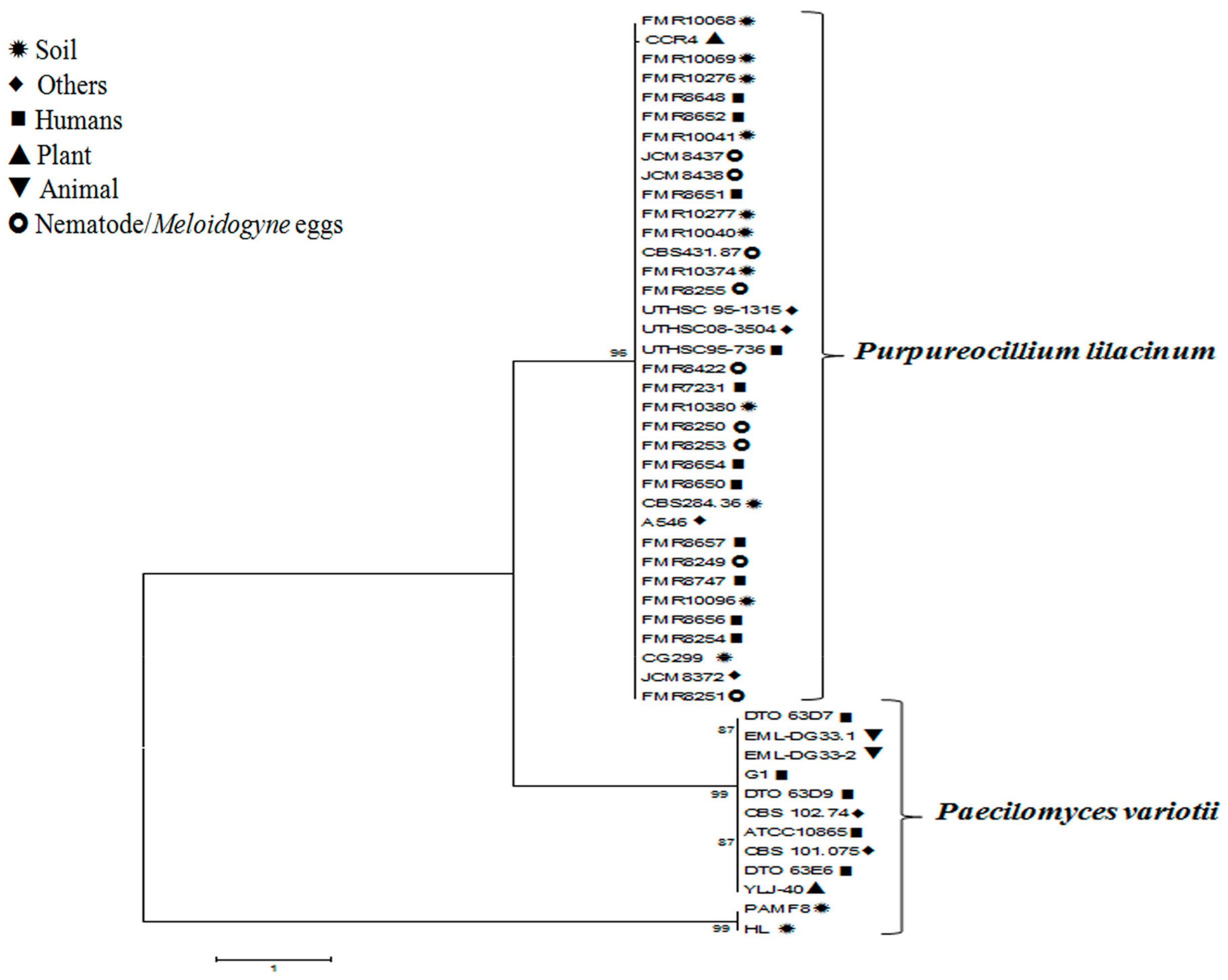
Molecular data presented by Luangsa-ard et al. [55] suggest that it is impossible to differentiate dangerous from beneficial isolates of Purp. lilacinum. We present in Figure 1 a comparison of nucleotide sequences of clinical isolates used in the papers referenced in this review with strains of P. variotii and Purp. lilacinum isolated from animals, plants, and soil (the sequences were obtained from GenBank). Figure 1 shows that isolates of P. variotii are distributed in two distinct clades with human and animal isolates in clade 1 and soil isolates in clade 2 and isolates of Purp. lilacinum show great similarity between clinical and environmental isolates. These phylogenetic trees demonstrate that it is difficult to separate Purp. lilacinum pathogenic from environmental isolates, which makes it a fungus that deserves greater attention.
4. Concluding Remarks
Paecilomyces spp. and Byssochlamys spp. represent some of the fungi that cause problems to the food and beverage industry and because these species are pathogenic to humans the contaminated food and beverage may become a probable source of infection. A better comprehension of the heat-resistance mechanisms of these species may provide insights for the development of new treatments to avoid food colonization and further investigations are necessary to understand the association of fungi with foods and the risks involved for humans.
Author Contributions
C.M.B.; M.M.E.O. and D.C.M. conceived the manuscript; C.M.B. wrote the paper; M.M.E.O. and D.C.M revised the paper; M.M.E.O. compared the nucleotide sequences deposited in the GenBank.
Funding
This research received no external funding.
Conflicts of Interest
The authors declare no conflict of interest.
References
- Bainier, G. Mycothèque de l’école de Pharmacie. XI. Paecilomyces, genre nouveau de Mucédinées. Soc. Mycol. Fr. 1907, 23, 26–27. [Google Scholar]
- Hughes, S.J. Studies on micro-fungi. XI. Some Hyphomycetes, which produce phialides. Mycol. Pap. 1951, 45, 1–36. [Google Scholar]
- Brown, A.H.S.; Smith, G. The genus Paecilomyces Bainier and its perfect stage Byssochlamys Westiling. Trans. Br. Mycol. Soc. 1957, 40, 17–89. [Google Scholar] [CrossRef]
- Morris, E.F. The Synnematous Genera of the Fungi Imperfecti; Western Illinois University: Macomb, IL, USA, 1963; p. 137. [Google Scholar]
- Onions, A.H.S.; Barron, G.L. Monophialidic species of Paecilomyces. Mycol. Pap. 1967, 107, 1–5. [Google Scholar]
- De Hoog, G.S. The genera Beauveria, Isaria, Tritirachium and Acrodontium gen. nov. Stud. Mycol. 1972, 1, 1–41. [Google Scholar]
- Samson, R.A. Paecilomyces and some allied Hyphomycetes. Stud. Mycol. 1974, 6, 1–119. [Google Scholar]
- He, J.; Kang, J.; Lei, B.; Wen, T. Paecilomyces wawuensis, a new species isolated from soil in China. Mycotaxon 2011, 115, 303–310. [Google Scholar] [CrossRef]
- Borba, C.M.; Brito, M.M.S. Paecilomyces: Mycotoxin production and human infection. In Molecular Biology of Food and Water Borne Mycotoxigenic and Mycotic Fungi; Paterson, R.R.M., Lima, N., Eds.; CRC Press: Boca Raton, FL, USA, 2015; pp. 401–421. ISBN 9781466559868. [Google Scholar]
- Stolk, A.C.; Samson, R.A. The genus Talaromyces Studies on Talaromyces and related genera II. Stud. Mycol. 1972, 2, 1–46. [Google Scholar]
- Raja, H.A.; Miller, A.N.; Pearce, C.J.; Oberlies, N.H. Fungal identification using molecular tools: A primer for the natural products research community. J. Nat. Prod. 2017, 80, 756–770. [Google Scholar] [CrossRef] [PubMed]
- Luangsa-ard, J.J.; Hywel-Jones, N.L.; Samson, R.A. The polyphyletic nature of Paecilomyces sensu lato based on 18S-generated rDNA phylogeny. Mycologia 2004, 96, 773–780. [Google Scholar] [CrossRef] [PubMed]
- Samson, R.A.; Houbraken, J.; Varga, J.; Frisvad, J.C. Polyphasic taxonomy of the heat resistant ascomycete genus Byssochlamys and its Paecilomyces anomorphs. Persoonia 2009, 22, 14–27. [Google Scholar] [CrossRef] [PubMed]
- Paterson, R.R.M.; Lima, N. Filamentous fungal human pathogens from food emphasizing Aspergillus, Fusarium and Mucor. Microorganisms 2017, 5, 44. [Google Scholar] [CrossRef] [PubMed]
- Dijksterhuis, J.; Houbraken, J.; Samson, R.A. Fungal spoilage of crops and food. In Agricultural Applications, 2nd ed.; Kempken, F., Ed.; Springer-Verlag: Berlin/Heidelberg, Germany, 2013; Volume 11, pp. 35–56. ISBN 978-3-642-36821-9. [Google Scholar]
- Leong, S.L.; Halliday, C.; Chen, C.A.S. Proposed strategy for routine screening of pathogenic fungi in foods. In Proceedings of the International Commission on Food Mycology Workshop 2016 Current and Future Trends in Food Mycology—Methods, Taxonomy, and Emerging Problems, Freising, Germany, 13–15 June 2016. [Google Scholar]
- Tournas, V. Heat-resistant fungi of importance to the food and beverage industry. Crit. Rev. Microbiol. 1994, 20, 243–263. [Google Scholar] [CrossRef] [PubMed]
- Sant’Ana, A.S.; Rosenthal, A.; Massaguer, P.R. Heat resistance and the effects of continuous pasteurization on the inactivation of Byssochlamys fulva ascospores in clarified apple juice. J. Appl. Microbiol. 2009, 107, 197–209. [Google Scholar] [CrossRef] [PubMed]
- Hosoya, K.; Nakayama, M.; Tomiyama, D.; Matsuzawa, T.; Imanishi, Y.; Ueda, S.; Yaguchi, T. Risk analysis and rapid detection of the genus Thermoascus, food spoilage fungi. Food Control 2014, 41, 7–12. [Google Scholar] [CrossRef]
- Garnier, L.; Valence, F.; Mounier, J. Diversity and control of spoilage fungi in dairy products: An update. Microorganisms 2017, 5, 42. [Google Scholar] [CrossRef] [PubMed]
- Ham, H.; Kim, S.; Kim, M.; Lee, S.; Hong, S.K.; Ryu, J.; Lee, T. Mycobiota of ground red pepper and their aflatoxigenic potential. J. Microbiol. 2016, 54, 832–837. [Google Scholar] [CrossRef] [PubMed]
- Minamor, A.A.; Odamtten, G.T. Radial growth of three Paecilomyces species isolated from two Ghanaian maize varieties Abeleehi and Obaatanpa on five different media and the effects of their culture filtrate on seed germination and radicle elongation of Abeleehi and Obaatanpa. Int. J. Curr. Microbiol. Appl. Sci. 2016, 5, 604–617. [Google Scholar] [CrossRef]
- Minamor, A.A.; Odamtten, G.T. Influence of the culture filtrate of three Paecilomyces species on some growth parameters, chlorophyll content, and root anatomy of two Ghanaian maize varieties (Abeleehi and Obaatanpa) and on germination capacity of tomato and pepper seeds. Am. J. Microbiol. Res. 2017, 5, 51–58. [Google Scholar] [CrossRef]
- Pitt, J.I.; Hocking, A.D. Fungi and Food Spoilage; Springer: Heidelberg, Germany, 1997; ISBN 978-1-4615-6391-4. [Google Scholar]
- Roussos, S.; Zaouia, N.; Salih, G.; Tantaoui-Elaraki, A.; Lamrani, K.; Cheheb, M.; Hassouni, H.; Verh, F.; Perraud-Gaime, I.; Augur, C.; et al. Characterization of filamentous fungi isolated from Moroccan olive and olive cake: Toxinogenic potential of Aspergillus strains. Mol. Nutr. Food Res. 2006, 50, 500–506. [Google Scholar] [CrossRef] [PubMed]
- Alkenz, S.; Sassi, A.A.; Abugnah, Y.S.; Alryani, M.B. Isolation and identification of fungi associated with some Libyan foods. Afr. J. Food Sci. 2015, 9, 406–410. [Google Scholar] [CrossRef]
- Rice, S.L.; Beuchat, L.R.; Worthington, R.E. Patulin production by Byssochlamys spp. in fruit juices. Appl. Environ. Microbiol. 1977, 34, 791–796. [Google Scholar] [PubMed]
- Beuchat, L.; Rice, S. Byssochlamys spp. and processed fruits. Adv. Food Res. 1979, 25, 237–288. [Google Scholar]
- Kotzekidou, P. Heat resistance of Byssochlamys nivea, Byssochlamys fulva and Neosartorya fischeri isolated from canned tomato paste. J. Food Sci. 1997, 62, 410–413. [Google Scholar] [CrossRef]
- Evelyn; Silva, F.V.M. Inactivation of Byssochlamys nivea ascospores in strawberry puree by high pressure, power ultrasound and thermal processing. Int. J. Food Microbiol. 2015, 214, 129–136. [Google Scholar] [CrossRef]
- Rawat, S. Food spoilage: Microorganisms and their prevention. Asian J. Plant Sci. Res. 2015, 5, 47–56. [Google Scholar]
- Engel, G.; Teuber, M. Heat resistance of ascospores of Byssochlamys nivea in milk and cream. Int. J. Food Microbiol. 1991, 12, 225–234. [Google Scholar] [CrossRef]
- Rosenthal, A.; Guerra Filho, D.; Xavier, A.; Duarte, S. Fungos filamentosos termorresistentes em linha de suco de abacaxi envasado assepticamente. In Congresso Brasileiro de Ciência e Tecnologia de Alimentos; SBCTA: Porto Alegre, Brazil, 2002. [Google Scholar]
- Welke, J.E.; Hoeltz, M.; Dottoni, H.A.; Noll, I.B. Ocorrência de fungos termorresistentes em suco de maçã. Braz. J. Food Technol. 2009, 2, 78–83. [Google Scholar]
- Salomão, B.M.C.; Massaguer, P.; Aragão, G.M.C. Isolamento e seleção de fungos termorresistentes em etapas do processo produtivo de maçã. Ciênc. Tecnol. Aliment. 2008, 28, 116–121. [Google Scholar] [CrossRef]
- Mattietto, R.A.; Lopes, A.S.; Menezes, H.C. Estabilidade do néctar misto de cajá e umbu. Ciênc. Tecnol. Aliment. 2007, 27, 456–463. [Google Scholar] [CrossRef] [Green Version]
- Ferreira, E.H.R.; Masson, L.M.P.; Rosenthal, A.; Souza, M.L.; Tashima, L.; Massaguer, P.R. Thermoresistance of filamentous fungi isolated from aseptically packaged fruit nectars. Braz. J. Food Technol. 2011, 14, 164–171. [Google Scholar] [CrossRef]
- Rico-Munoz, E. Heat-resistant mold ascospores elimination in the beverage and food processing environment: Is it possible? In Proceedings of the International Commission on Food Mycology Workshop 2016 Current and Future Trends in Food Mycology—Methods, Taxonomy, and Emerging Problems, Freising, Germany, 13–15 June 2016. [Google Scholar]
- Darke, C.S.; Knowelden, J.; Lacey, J.; Ward, M.A. Respiratory disease of workers harvesting grain. Thorax 1976, 31, 294. [Google Scholar] [CrossRef] [PubMed]
- Tribst, A.A.L.; Sant’Ana, A.S.; Massaguer, P.R. Review: Microbiological quality and safety of fruit juices—Past, present and future perspectives. Crit. Rev. Microbiol. 2009, 35, 310–339. [Google Scholar] [CrossRef] [PubMed]
- Cast (Council for Agricultural Science and Technology). Mycotoxins: Risks in plant, animal and human systems. Task Force Rep. Am. 2003, 139, 1–196. [Google Scholar]
- Houbraken, J.; Verweij, P.E.; Rijs, A.J.M.M.; Borman, A.M.; Samson, R.A. Indetification of Paecilomycesvariotii in clinical samples and settings. J. Clin. Microbiol. 2010, 48, 2754–2761. [Google Scholar] [CrossRef] [PubMed]
- Atencia, S.; Papakonstantinou, S.; Leggett, B.; McAllister, H.; Mooney, C.T. Systemic fungal infection in a dog: A unique case in Ireland. Irish Vet. J. 2014, 67, 17. [Google Scholar] [CrossRef] [PubMed]
- Bellanger, A.P.; Cervoni, J.P.; Faucher, J.F.; Weil-Verhoeven, D.; Ginet, M.; Deconinck, E.; Grenouillet, F. Paecilomycesvariotii fungemia in a patient with lymphoma needing liver transplant. Mycopathologia 2017, 182, 761–765. [Google Scholar] [CrossRef] [PubMed]
- Uzunoglu, E.; Sahin, A.M. Paecilomyces variotii peritonitis in a patient on continuous ambulatory peritoneal dialysis. J. Mycol. Med. 2017, 27, 277–280. [Google Scholar] [CrossRef] [PubMed]
- Polat, M.; Kara, S.S.; Taplslz, A.; Demirtas, Z.; Sarl, S.; Kalkancl, A.; Tezer, H.; Dalglc, B. Successful treatment of Paecilomyces variotii peritonitis in a liver transplant patient. Mycopathologia 2015, 179, 317–320. [Google Scholar] [CrossRef] [PubMed]
- Torres, R.; Gonzalez, M.; Sanhueza, M.; Segovia, E.; Alvo, M.; Passalacqua, W.; Saffie, A.; Elgueta, L.; Diaz, M.; Silva, F. Outbreak of Paecilomyces variotii peritonitis in peritoneal dialysis patients after the 2010 Chilean earthquake. Per. Dial. Int. 2014, 34, 322–325. [Google Scholar] [CrossRef] [PubMed]
- Feldman, R.; Cockerham, L.; Buchan, B.W.; Lu, Z.; Huang, A.M. Treatment of Paecilomyces variotii pneumonia with posaconazole: Case report and literature review. Mycoses 2016, 59, 746–750. [Google Scholar] [CrossRef] [PubMed]
- Abolghasemi, S.; Tabarsi, P.; Adimi, P.; Kiani, A.; Dolatshahi, S.; Mansouri, D. Pulmonary Paecilomyces in a diabetic patient. Tanaffos 2015, 14, 268–271. [Google Scholar] [PubMed]
- Kim, J.H.; Williams, K. Posaconazole salvage treatment for invasive fungal infection. Mycopathologia 2014, 178, 259–265. [Google Scholar] [CrossRef] [PubMed]
- Steiner, B.; Aquino, V.R.; Paz, A.A.; Silla, L.M.R.; Zavascki, A.; Goldani, L.Z. Paecilomycesvariotii as an emergent pathogenic agent of pneumonia. Case Rep. Inf. Dis. 2013, 3. [Google Scholar] [CrossRef]
- Akhunov, V.M.; Akhunova, A.M.; Lavrent’eva, T.P. Hypereosinophilic syndrome associated with sepsis due to Paecilomyces fungi disseminated into the liver. Klinmed 2016, 94, 149–152. [Google Scholar]
- Swami, T.; Pannu, S.; Kumar, M.; Gupta, G. Chronic invasive fungal rhinosinusitis by Paecilomycesvariotii: A rare case report. Indian J. Med. Microbiol. 2016, 34, 103–106. [Google Scholar] [CrossRef] [PubMed]
- Vasudevan, B.; Hazra, N.; Verma, R.; Srinivas, V.; Vijendran, P.; Badad, A. First reported case of subcutaneous hyalohyphomycosis caused by Paecilomyces variotii. Int. J. Dermatol. 2013, 52, 711–713. [Google Scholar] [CrossRef] [PubMed]
- Luangsa-ard, J.; Houbraken, J.; van Doorn, T.; Hong, S.B.; Borman, A.M.; Hywel-Jones, N.L.; Samson, R.A. Purpureocillium, a new genus for the medically important Paecilomyces lilacinus. FEMS Microbiol. Lett. 2011, 321, 141–149. [Google Scholar] [CrossRef] [PubMed]
- Naik, A.U.; Gadewar, S.B. Paecilomyces keratitis in western India: A case report. J. Clin. Diag. Res. 2017, 11, ND01–ND02. [Google Scholar] [CrossRef] [PubMed]
- Monno, R.; Alessio, G.; Guerriero, S.; Capolongo, C.; Calia, C.; Fumarola, L.; Pazzani, C.; Di Taranto, A.; Miragliotta, G. Paecilomyces lilacinus keratitis in a soft contact lens wearer. Eye Contact Lens 2016, 29. [Google Scholar] [CrossRef] [PubMed]
- Pastor, F.J.; Guarro, J. Clinical manifestations, treatment and outcome of Paecilomyces lilacinus infections. Clin. Microbiol. Infect. 2006, 12, 948–960. [Google Scholar] [CrossRef] [PubMed]
- De Hoog, G.S.; Guarro, J.; Gené, J.; Figueras, M.J. Atlas of Clinical Fungi; Centraalbureau Voor Schimmelcultures: Baarn, The Netherlands, 2004. [Google Scholar]
- De Sequeira, D.C.M.; Menezes, R.C.; Oliveira, M.M.E.; De Luca, P.M.; Antas, P.R.Z.; Oliveira-Ferreira, J.; Borba, C.M. Experimental Hyalohyphomycosis by Purpureocillium lilacinum: Outcome of the Infection in C57BL/6 Murine Models. Front. Microbiol. 2017, 8, 1617. [Google Scholar] [CrossRef] [PubMed]
- Walkin, W.; Ghazanfar, M.U.; Yasin, M. Naturally occurring entomopathogenic fungi infecting stored grain insect species in Punjab, Pakistan. J. Insect Sci. 2014, 14. [Google Scholar] [CrossRef]
- Huis, A.V.; Itterbeeck, J.V.; Klunder, H.; Mertens, E.; Halloran, A.; Muir, G.; Vantomme, P. Edible Insects: Future Prospects for Food and Feed Security; Food and Agriculture Organization of the United Nations: Rome, Italy, 2013; 190p. [Google Scholar]
- Trinh, S.A.; Angarone, M.P. Purpureocillium lilacinum tattoo-related skin infection in a kidney transplant recipient. Transpl. Infect. Dis. 2017, 19. [Google Scholar] [CrossRef] [PubMed]
- Sotell, D.; Cappel, M.; Huff, T.; Meza, D.; Alvarez, S.; Libertin, C.R. Cutaneous fungal infection in a immunocompromised host. JMM Case Rep. 2017, 4. [Google Scholar] [CrossRef] [PubMed]
- Demitsu, T.; Nagashima, K.; Okabe, T.; Morisawa, Y.; Ishikawa, N.; Yagisawa, T.; Fukuta, H.; Ohtsuki, M.; Kano, R.; Harada, K. Subcutaneous hyalohyphomycosis due to Purpureocillium lilacinum in an immunocompromised patient after renal transplantation. J. Dermat. 2017, 44, 725–726. [Google Scholar] [CrossRef] [PubMed]
- Saghrouni, F.; Saidi, W.; Said, Z.B.; Gheith, S.; Said, M.B.; Ranque, S.; Denguezli, M. Cutaneous hyalohyphomycosis caused by Purpureocillium lilacinum in an immunocompetent patient: Case report and review. Med. Mycol. 2013, 51, 664–668. [Google Scholar] [CrossRef] [PubMed]
- Ali, T.K.; Amescua, G.; Miller, D.; Suh, L.H.; Delmonte, D.W.; Gibbons, A.; Alfonso, E.C.; Forster, R.K. Contact-lens-associated Purpureocillium keratitis: Risk factors, microbiologic characteristics, clinical course, and outcomes. Sem. Ophthal. 2017, 32, 157–162. [Google Scholar] [CrossRef] [PubMed]
- Yoshida, M.; Yokokura, S.; Kunikata, H.; Takada, N.; Maruyama, K.; Toyokawa, M.; Kashio, K.; Kaku, M.; Nakazawa, T. Endophthalmitis associated with Purpureocillium lilacinum during infliximab treatment for surgically induced necrotizing scleritis, successfully treated with 27-gauge vitrectomy. Int. Ophthalmol. 2017. [Google Scholar] [CrossRef] [PubMed]
- Mihailovic, N.; Alnawaiseh, M.; Zumhagen, L.; Eter, N. Contact lens-associated Paecilomyces lilacinus keratitis. Ophthalmologe 2017, 114, 57–59. [Google Scholar] [CrossRef] [PubMed]
- Chew, R.; Dorman, A.; Woods, M.L. Purpureocillium lilacinum keratitis: A case series and review of the literature. Can. J. Ophthalmol. 2016, 51, 382–385. [Google Scholar] [CrossRef] [PubMed]
- Narita, A.; Seguchi, J.; Shiraga, F. Paecilomyces lilacinus-induced scleritis following bleb-associated endophthalmitis after trabeculectomy. Acta Med. Okayama 2015, 69, 313–318. [Google Scholar] [PubMed]
- Turner, L.D.; Conrad, D. Retrospective case-series of Paecilomyces lilacinus ocular mycoses in Queensland, Australia. BMC Res. Notes 2015, 8, 627. [Google Scholar] [CrossRef] [PubMed]
- Sharma, V.; Angrup, A.; Panwar, P.; Verma, S.; Singh, D.; Kanga, A. Keratitis by Paecilomyces lilacinus: A case report from Sub-Himalayan region. Indian J. Med. Microbiol. 2015, 33, 585–587. [Google Scholar] [CrossRef] [PubMed]
- Evans, J.M.; Wang, A.L.; Elewski, B.E. Successful treatment of Paecilomyces lilacinus onychomycosis with efinaconazole and tavaborole. Skin Appendage Disord. 2015, 1, 169–171. [Google Scholar] [CrossRef] [PubMed]
- Pontini, P.; Gorani, A.; Veraldi, S. Onychomycosis by Paecilomyces lilacinus. Giornale Italiano di Dermatologia e Venereologia 2016, 151, 706–709. [Google Scholar] [PubMed]
- Shivaprasad, A.; Ravi, G.C.; Shivapriya, R. A rare case of nasal septal perforation due to Purpureocillium lilacinum: Case report and review. Indian J. Otolaryngol. Head Neck Surg. 2013, 65, 184–188. [Google Scholar] [CrossRef] [PubMed]
- Atkins, S.D.; Hidalgo-Diaz, L.; Kalisz, H.; Mauchline, T.H.; Hirsch, P.R.; Kerry, B.R. Development of a new management strategy for the control of root-knot nematodes (Meloidogyne spp.) in organic vegetable production. Pest. Manag. Sci. 2003, 59, 183–189. [Google Scholar] [CrossRef] [PubMed]
- Baidoo, R.; Mengistu, T.; McSorley, R.; Stamps, R.H.; Brito, J.; Crow, W.T. Management of root-knot nematode (Meloidogyne incognita) on Pittosporum tobira under greenhouse, field, and on-farm conditions in Florida. J. Nematol. 2017, 49, 133–139. [Google Scholar] [CrossRef] [PubMed]
- Blaszkowska, J.; Kurnatowski, P.; Wojcik, A.; Goralska, K.; Szwabe, K. In vitro evaluation of the ovistatic and ovicidal effect of the cosmopolitan filamentous fungi isolated from soil on Ascaris suum eggs. Vet. Parasitol. 2014, 199, 165–171. [Google Scholar] [CrossRef] [PubMed]
- Antas, P.R.Z.; Brito, M.M.S.; Peixoto, E.; Ponte, C.G.G.; Borba, C.M. Neglected and emerging fungal infections: Review of hyalohyphomycosis by Paecilomyces lilacinus focusing in disease burden, in vitro antifungal susceptibility and management. Microbes Infect. 2012, 14, 1–8. [Google Scholar] [CrossRef] [PubMed]
- Saitou, N.; Nei, M. The neighbor-joining method: A new method for reconstructing phylogenetic trees. Mol. Biol. Evol. 1987, 4, 406–425. [Google Scholar] [PubMed]
- Felsenstein, J. Confidence limits on phylogenies: An approach using the bootstrap. Evolution 1985, 39, 783–791. [Google Scholar] [CrossRef] [PubMed]
- Tamura, K.; Dudley, J.; Nei, M.; Kumar, S. MEGA4: Molecular Evolutionary Genetics Analysis (MEGA) software version 4.0. Mol. Biol. Evol. 2007, 24, 1596–1599. [Google Scholar] [CrossRef] [PubMed]
Figure 1.
Phylogenetic tree of Purpureocillium lilacinum and Paecilomyces variotii isolates. Similarity between clinical and environmental Purp. lilacinum isolates is shown. P. variotii isolates show one possible differentiation between human and animal (clinical) isolates from soil isolates (environmental). The evolutionary history was inferred using the Neighbor–Joining method [81] among 48 taxa. The percentage of replicate trees in which the associated taxa clustered together in the bootstrap test (500 replicates) is shown next to the branches [82]. There were a total of 382 positions in the final dataset. Phylogenetic analyses were conducted in MEGA4 [83].


Table 1.
Reported human cases of hyalohyphomycosis caused by Paecilomyces variotii between 2013 and 2017.
Table 1.
Reported human cases of hyalohyphomycosis caused by Paecilomyces variotii between 2013 and 2017.
| Infection | Number of Cases | Reference |
|---|---|---|
| Fungemia | 1 | Bellanger et al. [44] |
| Peritonitis | 5 | Uzunoglu and Sahin [45] Polat et al. [46] Torres et al. [47] |
| Pneumonia | 4 | Feldman et al. [48] Abolghasemi et al. [49] Kim and Williams [50] Steiner et al. [51] |
| Sepsis | 1 | Akhunov et al. [52] |
| Sinusitis | 1 | Swami et al. [53] |
| Subcutaneous infection | 1 | Vasudevan et al. [54] |
© 2018 by the authors. Licensee MDPI, Basel, Switzerland. This article is an open access article distributed under the terms and conditions of the Creative Commons Attribution (CC BY) license (http://creativecommons.org/licenses/by/4.0/).
Share and Cite
MDPI and ACS Style
Moreira, D.C.; Oliveira, M.M.E.; Borba, C.M. Human Pathogenic Paecilomyces from Food. Microorganisms 2018, 6, 64. https://0-doi-org.brum.beds.ac.uk/10.3390/microorganisms6030064
AMA Style
Moreira DC, Oliveira MME, Borba CM. Human Pathogenic Paecilomyces from Food. Microorganisms. 2018; 6(3):64. https://0-doi-org.brum.beds.ac.uk/10.3390/microorganisms6030064
Chicago/Turabian StyleMoreira, Danielly C., Manoel M. E. Oliveira, and Cintia M. Borba. 2018. "Human Pathogenic Paecilomyces from Food" Microorganisms 6, no. 3: 64. https://0-doi-org.brum.beds.ac.uk/10.3390/microorganisms6030064
Note that from the first issue of 2016, this journal uses article numbers instead of page numbers. See further details here.