Magnetic Marking and Intraoperative Detection of Primary Draining Lymph Nodes in High-Risk Prostate Cancer Using Superparamagnetic Iron Oxide Nanoparticles: Additional Diagnostic Value

Abstract
:1. Introduction
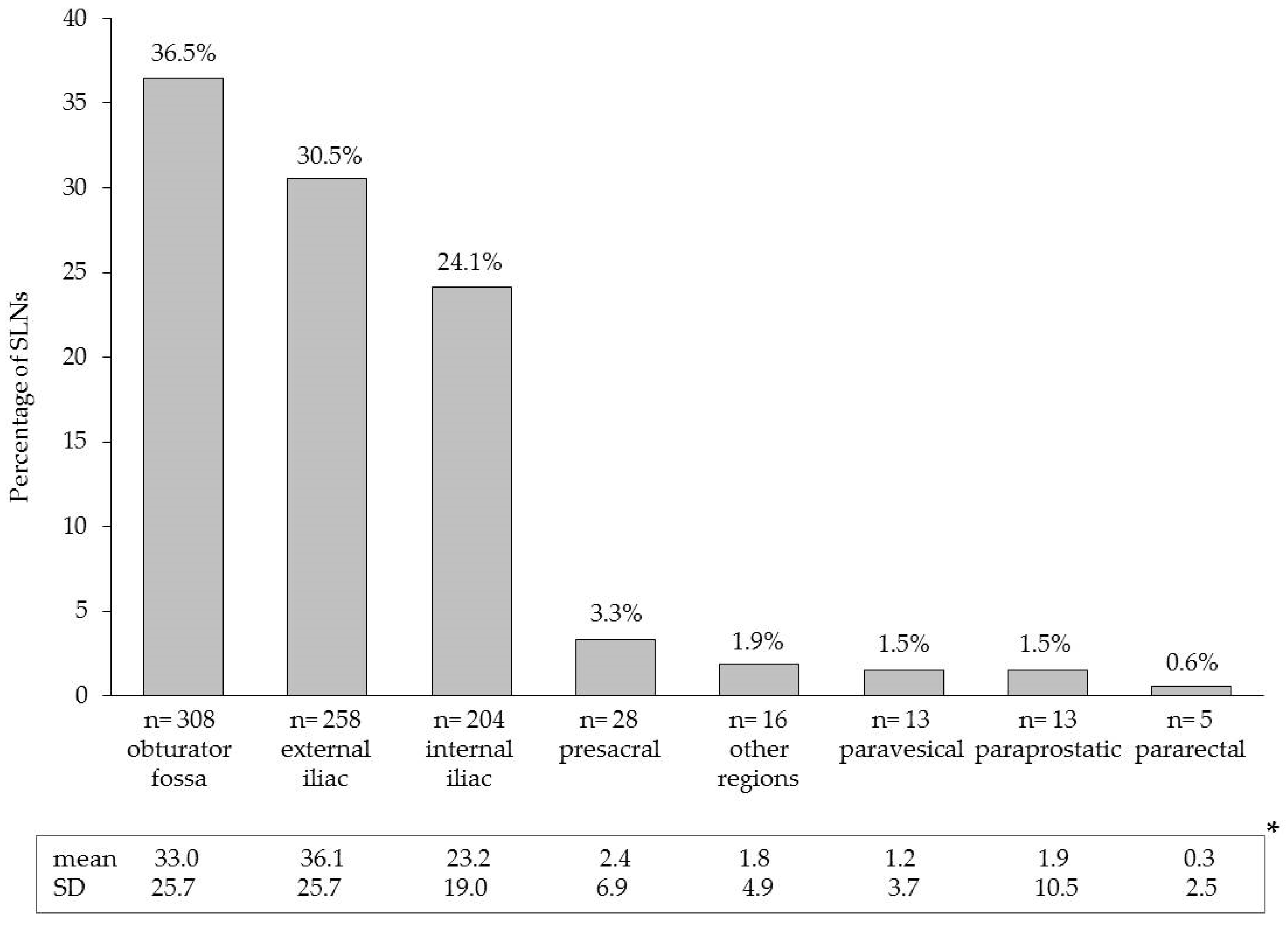
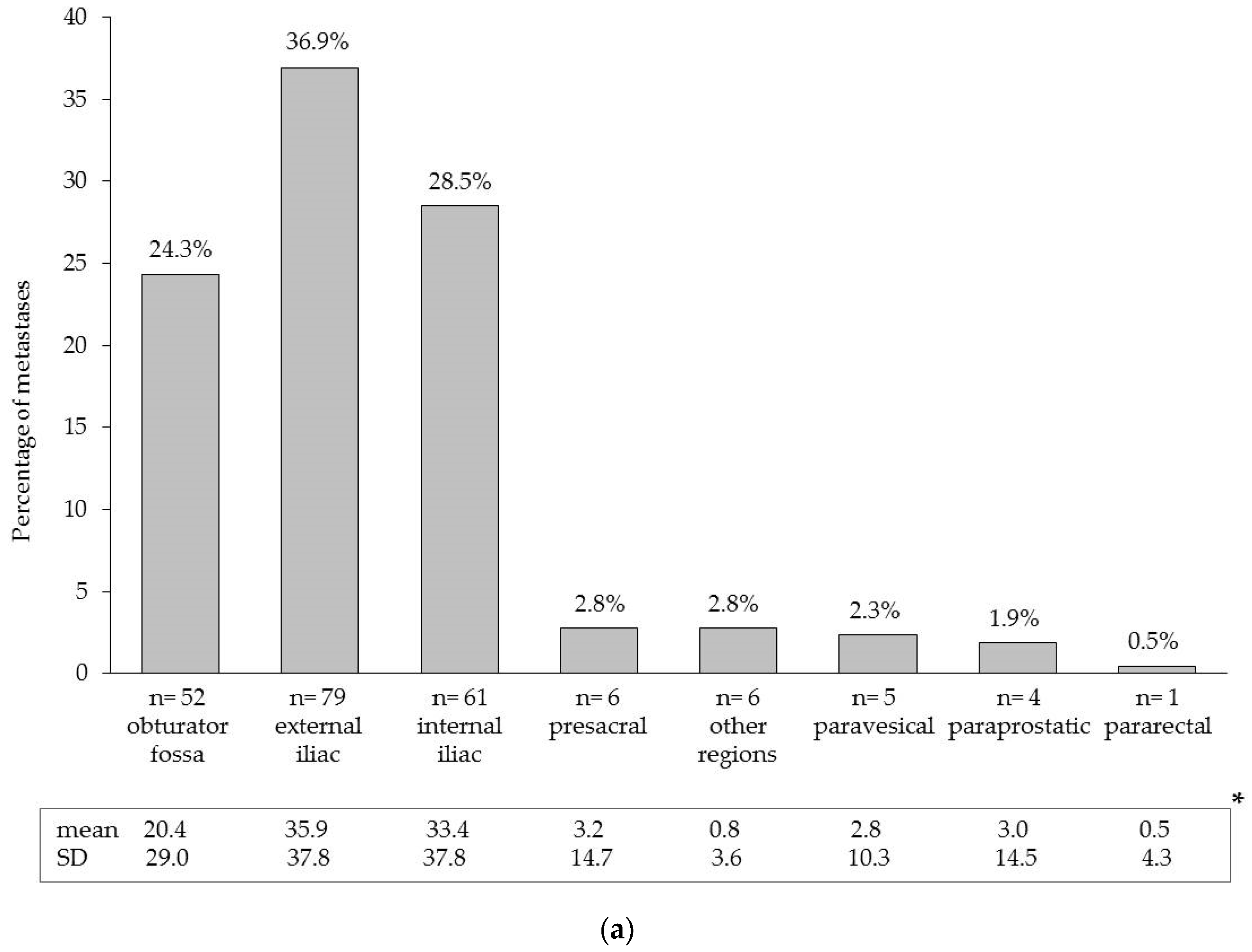
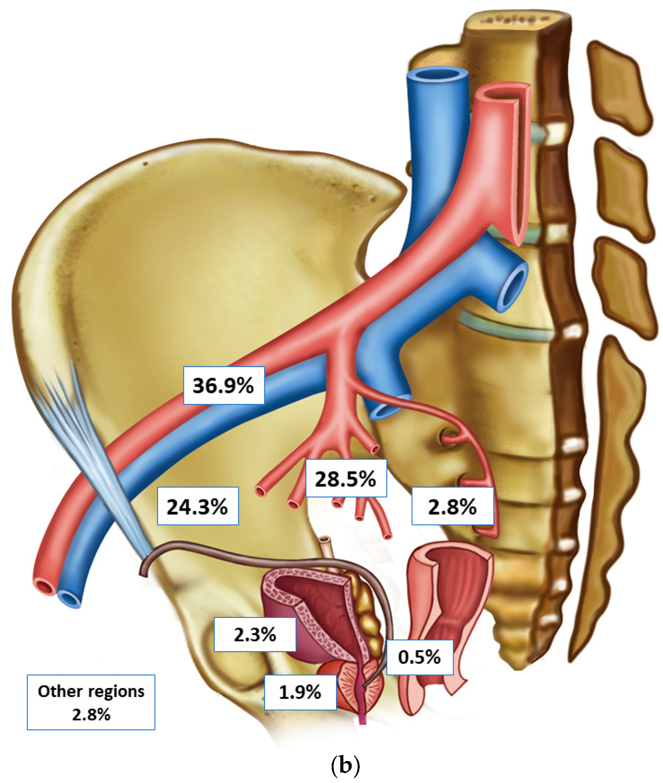
2. Results
3. Discussion
4. Materials and Methods
4.1. Patients
4.2. Magnetic SPION Tracer
4.3. Tracer Injection
4.4. Magnetometer-Guided sLND, ePLND, and Histopathological Examination
4.5. Outcome Measures of Magnetometer-Guided sLND
4.6. Ethical Approval
5. Conclusions
Acknowledgments
Author Contributions
Conflicts of Interest
Abbreviations
| ePLND | extended pelvic lymph node dissection |
| IQR | interquartile range |
| LN | lymph node |
| LNI | lymph node invasion |
| PCa | prostate cancer |
| PLND | pelvic lymph node dissection |
| PSA | prostate-specific antigen |
| SLN | sentinel lymph node |
| sLND | sentinel lymph node dissection |
| SPION | superparamagnetic iron oxide nanoparticles |
| SD | standard deviation |
References
- Choo, M.S.; Kim, M.; Ku, J.H.; Kwak, C.; Kim, H.H.; Jeong, C.W. Extended Versus Standard Pelvic Lymph Node Dissection in Radical Prostatectomy on Oncological and Functional Outcomes: A Systematic Review and Meta-Analysis. Ann. Surg. Oncol. 2017, 24, 2047–2054. [Google Scholar] [CrossRef] [PubMed]
- Withrow, D.R.; DeGroot, J.M.; Siemens, D.R.; Groome, P.A. Therapeutic value of lymph node dissection at radical prostatectomy: A population-based case-cohort study. BJU Int. 2011, 108, 209–216. [Google Scholar] [CrossRef] [PubMed]
- Winter, A.; Henke, R.P.; Wawroschek, F. Targeted salvage lymphadenectomy in patients treated with radical prostatectomy with biochemical recurrence: Complete biochemical response without adjuvant therapy in patients with low volume lymph node recurrence over a long-term follow-up. BMC Urol. 2015, 15, 10. [Google Scholar] [CrossRef] [PubMed]
- Seiler, R.; Studer, U.E.; Tschan, K.; Bader, P.; Burkhard, F.C. Removal of limited nodal disease in patients undergoing radical prostatectomy: Long-term results confirm a chance for cure. J. Urol. 2014, 191, 1280–1285. [Google Scholar] [CrossRef] [PubMed]
- Budäus, L.; Leyh-Bannurah, S.R.; Salomon, G.; Michl, U.; Heinzer, H.; Huland, H.; Graefen, M.; Steuber, T.; Rosenbaum, C. Initial experience of (68)Ga-PSMA PET/CT imaging in high-risk prostate cancer patients prior to radical prostatectomy. Eur. Urol. 2016, 69, 393–396. [Google Scholar] [CrossRef] [PubMed]
- Heidenreich, A.; Ohlmann, C.H.; Polyakov, S. Anatomical extent of pelvic lymphadenectomy in patients undergoing radical prostatectomy. Eur. Urol. 2007, 52, 29–37. [Google Scholar] [CrossRef] [PubMed]
- Mottet, N.; Bellmunt, J.; Bolla, M.; Briers, E.; Cumberbatch, M.G.; de Santis, M.; Fossati, N.; Gross, T.; Henry, A.M.; Joniau, S.; et al. EAU-ESTRO-SIOG guidelines on prostate cancer. Part 1: Screening, diagnosis, and local treatment with curative intent. Eur. Urol. 2017, 71, 618–629. [Google Scholar] [CrossRef] [PubMed]
- Briganti, A.; Chun, F.K.; Salonia, A.; Suardi, N.; Gallina, A.; Da Pozzo, L.F.; Roscigno, M.; Zanni, G.; Valiquette, L.; Rigatti, P.; et al. Complications and other surgical outcomes associated with extended pelvic lymphadenectomy in men with localized prostate cancer. Eur. Urol. 2006, 50, 1006–1013. [Google Scholar] [CrossRef] [PubMed]
- Fossati, N.; Willemse, P.M.; van den Broeck, T.; van den Bergh, R.C.N.; Yuan, C.Y.; Briers, E.; Bellmunt, J.; Bolla, M.; Cornford, P.; de Santis, M.; et al. The Benefits and Harms of Different Extents of Lymph Node Dissection during Radical Prostatectomy for Prostate Cancer: A Systematic Review. Eur. Urol. 2017, 72, 84–109. [Google Scholar] [CrossRef] [PubMed]
- Wawroschek, F.; Vogt, H.; Weckermann, D.; Wagner, T.; Harzmann, R. The sentinel lymph node concept in prostate cancer—First results of gamma probe-guided sentinel lymph node identification. Eur. Urol. 1999, 36, 595–600. [Google Scholar] [CrossRef] [PubMed]
- Wit, E.M.K.; Acar, C.; Grivas, N.; Yuan, C.; Horenblas, S.; Liedberg, F.; Valdes Olmos, R.A.; van Leeuwen, F.W.B.; van den Berg, N.S.; Winter, A.; et al. Sentinel Node Procedure in Prostate Cancer: A Systematic Review to Assess Diagnostic Accuracy. Eur. Urol. 2017, 71, 596–605. [Google Scholar] [CrossRef] [PubMed]
- Winter, A.; Kneib, T.; Henke, R.P.; Wawroschek, F. Sentinel lymph node dissection in more than 1200 prostate cancer cases: Rate and prediction of lymph node involvement depending on preoperative tumor characteristics. Int. J. Urol. 2014, 21, 58–63. [Google Scholar] [CrossRef] [PubMed]
- Winter, A.; Kneib, T.; Wasylow, C.; Reinhardt, L.; Henke, R.P.; Engels, S.; Gerullis, H.; Wawroschek, F. Updated nomogram incorporating percentage of positive cores to predict probability of lymph node invasion in prostate cancer patients undergoing sentinel lymph node dissection. J. Cancer 2017, 8, 2692–2698. [Google Scholar] [CrossRef] [PubMed]
- Grivas, N.; Wit, E.; Tillier, C.; van Muilekom, E.; Pos, F.; Winter, A.; van der Poel, H. Validation and head-to-head comparison of three nomograms predicting probability of lymph node invasion of prostate cancer in patients undergoing extended and/or sentinel lymph node dissection. Eur. J. Nucl. Med. Mol. Imaging 2017. [Google Scholar] [CrossRef] [PubMed]
- Joniau, S.; van den Bergh, L.; Lerut, E.; Deroose, C.M.; Haustermans, K.; Oyen, R.; Budiharto, T.; Ameye, F.; Bogaerts, K.; van Poppel, H. Mapping of pelvic lymph node metastases in prostate cancer. Eur. Urol. 2013, 63, 450–458. [Google Scholar] [CrossRef] [PubMed]
- Douek, M.; Klaase, J.; Monypenny, I.; Kothari, A.; Zechmeister, K.; Brown, D.; Wyld, L.; Drew, P.; Garmo, H.; Agbaje, O.; et al. Sentinel node biopsy using a magnetic tracer versus standard technique: The SentiMAG Multicentre Trial. Ann. Surg. Oncol. 2014, 21, 1237–1245. [Google Scholar] [CrossRef] [PubMed]
- Thill, M.; Kurylcio, A.; Welter, R.; van Haasteren, V.; Grosse, B.; Berclaz, G.; Polkowski, W.; Hauser, N. The Central-European SentiMag study: Sentinel lymph node biopsy with superparamagnetic iron oxide (SPIO) vs. radioisotope. Breast 2014, 23, 175–179. [Google Scholar] [CrossRef] [PubMed]
- Winter, A.; Woenkhaus, J.; Wawroschek, F. A novel method for intraoperative sentinel lymph node detection in prostate cancer patients using superparamagnetic iron oxide nanoparticles and a handheld magnetometer: The initial clinical experience. Ann. Surg. Oncol. 2014, 21, 4390–4396. [Google Scholar] [CrossRef] [PubMed]
- Winter, A.; Kowald, T.; Paulo, T.; Goos, P.; Engels, S.; Gerullis, H.; Chavan, A.; Wawroschek, F. Magnetic resonance sentinel lymph node imaging in prostate cancer using intraprostatic injection of superparamagnetic iron oxide nanoparticles: The first in-human results. Eur. Urol. Suppl. 2016, 15 (Suppl. 3), 1060. [Google Scholar] [CrossRef]
- Winter, A.; Engels, S.; Goos, P.; Süykers, M.C.; Gudenkauf, S.; Henke, R.P.; Gerullis, H.; Wawroschek, F. Magnetometer guided sentinel lymphadenectomy after intraprostatic injection of super-paramagnetic iron oxide nanoparticles in intermediate and high risk prostate cancer patients. J. Urol. 2016, 195 (Suppl. 4), e987. [Google Scholar] [CrossRef]
- Holl, G.; Dorn, R.; Wengenmair, H.; Weckermann, D.; Sciuk, J. Validation of sentinel lymph node dissection in prostate cancer: Experience in more than 2000 patients. Eur. J. Nucl. Med. Mol. Imaging 2009, 36, 1377–1382. [Google Scholar] [CrossRef] [PubMed]
- Sadeghi, R.; Tabasi, K.T.; Bazaz, S.M.; Kakhki, V.R.; Massoom, A.F.; Gholami, H.; Zakavi, S.R. Sentinel node mapping in the prostate cancer. Meta-analysis. Nuklearmedizin 2011, 50, 107–115. [Google Scholar] [CrossRef] [PubMed]
- Morgan-Parkes, J.H. Metastases: Mechanisms, pathways, and cascades. AJR Am. J. Roentgenol. 1995, 164, 1075–1082. [Google Scholar] [CrossRef] [PubMed]
- Weckermann, D.; Dorn, R.; Holl, G.; Wagner, T.; Harzmann, R. Limitations of radioguided surgery in high-risk prostate cancer. Eur. Urol. 2007, 51, 1549–1556. [Google Scholar] [CrossRef] [PubMed]
- Mattei, A.; Fuechsel, F.G.; Bhatta Dhar, N.; Warncke, S.H.; Thalmann, G.N.; Krause, T.; Studer, U.E. The template of the primary lymphatic landing sites of the prostate should be revisited: Results of a multimodality mapping study. Eur. Urol. 2008, 53, 118–125. [Google Scholar] [CrossRef] [PubMed]
- Wawroschek, F.; Wagner, T.; Hamm, M.; Weckermann, D.; Vogt, H.; Märkl, B.; Gordijn, R.; Harzmann, R. The influence of serial sections, immunohistochemistry, and extension of pelvic lymph node dissection on the lymph node status in clinically localized prostate cancer. Eur. Urol. 2003, 43, 132–136. [Google Scholar] [CrossRef]
- Mottet, N.; Bellmunt, J.; Briers, E.; Bolla, M.; Bourke, L.; Cornford, P.; de Santis, M.; Henry, A.M.; Joniau, S.; Lam, T.B.; et al. Guidelines on Prostate Cancer 2017. Available online: http://uroweb.org/guideline/prostate-cancer/ (accessed on 19 October 2017).
- Weingärtner, K.; Ramaswamy, A.; Bittinger, A.; Gerharz, E.W.; Vöge, D.; Riedmiller, H. Anatomical basis for pelvic lymphadenectomy in prostate cancer: Results of an autopsy study and implications for the clinic. J. Urol. 1996, 156, 1969–1971. [Google Scholar] [CrossRef]
- Van der Poel, H.G.; Wit, E.M.; Acar, C.; van den Berg, N.S.; van Leeuwen, F.W.B.; Valdes Olmos, R.A.; Winter, A.; Wawroschek, F.; Liedberg, F.; Maclennan, S.; et al. Sentinel node prostate cancer consensus panel group members. Sentinel node biopsy for prostate cancer: Report from a consensus panel meeting. BJU Int. 2017, 120, 204–211. [Google Scholar] [CrossRef] [PubMed]
Sample Availability: Not available. |


| Overall n = 104 | Patients with Negative LNs n = 43 (41.35%) | Patients with Positive LNs n = 61 (58.65%) | |
|---|---|---|---|
| Age, years (median) | 69 | 69 | 68 |
| IQR | 63–72.5 | 63.5–72 | 62–73 |
| Total PSA, ng/mL (median) | 17.21 | 12.77 | 21.79 |
| IQR | 8.32–32.56 | 7.49–24.27 | 11–41.27 |
| No. of LNs removed (median) | 17 | 15 | 17 |
| IQR | 12–21 | 12–20 | 13–21 |
| No. of SLNs removed (median) | 8 | 9 | 7 |
| IQR | 5–11 | 6–12 | 5–11 |
| No. of positive LNs (median) | 2 | ||
| IQR | 1–4 | ||
| Tumor stage (%) | |||
| T1c | 23 (22.12) | 11 (25.58) | 12 (19.67) |
| T2a | 7 (6.73) | 3 (6.98) | 4 (6.56) |
| T2b | 6 (5.77) | 3 (6.98) | 3 (4.92) |
| T2c | 49 (47.12) | 19 (44.19) | 30 (49.18) |
| T3 | 19 (18.27) | 7 (16.28) | 12 (19.67) |
| Biopsy Gleason score (%) | |||
| 6 (3 + 3) | 10 (9.62) | 8 (18.60) | 2 (3.28) |
| 7 (3 + 4) | 25 (24.04) | 11 (25.58) | 14 (22.95) |
| 7 (4 + 3) | 20 (19.23) | 7 (16.28) | 13 (21.31) |
| ≥8 | 49 (47.12) | 17 (39.53) | 32 (52.46) |
| Postoperative Gleason score (%) | |||
| 6 (3 + 3) | 2 (1.94) * | 2 (4.65) | 0 ** |
| 7 (3 + 4) | 23 (22.33) * | 20 (46.51) | 3 (5.00) ** |
| 7 (4 + 3) | 35 (33.98) * | 11 (25.58) | 24 (40.00) ** |
| ≥8 | 43 (41.75) * | 10 (23.26) | 33 (55.00) ** |
| Pathologic stage (%) | |||
| pT2 | 28 (26.92) | 26 (60.47) | 2 (3.28) |
| pT3a | 22 (21.15) | 10 (23.26) | 12 (19.67) |
| pT3b | 50 (48.08) | 7 (16.28) | 43 (70.49) |
| pT4 | 4 (3.85) | 0 | 4 (6.56) |
© 2017 by the authors. Licensee MDPI, Basel, Switzerland. This article is an open access article distributed under the terms and conditions of the Creative Commons Attribution (CC BY) license (http://creativecommons.org/licenses/by/4.0/).
Share and Cite
Winter, A.; Engels, S.; Reinhardt, L.; Wasylow, C.; Gerullis, H.; Wawroschek, F. Magnetic Marking and Intraoperative Detection of Primary Draining Lymph Nodes in High-Risk Prostate Cancer Using Superparamagnetic Iron Oxide Nanoparticles: Additional Diagnostic Value. Molecules 2017, 22, 2192. https://0-doi-org.brum.beds.ac.uk/10.3390/molecules22122192
Winter A, Engels S, Reinhardt L, Wasylow C, Gerullis H, Wawroschek F. Magnetic Marking and Intraoperative Detection of Primary Draining Lymph Nodes in High-Risk Prostate Cancer Using Superparamagnetic Iron Oxide Nanoparticles: Additional Diagnostic Value. Molecules. 2017; 22(12):2192. https://0-doi-org.brum.beds.ac.uk/10.3390/molecules22122192
Chicago/Turabian StyleWinter, Alexander, Svenja Engels, Lena Reinhardt, Clara Wasylow, Holger Gerullis, and Friedhelm Wawroschek. 2017. "Magnetic Marking and Intraoperative Detection of Primary Draining Lymph Nodes in High-Risk Prostate Cancer Using Superparamagnetic Iron Oxide Nanoparticles: Additional Diagnostic Value" Molecules 22, no. 12: 2192. https://0-doi-org.brum.beds.ac.uk/10.3390/molecules22122192