
Chemical Structures of Lignans and Neolignans Isolated from Lauraceae

1
State Key Laboratory of Applied Organic Chemistry, College of Chemistry and Chemical Engineering, Lanzhou University, Lanzhou 730000, China
2
Lanzhou University High School, Lanzhou 730000, China
*
Authors to whom correspondence should be addressed.
Molecules 2018, 23(12), 3164; https://0-doi-org.brum.beds.ac.uk/10.3390/molecules23123164
Submission received: 9 November 2018
/
Revised: 27 November 2018
/
Accepted: 29 November 2018
/
Published: 30 November 2018
(This article belongs to the Special Issue Lignans)
Abstract
:Lauraceae is a good source of lignans and neolignans, which are the most chemotaxonomic characteristics of many species of the family. This review describes 270 naturally occurring lignans and neolignans isolated from Lauraceae.
1. Introduction
Lignans are widely distributed in the plant kingdom, and show diverse pharmacological properties and a great number of structural possibilities. The Lauraceae family, especially the genera of Machilus, Ocotea, and Nectandra, is a rich source of lignans and neolignans, and neolignans represent potential chemotaxonomic significance in the study of the Lauraceae. Lignans and neolignans are dimers of phenylpropane, and conventionally classified into three classes: lignans, neolignans, and oxyneolignans, based on the character of the C–C bond and oxygen bridge joining the two typical phenyl propane units that make up their general structures [1]. Usually, lignans show dimeric structures formed by a β,β’-linkage (8,8’-linkage) between two phenylpropanes units. Meanwhile, the two phenylpropanes units are connected through a carbon–carbon bond, except for the 8,8’-linkage, which gives rise to neolignans. Many dimers of phenylpropanes are joined together through two carbon–carbon bonds forming a ring, including an 8,8’-linkage and another carbon–carbon bond linkage; such dimers are classified as cyclolignans. When the two phenylpropanes units are linked through two carbon–carbon bonds, except for the 8,8’-linkage, this constitutes a cycloneolignan. Oxyneolignans also contain two phenylpropanes units which are joined together via an oxygen bridge. Herein, lignans and neolignans are classfied into five groups: lignans, cyclolignans, neolignans, cycloneolignans, and oxyneolignans on the basis of their carbon skeletons and cyclization patterns. The majority of lignans isolated from Lauraceae have shown only minor variations on well-known structures; for example, a different degree of oxidation in the side-chain and different substitutions in the aromatic moieties, including hydroxy, methoxy, and methylenedioxy groups. Since the nomenclature and numbering of the lignans and neolignans in the literature follow different rules, the trivial names or numbers of the compounds were used to represent them. Furthermore, the semi-systematic names of compounds and their corresponding names in the literature are summarized in the Supporting Information. Herein, we give a comprehensive overview of the chemical structures of lignans and neolignans isolated from Lauraceae.
2. Lignans
This section covers lignans formed by an 8,8’-linkage between two phenyl propane units, which are subclassified according to the pattern of the oxygen rings as depicted in Figure 1. The semi-systematic names of those lignans without trivial names and their corresponding names in found in the literature are given in Table SI-1 (Supporting Information).
2.1. Simple Lignans
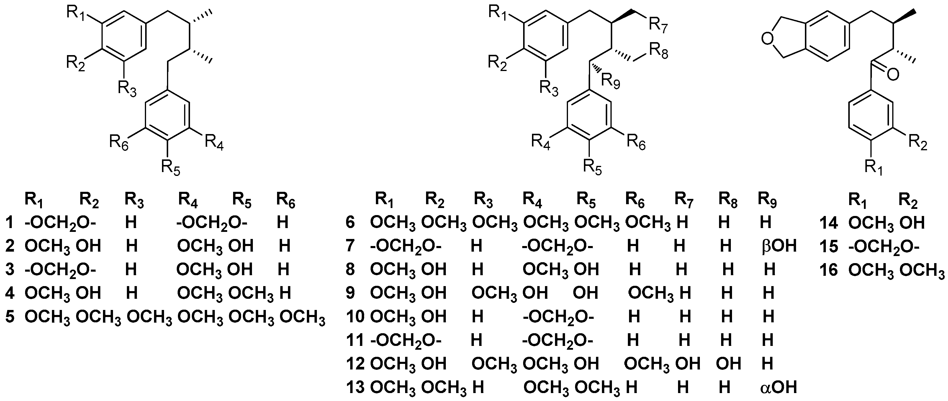
Machilin A (1) was first obtained from the CHCl3-soluble portion of the methanolic extract of the bark of Machilus thunbergii collected at Izu Peninsula, together with meso-dihydroguaiaretic acid (2). The absolute structure of machilin A (1) was determined to be 2S and 3R (meso-form) [2]. Yu and Ma et al. also reported that the bark of M. thunbergii contained machilin A (1) and meso-dihydroguaiaretic acid (2). Furthermore, meso-dihydroguaiaretic acid (2) was found to have significant neuroprotective activity against glutamate-induced neurotoxicity in primary cultures of rat cortical cells and exerted diverse hepatoprotective activity, perhaps by serving as a potent antioxidant [3,4]. Activity-guided fractionation of the dichloromethane extract of the bark of M. thunbergii not only led to the isolation of machilin A (1) and meso-dihydroguaiaretic acid (2), but also meso-austrobailignan-6 (3) and meso-monomethyl dihydroguaiaretic acid (4). It was reported that meso-dihydroguaiaretic acid (4) showed potent inhibitory activity against DNA topoisomerase I and II in vitro at a concentration of 100 μM with inhibition ratios of 93.6 and 82.1%, respectively. Furthermore, meso-austrobailignan-6 (3) was referred to as threo-austrobailignan-6 (10) in the article [5]. Two diastereomeric dibenzylbutane lignans ((5) and (6)) which exhibited selective inhibition against COX-2 (cyclooxygenase) were obtained from the leaves of Ocotea macrophylla Kunth, which were collected in Nocaima county, Colombia [6]. Besides machilin A (1) and meso-dihydroguaiaretic acid (2), oleiferin C (7) also were found in the stem bark of M. thunbergii collected at Ulleung-Do, Kyungbook, Korea. Moreover, meso-dihydroguaiaretic acid (2) and oleiferin C (7) induced an apoptotic effect in HL-60 cells via caspase-3 activation [7]. meso-Dihydroguaiaretic acid (2), threo-dihyidroguaiaretic acid (8), sauriol B (9), and threo-austrobailignan-6 (10) were isolated from the ethanolic extract of the bark of Nectandra turbacensis (Kunth) Nees [8]. The leaves and root bark of N. turbacensis (Kunth) Nees collected in the city of Santa Marta (Magdalena, Colombia) contained meso-monomethyl dihydroguaiaretic acid (4), threo-dihyidroguaiaretic acid (8), austrobailignan-5 (11), and schineolignin B (17) [9]. Lignan 12 was first obtained from the leaves of Apollonias barbujana collected in San Andrésy Sauces [10]. Compounds 13–16 were found to occur in the trunk wood of N. puberula. Proof of the absolute structure of compound 13 relied on its acid catalyzed cyclization into (-)-galbulin, a tetralin-type neolignan of known absolute stereochemistry [11] (Figure 2).
2.2. 7,7’-Epoxylignans
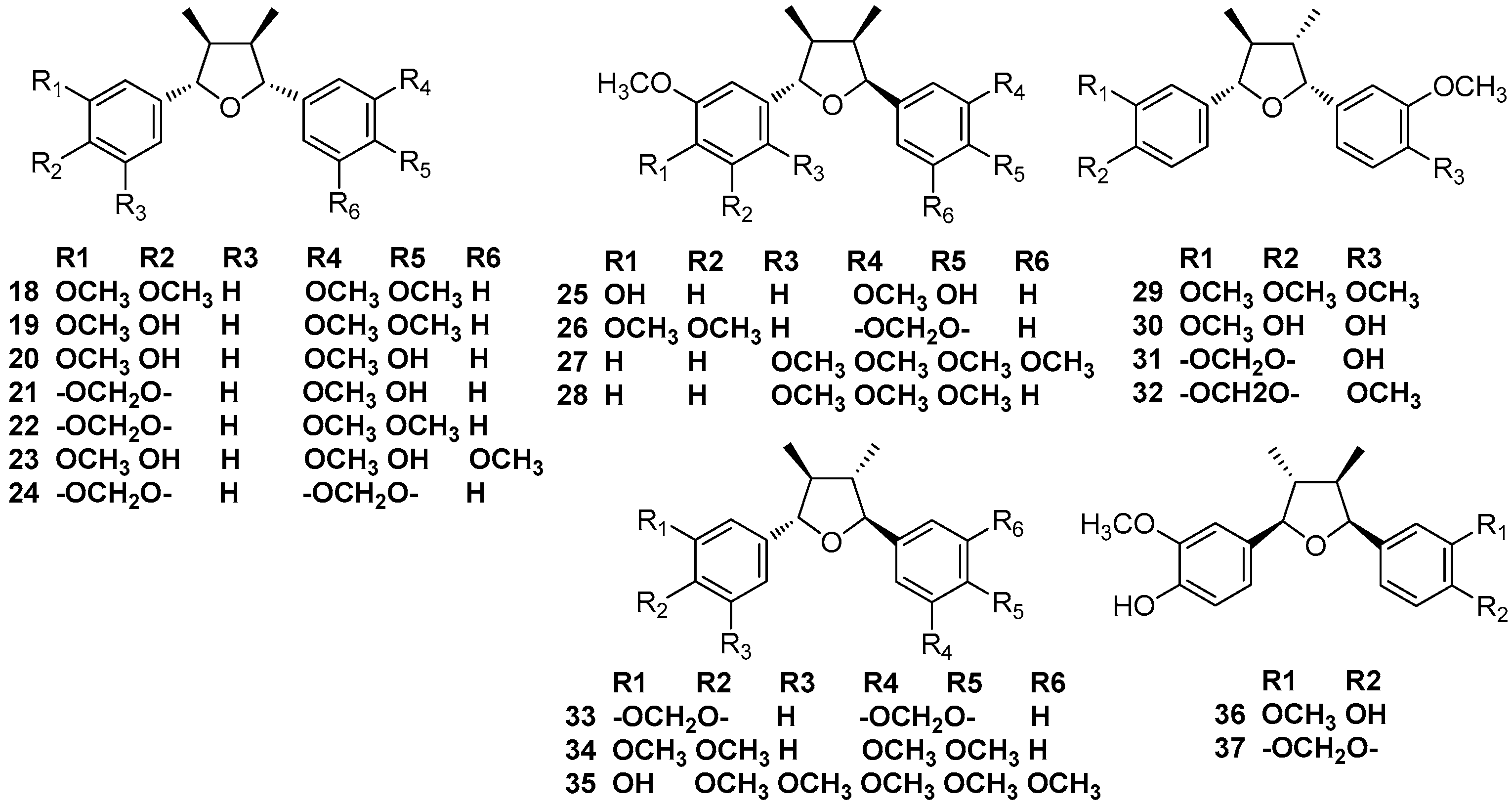
Nectandrin A (19) and nectandrin B (20) were first isolated from leaves and stems of Nectandra rigida Nees, along with galgravin (18) [12]. Nectandrin A (19) and nectandrin B (20), together with machilin F (21), machilin G (22), machilin H (23), and machilin I (25) were found to occur in the methanolic extract of the bark of M. thunbergii Sieb. et Zucc [13]. Galgravin (18), henricine (26), and veraguensin (29) were found in the leaves and root bark of N. turbacensis (Kunth) Nees [9]. Zuonin B (24), machilin F (30), and nectandrin B (20) were obtained from the stem bark of M. thunbergii [7]. Galgravin (18) and veraguensin (29), together with the 2,5-phenyl ring disubstituted lignans 27 and 28 were described for the first time from an ethanolic extract of the leaves Ocotea foetens [14]. Veraguensin (29) was first reported to be isolated from Ocotea veraguensis [15,16], and this compound was also found in N. puberula [11]. Verrucosin (30) was first gained from the benzene extract of branch wood of Urbanodendron verrucosum, together with austrobailignan-7 (31) and calopiptin (32). The structure of verrucosin (30) was established by comparison with the synthetic racemate and by the preparation of a dimethyl ether followed by a comparison of spectral data with published data to determine the absolute structure [17]. (+)-Galbacin (33), (+)-galbelgin (34), nectandrin A (19), nectandrin B (20), and machilin-G (22) were found to occur in the dichloromethane extract of the bark of M. thunbergii Sieb. et Zucc. Furthermore, nectandrin B (20) showed potent inhibitory activity against DNA topoisomerase I and II in vitro at a concentration of 100 μM, with inhibition ratios of 79.1 and 34.3%, respectively [3,4,5]. Beilschminol B (35) was first obtained from the roots of Beilschmiedia tsangii [18]. Odoratisol C (36), odoratisol D (37), and machilin-I (25) were obtained from the air-dried bark of the Vietnamese medicinal plant M. odoratissima Nees [19] (Figure 3).
2.3. 7,9’-Epoxylignans
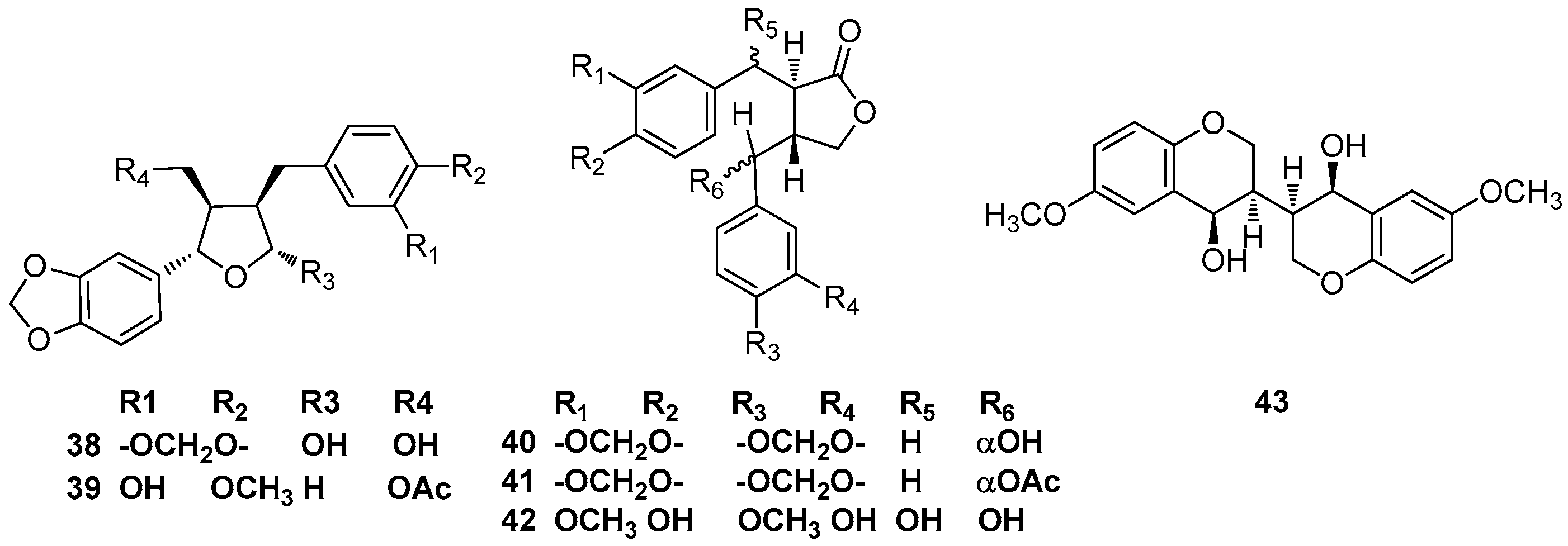
(-)-Parabenzoinol (38) was isolated from the fresh leaves of Parabenzoin trilobum Nakai, and its structure was elucidated by X-ray crystallographic analysis [20]. Actifolin (39) was identified in the stems of Lindera obtusiloba; moreover, its effect on tumor necrosis factor (TNF)-α and interleukin (IL)-6 as well as its inhibitory activity against histamine release were examined using human mast cells. Actifolin (39) suppressed the gene expressions of proinflammatory cytokines, TNF-α, and IL-6 in human mast cells [21,22] (Figure 4).
2.4. Lignan-9,9’-Olides
2.5. 2.9,2’.9’-Diepoxylignans
2.6. 7.9’,7’.9-Diepoxylignans
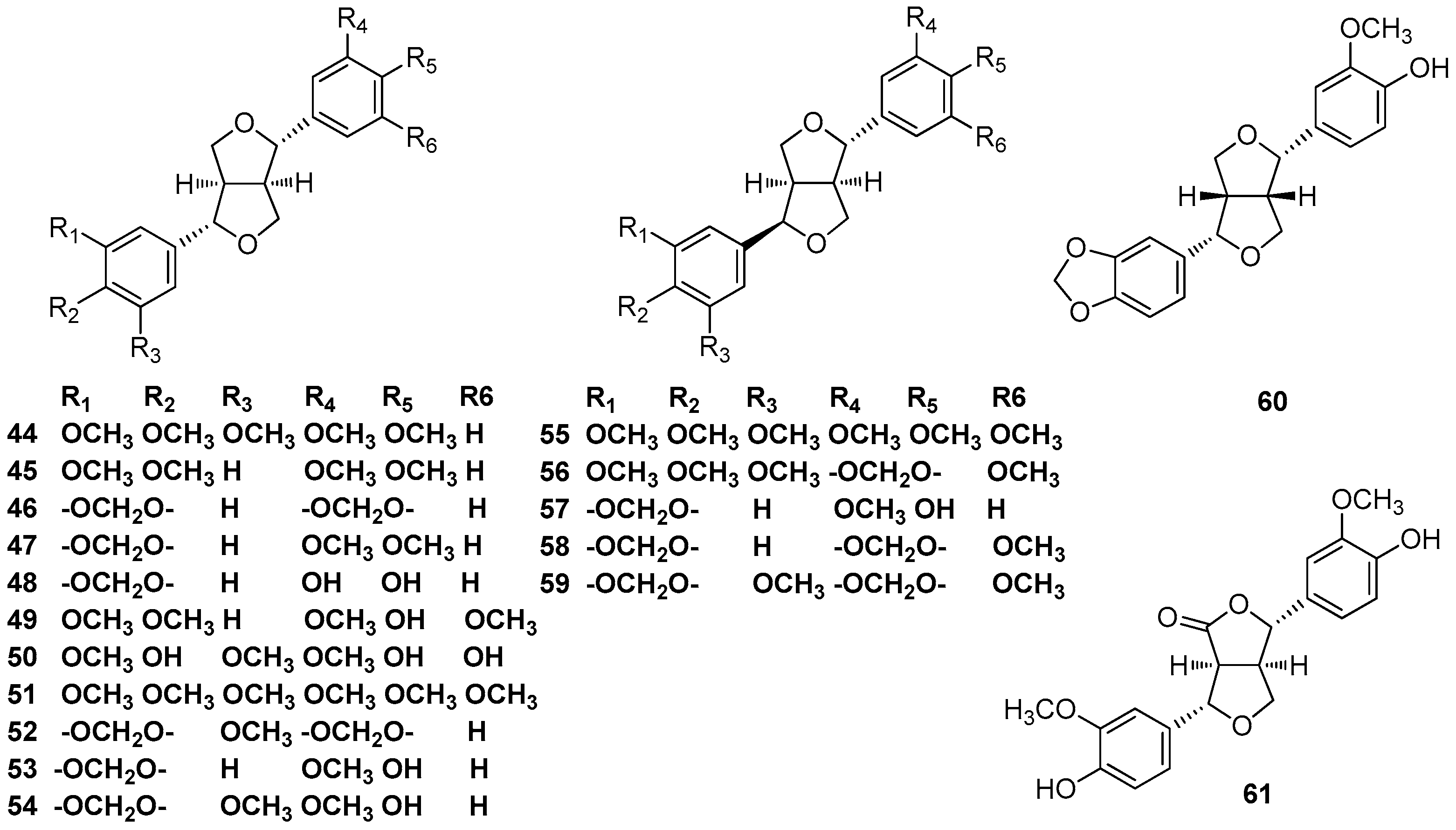
The ethanol/H2O (9:1) extract of the fruits of L. armeniaca contained magnolin (44) and eudesmin (45) [25]. Phytochemical studies revealed the presence of sesamin (46) and O-methylpiperitol (47) in the ethanolic extract of the fruit of calyces of N. amazonurn [26]. Sesamin (46) was also found to occur in the CH2Cl2 extract of the bark of M. thunbergii Sieb. et Zucc [3,4]. Magnolin (44), eudesmin (45), sesamin (46), and O-methylpiperitol (47) were all found to occur in Persea pyrifolia Nees and Mart. ex Nees [27]. Phytochemical investigations of the methanolic extract of the leaves of A. barbujana resulted in the isolation of demethylpiperitol (48) [10]. The ethanolic extract of Pleurothyrium cinereum also contained (+)-demethylpiperitol (48), as well as (+)-de-4’’-O-methylmagnolin (49), which was found to be a potent COX-2/5-LOX dual inhibitor and platelet-activating factor (PAF)-antagonist (COX-2: IC50 = 2.27 μM; 5-LOX: IC50 = 5.05 μM; PAF: IC50 = 2.51 μM) (49) [6,28]. (+)-Syringaresinol (50) was isolated from the stems of C. reticulatum Hay [29]. (+)-De-4’’-O-methylmagnolin (49) and (+)-syringaresinol (50) both were found to occur in the methanolic extract of the stems of Actinodaphne lancifolia [30]. The leaves of C. macrostemon Hayata [31] and the stems of C. burmanii [32] both contained (+)-syringaresinol (50) and yangambin (51), which showed various pharmacological effects. Moreover, C. burmanii also contained (+)-sesamin (46) [32]. (+)-Demethoxyexcelsin (52), (+)-piperitol (53), and (+)-methoxypiperitol (54) were obtained from the bark and wood of N. turbacensis, together with (+)-sesamin (46) [33]. Epiyangambin (55), episesartemin (56), and yangambin (51) were isolated from the leaves of O. duckei, and yangambin (51) represented the major constituent [34]. Kwon et al. reported the isolation of (+)-syringaresinol (50) and pluviatilol (57) from the stems of L. obtusilob, and pluviatilol (57) showed cytotoxicity against a small panel of human tumor cell lines [21]. (+)-5-Demethoxyepiexcelsin (58) and (+)-epiexcelsin (59) were reported to be found in Litsea verticillata Hance, and (+)-5- demethoxyepiexcelsin (58) showed moderate anti-HIV activity with an IC50 value of 16.4 μg/mL (42.7 μM) [35]. (+)-Xanthoxyol (60), (+)-syringaresinol (50), and pluviatilol (57) were obtained from the stems of L. obtusiloba Blume. The effect of these compounds on tumor necrosis factor (TNF)-α and interleukin (IL)-6 as well as their inhibitory activity against histamine release were examined using human mast cells. Pluviatilol (57) inhibited the release of histamine from mast cells [22]. 4-Keto-pinoresinol (61) was isolated from the ethanolic extract of the leaves and twigs of Litsea chinpingensis [36] (Figure 5).
3. Cyclolignans
There are three main types of cyclolignans isolated from nature, including 2,7’-cyclolignans, 2,2’-cyclolignans, and 7,7’-cyclolignans. Cyclolignans are not so common in Lauraceae. We have only retrieved less than 10 2,7’-cyclolignans isolated from Lauraceae. The semi-systematic names of those cyclolignans without trivial names and their corresponding names in the literature are given in Table SI-1 (Supporting Information).
2,7.’-Cyclolignans
(-)-Isoguaiacin (62) and (+)-guaiacin (63) were isolated from the extract of the bark of M. thunbergii Sieb. et Zucc. These two compounds showed significant neuroprotective activities against glutamate-induced neurotoxicity in primary cultures of rat cortical cells [3,4]. (+)-Otobaphenol (64) and cyclolignans 65 and 66 were isolated from the ethanolic extract of P. cinereum [28]. Cinnamophilin A (67) was first reported to be obtained from the methanolic extract of roots of Cinnamomum philippinense (Merr.) Chang [37]. (-)-Aristoligone (68), (-)-aristotetralone (69), and (-)-cagayanone A (70) were obtained from the ethanolic extract of the leaves and twigs of L. chinpingensis [36] (Figure 6).
4. Neolignans
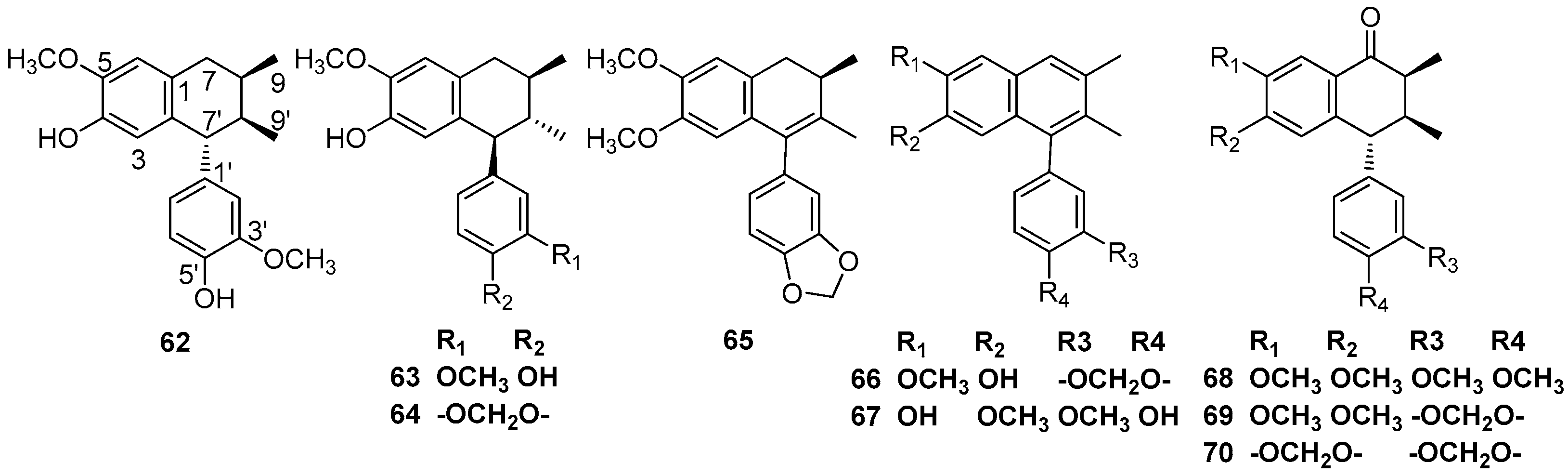
Neolignans are widely distributed in the Lauraceae family, especially in the genera of Aniba, Nectandra, and Ocotea. The types of neolignans isolated from Lauraceae include 8,1’-neolignans, 8,3’-neolignans, 7,1’-neolignans, and 7,3’-neolignan (Figure 7). 3,3’-neolignans, which also exist in nature, have not been isolated from Lauraceae. The semi-systematic names of the abovementioned neolignans without trivial names and their corresponding names in the literature are given in Table SI-2 and SI-3 (Supporting Information).
4.1. 8,1’-Neolignans
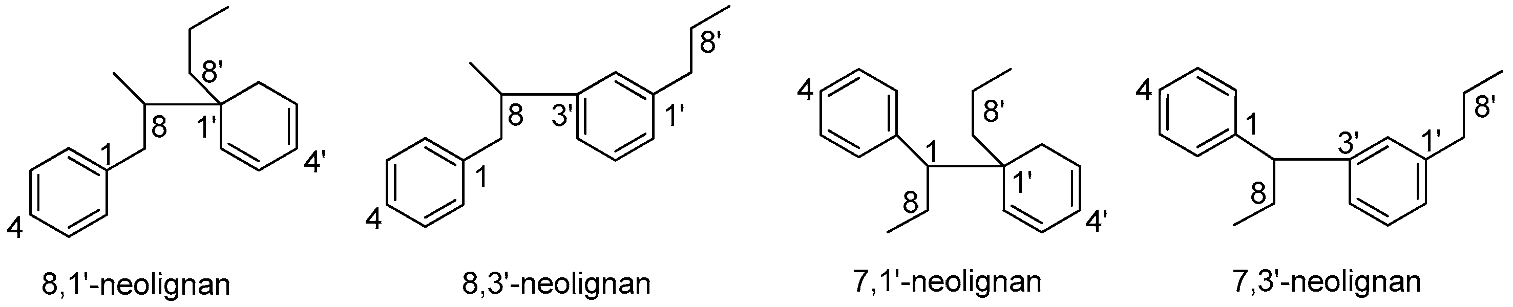
Burchellin (71) was first isolated from the trunk wood of Aniba burchellii Kosterm [38]. Burchellin (71) has also been found in the benzene extract of the trunk of an unclassified Aniba species collected in the vicinity of Manaus, Amazonas, along with compounds 72 and 73 [39,40]. Compounds 72 and 74 were obtained as a mixture from an unclassified Amazonian Nectandra species. As the analogous values for the mixture of compounds 72 and 74 were substantially identical to those of pure compound 72, including the ORD (optical rotatory dispersion) curves, then the two compounds should have the same absolute configuration [41]. 3’-Methoxyburchellin (75) was first isolated from the stem bark of O. veraguensis [16]. Benzene extract of the trunk wood of Aniba terminalis [42] and ethanolic extract of the trunk wood of an Aniba species collected 130 km north of Manaus, Amazonas [43] both contained burchellin (71) and compound 76. The trunk wood of Ocotea catharinensis yielded compound 77 [44,45]. Inspection of Aniba simulans revealed the occurrence of compounds 78–80 [46,47]. Armenin A (81) and armenin B (82) were first obtained from the benzene extract of the trunk wood of Licaria armeniaca [48]. The fruits of L. armeniaca yielded compounds 76 and 78 [25]. Compounds 74, 78 and armenin C (83) were isolated from the fruits of Aniba riparia [49]. Compound 85 was isolated from the benzene extract of trunk wood of an unclassified Aniba species [50]. Canellin B (86) was first obtained from the benzene extract of the trunk wood of Licaria canella [51]. The trunk wood of an Amazonian Aniba species contained armenin A (81), armenin B (82), C (83), canellin B (86), canellin D (87), canellin E (88), porosin (90), and porosin B (91) [52]. Porosin (90) was first obtained from the wood of Ocotea porosa [53]. Porosin B (91) was first obtained from the branch wood of U. verrucosum, and porosin (90) also were found to exist in the same species [17]. The wood of Ocotea catharinensis yielded armenin B (82), canellin B (86), ferrearin C (95), ferrearin E (96), and compounds 92 and 93. Moreover, the structures of compound 92 and ferrearin C (95) were certified by single-crystal X-ray analysis [45]. Ferrearin A (99) and ferrearin B (100) were first isolated from the trunk wood of the Amazonian Aniba ferra Kubitzki, together with compounds 85 and 92. The relative structures of ferrearin A (99) and ferrearin B (100) were elucidated as structures of 97 and 98 [54], then revised as ferrearin A (99) and ferrearin B (100). Besides these two compounds, 3’-methoxyburchellin (75), compound (77), ferrearin C (101), and ferrearin D (102) were found to occur in the trunk wood of Ocotea aciphylla [55,56]. Burchelin (71), porosin (90), porosin B (91), and compounds 76, 89, 94, 103–106, 112, and 113 all were identified in the trunk wood of O. porosa, collected from the Forest Reserve of the Botanical Institute, Sâo Paulo, Brazil [57,58]. Fifteen 8,1’-neolignans have been reported to be found in the bark and leaves of O. porosa harvested near Santa Maria, State of Rio Grande do Sul, Brazil, including burchellin (71), porosin (90), porosin B (91), and compounds 76, 89, 94, and 105–113 [59] (Figure 8).
4.2. 8,3’-Neolignans
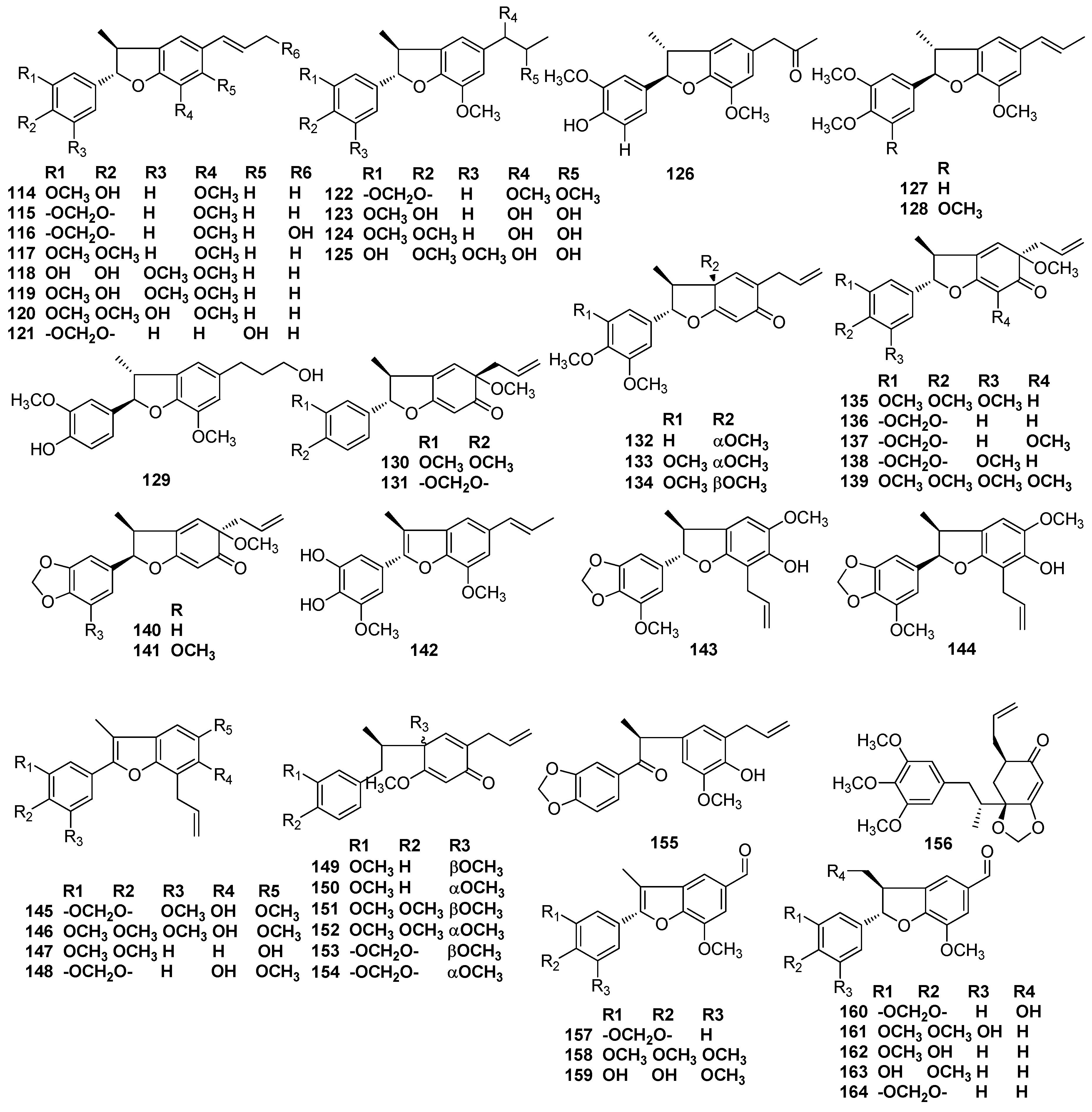
The benzene extract of trunk wood of Licaria aritu Ducke [60] and the EtOH/H2O (9:1) extract of the fruits of N. glabrescens contained licarin A (114) and licarin B (115) [26]. Licarin A (114) was also isolated from N. rigida Nees and is responsible for the major cytotoxic activity of crude extract of N. rigida Nees, displaying ED50 vs. KB cancer cell line at 7.0 μg/mL [12]. Machilin B (116) was obtained from the methanolic extract of the bark of M. thunbergii [2], as well as licarin A (114) and licarin B (115). Licarin A (114) showed significant neuroprotective activities against glutamate-induced neurotoxicity in primary cultures of rat cortical cells and induced an apoptotic effect in HL-60 cells via caspase-3 activation [4,7]. Licarin A (114) and licarin D (117) were found in branch wood of the shrub U. verrucosum [17]. Obovatifol (118), odoratisol-A (119), and (-)-licarin A (114) were obtained from the air-dried bark of the Vietnamese medicinal plant Machilius odoratissima Nees [19]. Besides licarin A (114) and licarin B (115), machilusol A (120), machilusol B (122), machilusol C (123), machilusol D (124), machilusol E (125), machilusol F (126), and acuminatin (127) were isolated from the stem wood of Machilus obovatifolia. Machilusols A–F showed moderate cytotoxic activity [61]. The dichloromethane extract of the bark of M. thunbergii Sieb. et Zucc also contained (-)-acuminatin (127), together with licarin A (114). (-)-Acuminatin exerted diverse hepatoprotective activities, perhaps by serving as a potent antioxidant [3,5]. Dihydrodehydrodiconifery alcohol (129) was found in the ethanolic extract of the leaves and twigs of L. chinpingensis [36]. Compound 130 was first obtained from the benzene extract of the trunk of an Aniba species collected in the vicinity of Manaus, Amazonas, along with acuminatin (127) and licarin D (116) [40]. A. burchellii Kosterm contains compounds 130 and 131. The determination of their absolute stereochemistry relied on spectra and a preparation by thermolysis as well as the acid isomerization of burchellin (71) [62]. Denudatin B (132), as well as (+)-licarin A (114), liliflol B (121), and (+)-acuminatin (127), have been found in leaves of Nectandra amazonum Nees [63]. Mirandin A (133) was proved to be the major neolignan of an unclassified Nectandra species, which grew at Rosa de Maio, a locality on the Manaus-Itacoatiara highway, Amazonas [41]. (+)-Mirandin A (133), (-)-licarin A (114), and (-)-licarin B (115) also were found to occur in the ethanolic extract of P. cinereum [28]. Licarin C (128), mirandin A (133), mirandin B (134), and compounds 135 and 146 were obtained from the benzene extract of Nectundru mirunda trunk wood [64]. Compounds 136 and 137 were found in the stem bark of O. veraguensis [16]. Furthermore, compound 136 also was found in the trunk wood of an Aniba species collected 130 km north of Manaus, Amazonas [43], and compound 137 was obtained from the wood of O. catharinensis [44] and the fruits of O. veraguensis [65]. Compounds 135, 138, 139, 141, and 143–145 were isolated from the benzene extract of Anibu simulans trunk wood [46]. The extract of EtOH/H2O (9:1) of fruits of L. armeniac also provided compounds 136 and 138 [25]. Obovaten (142), perseal D (159), perseal C (160), and obovatinal (161) were first obtained from the leaves of Persea obovatifolia; together with obovatifol (118), these compounds showed significant cytotoxicity against P-388, KB16, A549, and HT-29 cancer cell lines in vitro [66,67]. Compound 148 was isolated from the benzene extract of the trunk wood of A. terminalis [42]. Lancifolins A–F (149–154) were obtained from branches of the shrub Aniba lancifolia Kubitzki et Rodrigues [68]. Neolignan ketone 156 was found to exist in the chloroform extract of the bark of Ocotea bullata [69]. Ocophyllals A (157) and ocophyllals B (158), which have a C-1’ formyl side chain instead of a propenyl group, as well as (+)-licarin B (115) were observed to occur in the ethanolic extract from leaves of O. macrophylla [70]. Licarin A (114), licarin B (115), (7R,8S,1’R)-7,4’-epoxy-1’-methoxy-3,4-methylenedioxy-8,3’-neolign-8’-ene-6’(1’H)-one (140), and compounds 130, 147, 155, 162, and perseal F (163) were obtained from O. porosa [57,58,59]. Compound 162 also was found to occur in the dichloromethane extract of the bark of M. thunbergii SIEB. et ZUCC [5]. Meanwhile, perseal F (163) and perseal G (164) were present in the chloroform-soluble portion of the stem wood of M. obovatifolia [71] (Figure 9).
4.3. 7,1’-Neolignans
Licaria chrysophylla gave a considerable proportion of chrysophyllin A (165), which was the first type of 7,1’-neolignan to be obtained. Chrysophyllin B (166), chrysophyllon I-A (167), and chrysophyllon I-B (168) were also identified in L. chrysophylla [72,73]. The trunk wood of an Amazonian Aniba species collected in the vicinity of Manaus, Amazonas also contained chrysophyllin A (165) and chrysophyllin B (166) [52] (Figure 10).
4.4. 7,3’-Neolignans
5. Cycloneolignans
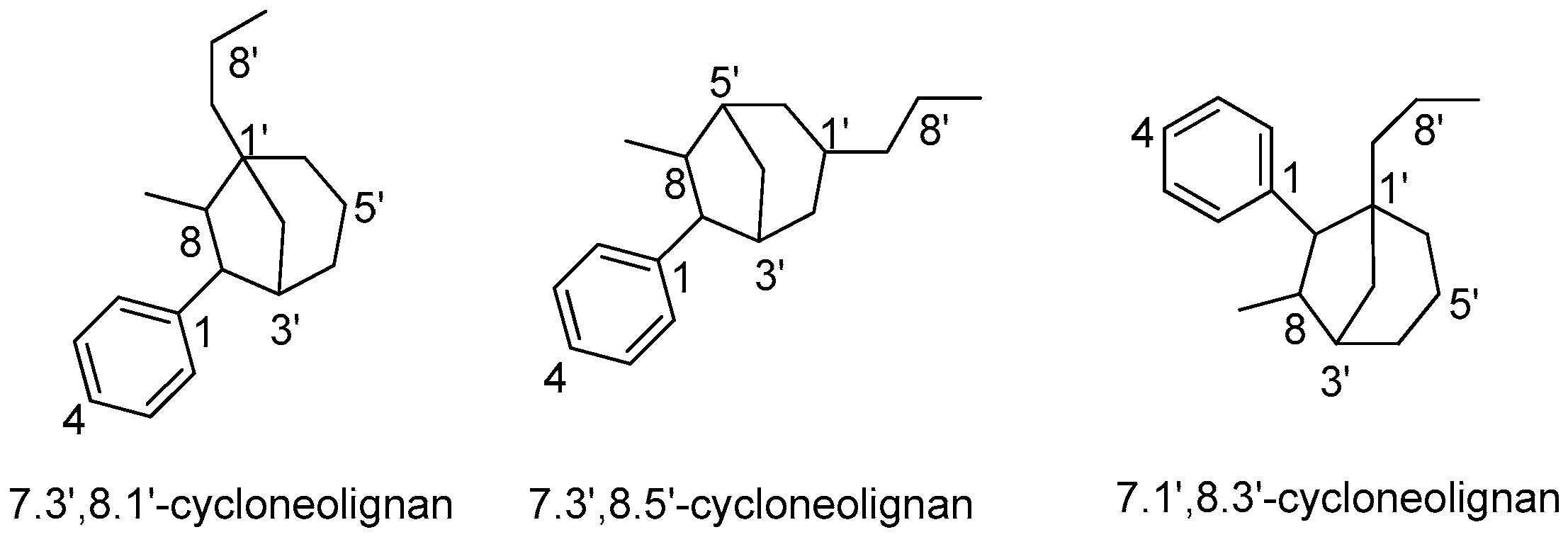
Cycloneolignans are responsible for the chemotaxonomic characteristics of some genera in the Lauraceae family, such as Aniba, Licaria, and Nectandra. Most cycloneolignans isolated from Lauraceae belong to the categories of 7.3’,8.1’-cycloneolignans and 7.3’,8.5’-cycloneolignans (Figure 11). Only two 7.1’,8.3’-cycloneolignans have been reported to be obtained from O. bullata. The semi-systematic names of those cycloneolignans without trivial names and their corresponding names in the literature are given in Table SI-4 (Supporting Information).
5.1. 7.3’,8.1’-Cycloneolignans
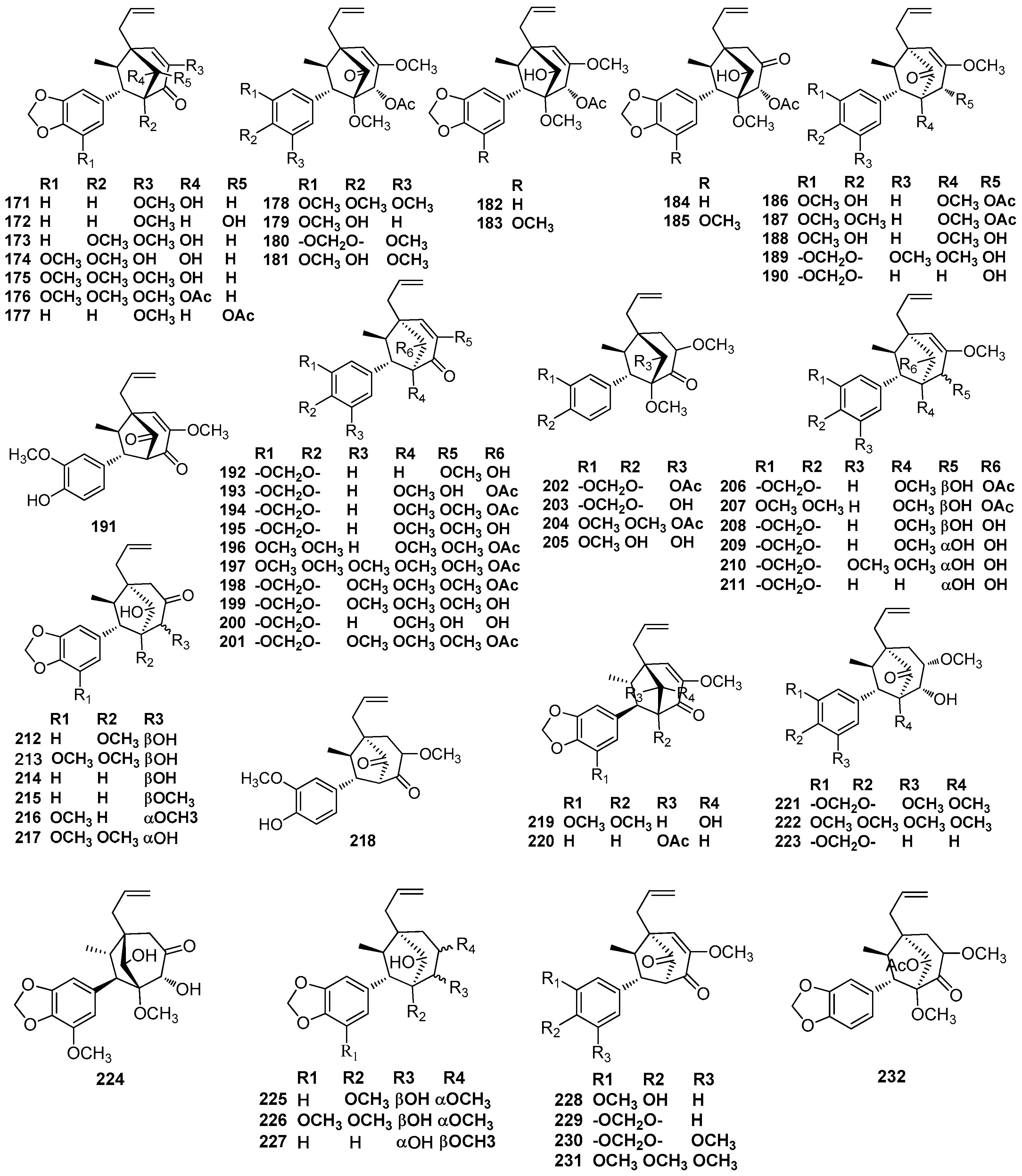
Guianin (171) was first obtained from the wood of Aniba guianensis Aubl [74]. Meanwhile, 2’-epiguianin (172) was isolated from the leaves of O. macrophylla Kunth, which showed inhibition activity against the platelet-activating factor (PAF)-induced aggregation of rabbit platelets with an IC50 value of 1.6 μM [70]. Fourteen 7.3’,8.1’-cycloneolignans, including 3’-methoxyguianin (173) and compounds 174–176 and 178–187, were isolated from the trunk wood of O. porosa collected from the mountainous Atlantic forest region of São Paulo State, where it is known as ‘canela parda’ [75,76]. Compound 192 was first obtained from the trunk wood of O. porosa collected from the Forest Reserve of Instituto BottInico (SHo Paulo, SP), along with guianin (171) and 2’-epiguianin (172) [58]. Compound 191 was first obtained from the benzene extract of A. burchellii Kosterm, together with guianin (171) [62]. Compounds 193–195 and 229 were found to be present in the benzene extract of the trunk of a unclassified Aniba species collected from the Ducke Forest Reserve, Manaus, Amazonas, as well as guianin (171) and 3’-methoxyguianin (173) [40]. Compounds 184, 194–198, and 202–207 were obtained from seed coat and dried fruit pulp of O. veraguensis. Since the author did not describe how to determine the absolute configuration of these compounds, their name should contain the addition of the prefix ‘rel’ [65]. Otherwise, compounds 188, 191, and 208–210 were isolated from the petrol and chloroform extract of the stem bark of O. veraguensis [16]. The trunk bark of O. catharinensis contained compounds 200, 208, 209, as well as canellin-C (212) and 5-methoxycanellin-C (213), and the contents of all these compounds in the bark were over 0.01% [44,45]. Compounds 214–216, 218, and 228 were found to occur in the trunk wood of an Aniba species collected 130 km north of Manaus, Amazonas [43]. The benzene extract of trunk wood pertaining to an unclassified Aniba species collected from the Ducke Forest Reserve, Manaus yielded 217, 219, 221, 222, 224, and methoxycanellin A (226) [50,77]. Compound 189 was first obtained from the ethanolic extract of wood of Ocotea costulatum, along with compound 221 [78]. The trunk wood of the Amazonian A. ferra Kubitzki contained rel-(7S,8R,1’S,2’S,3’R)-1’,2’-dihydro-2’-hydroxy-3,3’,5’- trimethoxy-4,5-methylenedioxy-7.3’,8.1’-cycloneolign-8’-ene- 4’(3’H)-one (199) and methoxycanellin A (226) [54]. Canellin A (225) and canellin C (212) were first obtained from the trunk wood of L. canella [51]. These two compounds have also been reported to be found in the trunk wood of L. rigida Kosterm. However, the relative structures of the compounds shown in the abovementioned article were different from those shown in other articles—the methyl occupied an exo-configuration and the aryl adopted an endo-configuration [79]. The trunk wood of the central Brazilian O. aciphylla also yielded canellin-A (225), as well as compound 208 and 3’-methoxyguianin (173) [55,56]. 2’-Epiguianin (172), compounds 177, 190–192, 211, 220, 223, 227, and rel-(7R,8R,1’S,3’S)-5’-methoxy-3,4-methylenedioxy-7.3’,8.1’-cycloneolign-8’-ene-2’,4’(1’H,3’H)-dione (229) were obtained from the bark and leaves of O. porosa [59]. Compounds 194, 201, 230, and 231 were found in the extract of EtOH/H2O (9:1) of the fruits of L. armeniaca [25]. The chloroform extract of the trunk wood of L. armeniaca yielded compound 232 [80] (Figure 12).
5.2. 7.3’,8.5’-Cycloneolignans
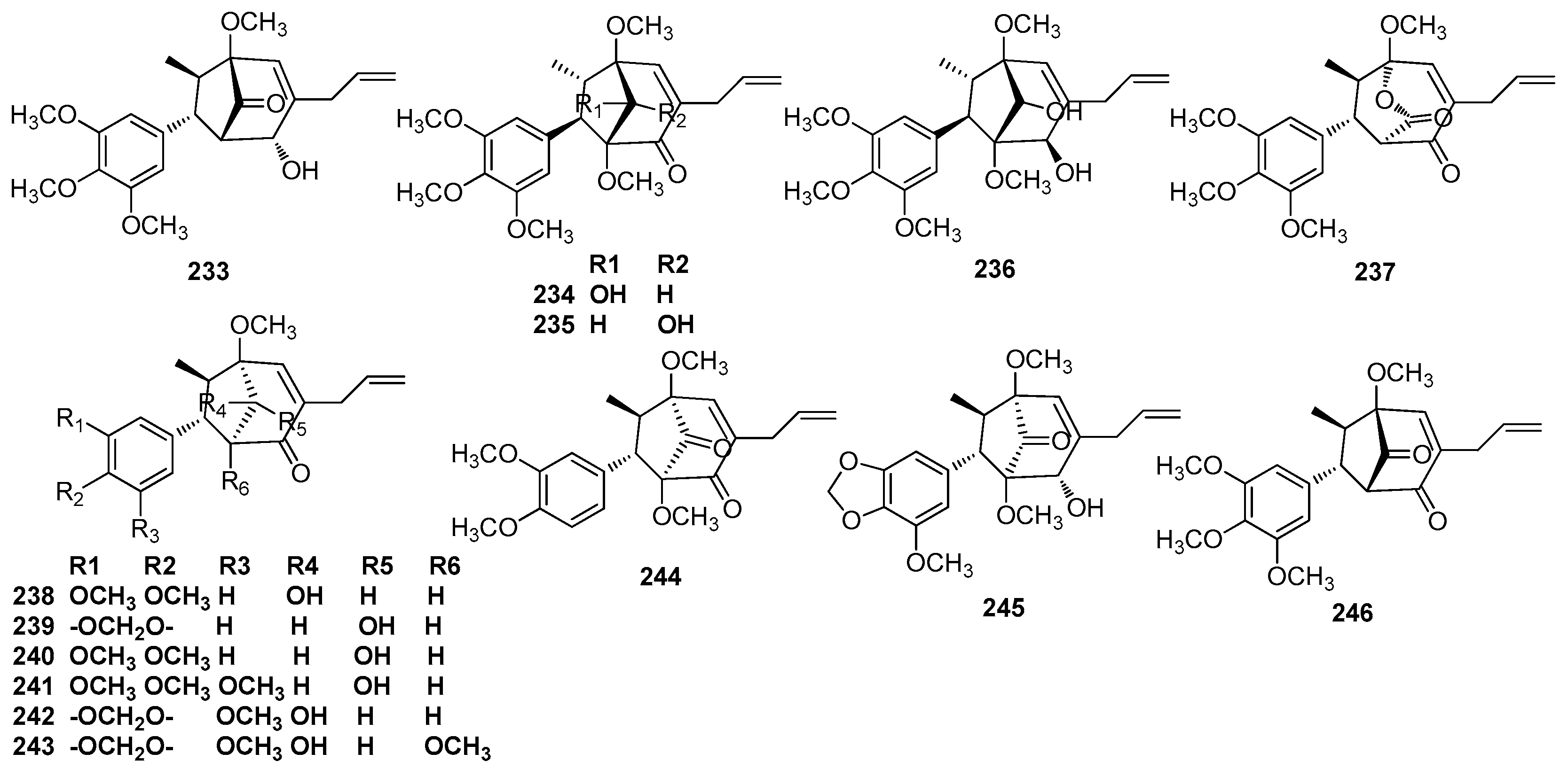
Macrophyllin B (233) was purified from an unclassified Nectandra species collected at Rosa de Maio, a locality on the Manaus-Itacoatiara highway (8 km), Amazonas [41]. Nectamazins A–C (234–236), macrophyllin B (233), denudanolide D (237), and kadsurenin C (238) isolated from leaves of N. amazonum Nees showed inhibition activity against the platelet-activating factor (PAF)-induced aggregation of rabbit platelets [63]. A phytochemical exploration of the leaves of O. macrophylla afforded ocophyllols A–C (239–241). Their absolute configurations were established by derivatizing them with (R)-and (S)-MTPA, and then analyzing the NMR data, as well as by a comparison of their circular dichroism (CD) data with that of a related compound whose absolute configuration was previously established by single-crystal X-ray analysis. Moreover, ocophyllols A–C (239–241) showed some inhibition activity against the platelet-activating factor (PAF)-induced aggregation of rabbit platelets [70]. Cinerin B (242), cinerin C (243), cinerin A (244), and cinerin D (245) were isolated from the leaves of P. cinereum. Again, their CD data was used to determine the absolute configuration of these compounds. Cinerin C (243) was the first known macrophyllin-type cycloneolignan, which was isolated from the trunk wood of Licaria macrophylla Kosterm and named as macrophyllin A [81]. Cinerin A–D also showed some inhibition activity against the platelet-activating factor (PAF)-induced aggregation of rabbit platelets [82]. Compound 246 and macrophyllin B (233) were identified in the ethanolic extract of leaves of P. cinereum [28] (Figure 13).
5.3. 7.1’,8.3’-Cycloneolignans
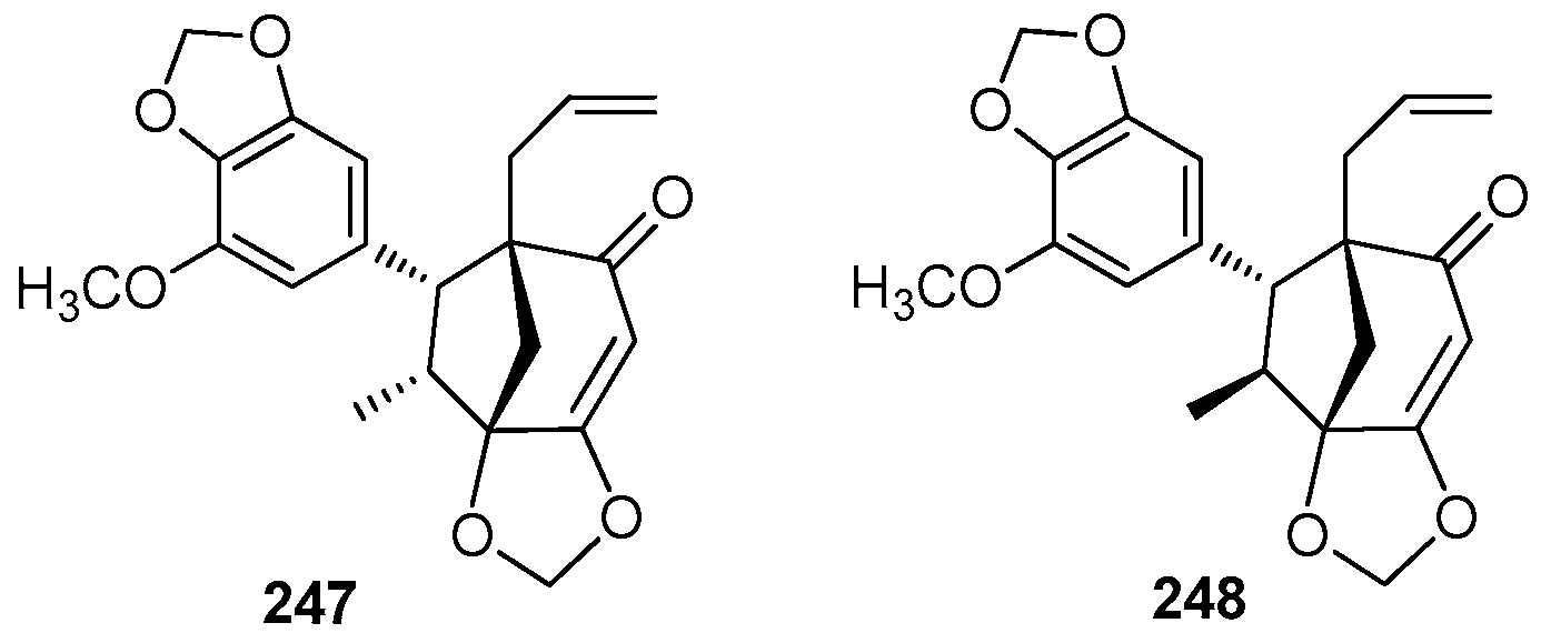
Ocobullenone (247) was the first naturally occurring bicyclooctanoid found to exhibit the 7.1’, 8.3’ linkage, and it was isolated from the chloroform extract of the bark of O. bullata [83]. Iso-ocobullenone (248) was also isolated from the chloroform extract of the bark of O. bullata, and its structure was confirmed by single-crystal X-ray analysis [69] (Figure 14).
6. Oxyneolignans
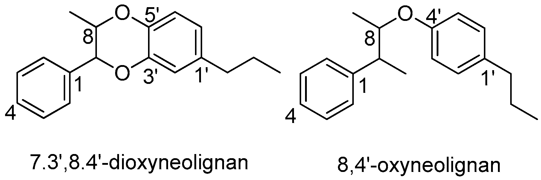
An ether oxygen atom provides the linkage between the two phenylpropane units, giving rise to oxyneolignans. Oxyneolignans are rarely distributed in Lauraceae. Less than 10 oxyneolignans have been found to occur in the Lauraceae family, belonging to two categories: 7.3’,8.4’-dioxyneolignans and 8,4’-oxyneolignans (Figure 15).
6.1. 7.3’,8.4’-Dioxyneolignans
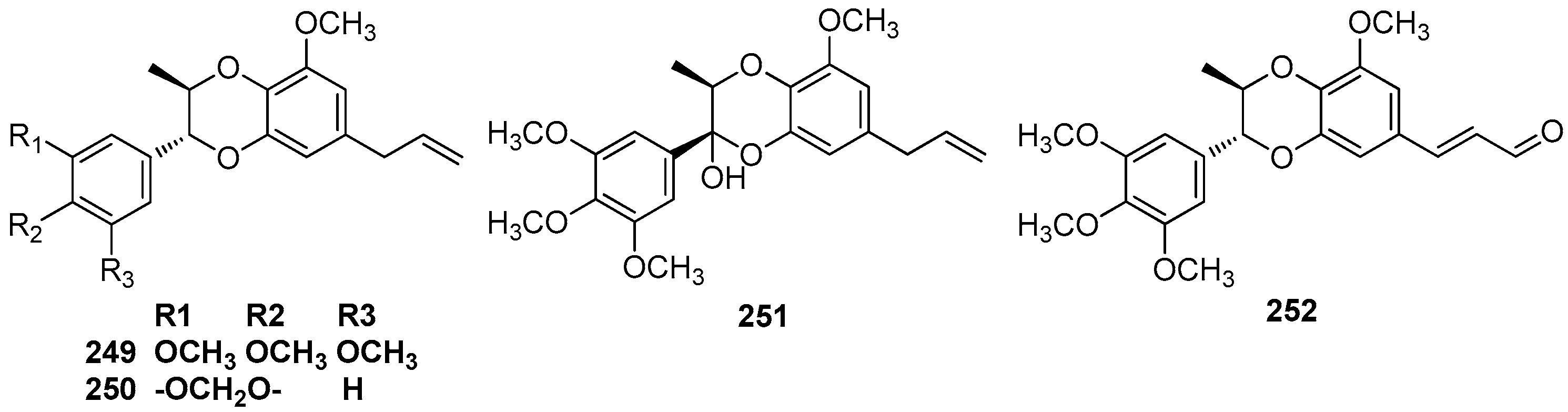
The trunk wood of L. rigida Kosterm contained eusiderin (249) and eusiderin B (250) [79]. The trunk wood of an unclassified Aniba species collected at the Ducke Forest Reserve, Manaus also yielded the benzodioxane-type neolignan eusiderin (249), eusiderin-F (251), and eusiderin-G (252) [50,77]. Eusiderin (249) was also found to be present in the ethanolic extract of wood of O. costulatum [78] (Figure 16).
6.2. 8,4’-Oxyneolignans
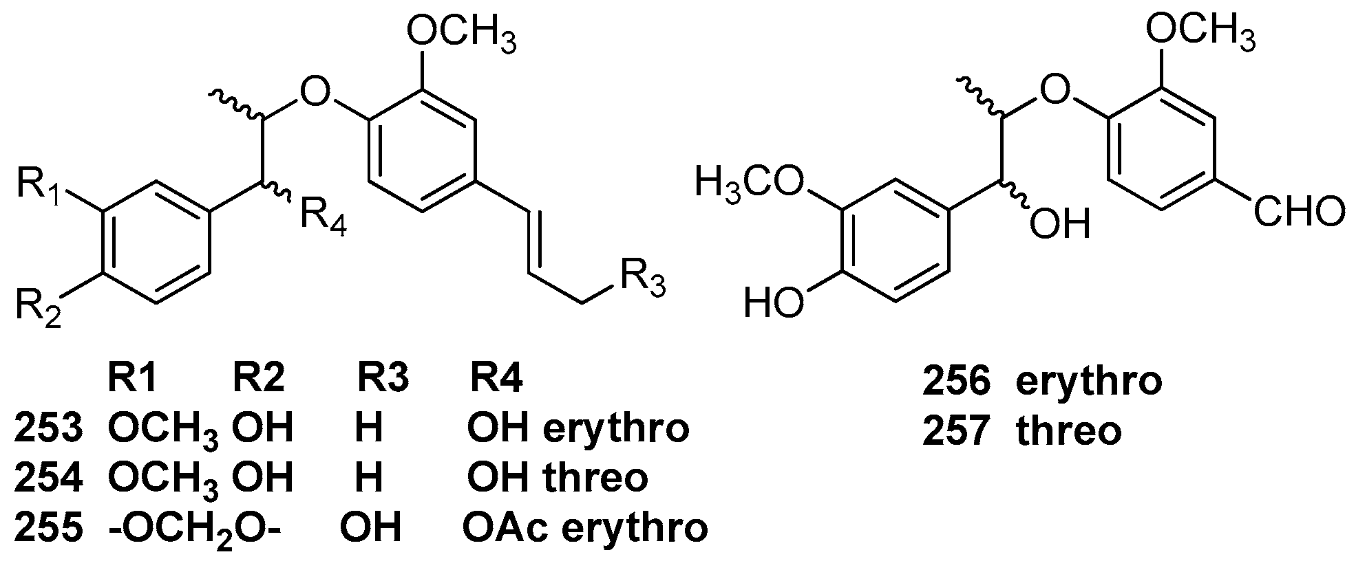
Machilin C (253), D (254), and E (255) were first obtained from the methanolic extract of the bark of M. thunbergii [2]. Odoratisol B was obtained from the air-dried bark of the Vietnamese medicinal plant M. odoratissima Nees. This compound showed the same relative structure as machilin C (253), but was termed odoratisol B in the article [19]. Perseal A (256) and perseal B (257), which have a C-1’ formyl side chain instead of a propenyl group, were isolated from the chloroform-soluble fraction of the leaves of P. obovatifolia. They showed significant cytotoxicity against P-388, KB 16, A549, and HT-29 cancer cell lines [67] (Figure 17).
7. Uncommon Lignans
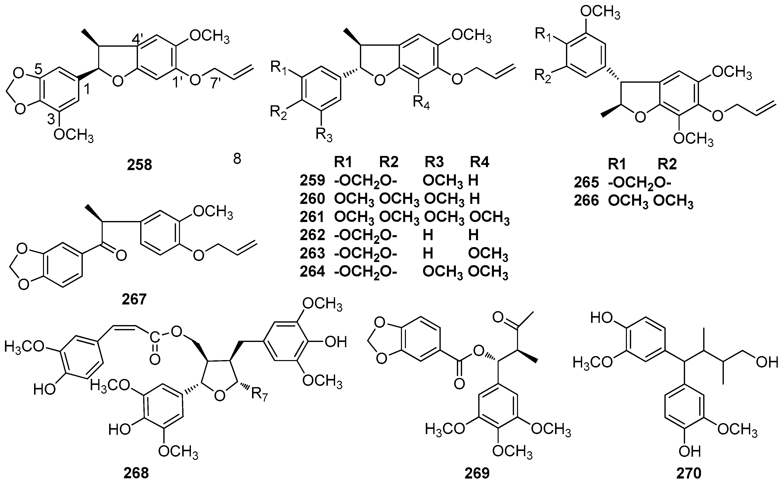
This section covers lignans and neolignans that contain uncommon skeletons. The molecular backbone of compounds 258–267 consists of a unique C6–C3 unit, and an ether oxygen atom provides the linkage between the phenyl and propyl groups. The semi-systematic names of those uncommon lignans without trivial names and their corresponding names in the literature are given in Table SI-5 (Supporting Information).
Compounds 258–261 were isolated from the benzene extract of A. simulans trunk wood [46]. Compounds 259 and 260 were also found in the fruits of L. armeniaca [25] and the trunk wood of N. mirunda, respectively [64]. Compound 262 was isolated from the benzene extract of the trunk wood of A. terminalis [42]. Compounds 263 and 264 have been found in the trunk bark of O. catharinensis [45]. The stem bark of O. veraguensis also yielded compound 263 [16]. Chrysophyllon III-A (265) and chrysophyllon III-B (266) have been found in trunk wood, bark, and fruit calyces of L. chrysophylla [73]. Compound 267 was obtained from the bark and leaves of O. porosa [59]. (+)-9’-O-trans-feruloyl-5,5’- dimethoxylariciresinol (268), which showed cytotoxicity against a small panel of human tumor cell lines with ED50 values around 10 μg/mL, was isolated from the stems of L. obtusiloba Blume [21,22]. Turbacenlignan A (269), a 7,8-secolignan, was isolated from the leaves and root bark of N. turbacensis (Kunth) Nees [9]. Cinnaburmanin A (270) was isolated from the roots of C. burmanii [84] (Figure 18).
8. Conclusions
A renewed interest in compounds isolated from natural resources has led to an enormous class of pharmacologically active compounds. Lignans and neolignans have been revealed to show significant pharmacological activities, including antitumor, anti-inflammatory, immunosuppression, cardiovascular, antioxidant, and antiviral activities [85,86]. The Lauraceae family, especially the genera of Machilus, Ocotea, and Nectandra, represents a rich source of lignans and neolignans. Moreover, neolignans are responsible for the potential chemotaxonomic significance found in the study of Lauraceae. Studies on lignans and neolignans in Lauraceae were mainly carried out in the 1980s. There have been more studies concerning the identification of lignans and neolignans in Lauraceae, but less on the biological activities of these compounds. Among the lignans and neolignans isolated from Lauraceae, the biological activities of sesamin and yangambin have been studied more, while there are relatively few articles published on other compounds. Sesamin, a 7.9’,7’.9-diepoxylignan present in many species in the Lauraceae family such as N. amazonurn, M. thunbergii, P. pyrifolia, C. burmanii, and N. turbacensis, showed significant anticancer properties [87]. Yangambin (51) which was the major constituent of O. duckei, showed diverse biological activities [88]. Therefore, it is extremely urgent to expand the scope of research on the lignans and neolignans in Lauraceae, with the aim of discovering all biological activities of these compounds.
Supplementary Materials
The supplementary materials are available online at https://0-www-mdpi-com.brum.beds.ac.uk/1420-3049/23/12/3164/s1.
Author Contributions
Investigation, S.X. and J.Y.; Writing-Original Draft Preparation, W.W.; Writing-Review & Editing, Y.L.; Funding Acquisition, K.G.
Funding
This work was funded by the National Natural Science Foundation of China (21402074).
Conflicts of Interest
The authors declare no conflict of interest.
References
- Teponno, R.B.; Kusari, S.; Spiteller, M. Recent advances in research on lignans and neolignans. Nat. Prod. Rep. 2016, 33, 1044–1094. [Google Scholar] [CrossRef] [PubMed]
- Shimomura, H.; Sashida, Y.; Oohara, M. Lignans from Machilus thunbergii. Phytochemistry 1987, 26, 1513–1515. [Google Scholar] [CrossRef]
- Yu, Y.U.; Kang, S.Y.; Park, H.Y.; Sung, S.H.; Lee, E.J.; Kim, S.Y.; Kim, Y.C. Antioxidant lignans from Machilus thunbergii protect CCl4–injured primary cultures of rat hepatocytes. J. Pharm. Pharmacol. 2000, 52, 1163–1169. [Google Scholar] [CrossRef] [PubMed]
- Ma, C.J.; Sung, S.H.; Kim, Y.C. Neuroprotective lignans from the bark of Machilus thunbergii. Planta Med. 2004, 70, 79–80. [Google Scholar] [PubMed]
- Li, G.; Lee, C.S.; Woo, M.H.; Lee, S.H.; Chang, H.W.; Son, J.K. Lignans from the bark of Machilus thunbergii and their DNA topoisomerases I and II inhibition and cytotoxicity. Biol. Pharm. Bull. 2004, 27, 1147–1150. [Google Scholar] [CrossRef]
- Coybarrera, E.D.; Cucasuarez, L.E. In vitro anti-inflammatory effects of naturally–occurring compounds from two Lauraceae plants. An. Acad. Bras. Cienc. 2011, 83, 1397–1402. [Google Scholar] [CrossRef]
- Park, B.Y.; Min, B.S.; Kwon, O.K.; Oh, S.R.; Ahn, K.S.; Kim, T.J.; Kim, D.Y.; Bae, K.; Lee, H.K. Increase of caspase-3 activity by lignans from Machilus thunbergii in HL-60 cells. Biol. Pharm. Bull. 2004, 27, 1305–1307. [Google Scholar] [CrossRef] [PubMed]
- Macías-Villamizar, V.; Cuca-Suárez, L. Diaryldimethylbutane lignans and other constituents isolated from Nectandra turbacensis (Kunth) Nees (Lauraceae). Rev. Colomb. Quim. 2014, 43, 12–16. [Google Scholar]
- Macías-Villamizar, V.; Cuca-Suárez, L.; González, F.V.; Rodríguez, S. Lignoids isolated from Nectandra turbacensis (Kunth) Nees (Lauraceae). Rec. Nat. Prod. 2016, 10, 654–658. [Google Scholar]
- Pérez, C.; Almonacid, L.N.; Trujillo, J.M.; González, A.G.; Alonso, S.J.; Navarro, E. Lignans from Apollonias barbujana. Phytochemistry 1995, 40, 1511–1513. [Google Scholar] [CrossRef]
- Moro, J.C.; Fernandes, J.B.; Vieira, P.C.; Yoshida, M.; Gottlieb, H.E. Neolignans from Nectandra puberula. Phytochemistry 1987, 26, 269–272. [Google Scholar] [CrossRef]
- Quesne, P.W.L.; Larrahondo, J.E.; Raffauf, R.F. Antitumor plants. X. constituents of Nectandra rigida. J. Nat. Prod. 1980, 43, 353–359. [Google Scholar] [CrossRef] [PubMed]
- Shimomura, H.; Sashida, Y.; Oohara, M. Lignans from Machilus thunbergii. Phytochemistry 1988, 27, 634–636. [Google Scholar] [CrossRef]
- López, H.; Valera, A.; Trujillo, J. Lignans from Ocotea foetens. J. Nat. Prod. 1995, 58, 782–785. [Google Scholar] [CrossRef]
- Crossley, N.S.; Djerassi, C. Naturally occurring oxygen heterocyclics. Part XI. Veraguensin. J. Chem. Soc. 1962, 1459–1462. [Google Scholar] [CrossRef]
- Khan, M.R.; Gray, A.I.; Waterman, P.G. Neolignans from stem bark of Ocotea veraguensis. Phytochemistry 1987, 26, 1155–1158. [Google Scholar] [CrossRef]
- Dias, A.D.F.; Giesbrecht, A.M. Neolignans from Urbanodendron verrucosum. Phytochemistry 1982, 21, 1137–1139. [Google Scholar] [CrossRef]
- Huang, Y.-T.; Chang, H.-S.; Wang, G.-J.; Lin, C.-H.; Chen, I.-S. Secondary metabolites from the roots of Beilschmiedia tsangii and their anti-inflammatory activities. Int. J. Mol. Sci. 2012, 13, 16430–16443. [Google Scholar] [CrossRef]
- Phan, M.G.; Phan, T.S.; Matsunami, K.; Otsuka, H. New neolignans and lignans from Vietnamese medicinal plant Machilus odoratissima Nees. Cheminform 2006, 54, 380–383. [Google Scholar]
- Takaoka, D.; Tani, H.; Nozaki, H. A new lignan, (-)-parabenzoinol, from Parabenzoin trilobum Nakai. Chem. Lett. 1995, 24, 915–916. [Google Scholar] [CrossRef]
- Kwon, H.C.; Sang, U.C.; Lee, J.O.; Bae, K.H.; Zee, O.P.; Kang, R.L. Two new lignans from Lindera obtusiloba Blume. Arch. Pharm. Res. 1999, 22, 417–422. [Google Scholar] [CrossRef] [PubMed]
- Choi, H.G.; Choi, Y.H.; Ji, H.K.; Kim, H.H.; Kim, S.H.; Kim, J.A.; Sang, M.L.; Na, M.K.; Lee, S.H. A new neolignan and lignans from the stems of Lindera obtusiloba Blume and their anti-allergic inflammatory effects. Arch. Pharm. Res. 2014, 37, 467–472. [Google Scholar] [CrossRef] [PubMed]
- Wada, K.; Munakata, K. (-)-Parabenzlactone, a new piperolignanolide isolated from Parabenzoin trilobum Nakai. Tetrahedron Lett. 1970, 11, 2017–2019. [Google Scholar] [CrossRef]
- Wei, X.; Li, G.H.; Wang, X.L.; He, J.X.; Wang, X.N.; Ren, D.M.; Lou, H.X.; Shen, T. Chemical constituents from the leaves of Cinnamomum parthenoxylon (Jack) Meisn. (Lauraceae). Biochem. Syst. Ecol. 2017, 70, 95–98. [Google Scholar] [CrossRef]
- Barbosa-Filho, J.M.; Yoshida, M. Neolignans from the fruits of Licaria armeniaca. Phytochemistry 1987, 26, 319–321. [Google Scholar] [CrossRef]
- Barbosa-Filho, J.M.; Yoshida, M. Lignoids from Nectandra amazonum and N. glabrescens. Phytochemistry 1989, 28, 1991. [Google Scholar] [CrossRef]
- Batista, A.N.D.L.; Batista Junior, J.M.; López, S.N.; Furlan, M.; Cavalheiro, A.J.; Silva, D.H.S.; Bolzani, V.D.S.; Nunomura, S.M.; Yoshida, M. Aromatic compounds from three Brazilian Lauraceae species. Quím. Nova 2010, 33, 321–323. [Google Scholar] [CrossRef] [Green Version]
- Barrera, E.D.C.; Suárez, L.E.C.; Suárez, L.E.C. Chemical constituents from Pleurothyrium cinereum (van der Werff) (Lauraceae) from Colombia. Biochem. Syst. Ecol. 2008, 36, 674–677. [Google Scholar] [CrossRef]
- Lin, I.J.; Lo, W.L.; Chia, Y.C.; Huang, L.Y.; Cham, T.M.; Tseng, W.S.; Yeh, Y.T.; Yeh, H.C.; Wang, Y.D.; Chen, C.Y. Isolation of new esters from the stems of Cinnamomum reticulatum Hay. Nat. Prod. Res. 2010, 24, 775–780. [Google Scholar] [CrossRef] [PubMed]
- Kim, M.R.; Jung, H.J.; Min, B.S.; Oh, S.R.; Kim, C.S.; Ahn, K.S.; Kang, W.S.; Lee, H.K. Constituents from the stems of Actinodaphne lancifolia. Phytochemistry 2002, 59, 861–865. [Google Scholar] [CrossRef]
- Li, C.T.; Kao, C.L.; Li, H.T.; Huang, S.T.; Huang, S.C.; Chen, C.Y. Secondary metabolites from the leaves of Cinnamomum macrostemon Hayata. Eur. J. Biomed. Pharm.Sci. 2015, 2, 38–51. [Google Scholar]
- Chen, C.; Hong, Z.; Yang, W.; Wu, M.; Huang, J.; Lee, J. A novel homosesquiterpenoid from the stems of Cinnamomum burmanii. Nat. Prod. Res. 2012, 26, 1218. [Google Scholar] [CrossRef] [PubMed]
- Carvalho, M.G.D.; Yoshida, M.; Gottlieb, H.E. Lignans from Nectandra turbacensis. Phytochemistry 1987, 26, 265–267. [Google Scholar] [CrossRef]
- Morais, L.C.S.L.; Almeida, R.N.; Dacunha, E.V.L.; Dasilva, M.S.; Barbosafilho, J.M.; Gray, A.I. Further lignans from Ocotea duckei. Pharm. Biol. 1999, 37, 144–147. [Google Scholar] [CrossRef]
- Hoang, V.D.; Tan, G.T.; Zhang, H.J.; Tamez, P.A.; Hung, N.V.; Cuong, N.M.; Soejarto, D.D.; Fong, H.H.; Pezzuto, J.M. Natural anti-HIV agents– Part I: (+)-demethoxyepiexcelsin and verticillatol from Litsea verticillata. Phytochemistry 2002, 59, 325–329. [Google Scholar] [CrossRef]
- Yang, L.J.; Wen, C.; Luo, Y.P.; Li, G.P.; Yang, X.D.; Liang, L. Lignans and ketonic compounds from Litsea chinpingensis (Lauraceae). Biochem. Syst. Ecol. 2009, 37, 696–698. [Google Scholar] [CrossRef]
- Chen, C.Y.; Yeh, Y.T.; Hsui, Y.R. A new lignan from the roots of Cinnamomum philippinense. Chem. Nat. Compd. 2011, 47, 519–520. [Google Scholar] [CrossRef]
- Lima, O.A.; Gottlieb, O.R.; Magalhães, M.T. Burchellin, a neolignan from Aniba burchellii. Phytochemistry 1972, 11, 2031–2037. [Google Scholar] [CrossRef]
- Mourão, J.C.; Yoshida, M.; Mascarenhas, Y.P.; Rodrigues, M.; Rosenstein, R.D.; Tomita, K. Absolute configuration of the benzofuranoid neolignans. Phytochemistry 1977, 16, 1003–1006. [Google Scholar]
- Fernandes, J.B.; Gottlieb, O.R.; Maia, J.G.S. Neolignans from an Aniba sp. Phytochemistry 1976, 15, 1033–1036. [Google Scholar] [CrossRef]
- Filho, R.B.; Figliuolo, R.; Gottlieb, O.R. Neolignans from a Nectandra species. Phytochemistry 1980, 19, 659–662. [Google Scholar] [CrossRef]
- Gottli, O.R.; Ferreira, S.Z.S. Neolignans from Aniba terminalis. Phytochemistry 1975, 14, 1825–1827. [Google Scholar] [CrossRef]
- Juan, C.M.V.; Maia, J.S.; Yoshida, M. Neolignans from an Aniba species. Phytochemistry 1980, 19, 474–476. [Google Scholar]
- Haraguchi, M.; Motidome, M.; Yoshida, M.; Gottlieb, O.R. Neolignans from Ocotea catharinensis. Phytochemistry 1983, 22, 561–563. [Google Scholar] [CrossRef]
- Ishige, M.; Motidome, M.; Yoshida, M.; Gottlieb, O.R. Neolignans from Ocotea catharinensis. Phytochemistry 1991, 30, 4121–4128. [Google Scholar] [CrossRef]
- Aiba, C.J.; Alvarenga, M.A.D.; Oscar, C.C.; Giesbrecht, A.M.; Pagliosa, F.M. Benzofuranoid neolignans from Aniba simulans. Phytochemistry 1977, 16, 741–743. [Google Scholar] [CrossRef]
- Aiba, C.J.; Fernandes, J.B.; Gottlieb, O.R.; Maia, J.G.S. Neolignans from an Aniba species. Phytochemistry 1975, 14, 1597–1604. [Google Scholar] [CrossRef]
- Aiba, C.J.; Maia, J.G.S.; Pagliosa, F.M.; Yoshida, M. Benzofuranoid neolignans from Licaria armeniaca. Phytochemistry 1978, 17, 2038–2039. [Google Scholar] [CrossRef]
- Barbosa-Filho, J.M.; Yoshida, M.; Barbosa, R. d. C.S.B.C.; Giesbrecht, A.M.; Young, M.C.M. Benzoyl esters and amides, styrylpyrones and neolignans from the fruits of Aniba riparia. Phytochemistry 1987, 26, 2615–2617. [Google Scholar] [CrossRef]
- Dias, S.M.C.; Fernandes, J.B.; Maia, J.G.S.; Gottlieb, H.E. Eusiderins and other neolignans from an Aniba species. Phytochemistry 1986, 25, 213–217. [Google Scholar] [CrossRef]
- Giesbrecht, A.M.; Franca, N.C.; Rocha, A.I.D. The neolignans of Licaria canella. Phytochemistry 1974, 13, 2285–2293. [Google Scholar] [CrossRef]
- Trevisan, L.M.V.; Yoshida, M. Hexahydrobenzofuranoid neolignans from an Aniba species. Phytochemistry 1984, 23, 661–665. [Google Scholar] [CrossRef]
- Aiba, C.J.; Filho, R.B.; Gottlieb, O.R. Porosin: A neolignan from Ocotea porosa. Phytochemistry 1973, 12, 413–416. [Google Scholar] [CrossRef]
- Andrade, C.H.S.; Filho, R.B. Neolignans from Aniba ferrea. Phytochemistry 1980, 19, 1191–1194. [Google Scholar] [CrossRef]
- Romoff, P.; Yoshida, M.; Gottlieb, O.R. Neolignans from Ocotea aciphylla. Phytochemistry 1984, 23, 2101–2104. [Google Scholar] [CrossRef]
- Felicio, J.D.A.; Motidome, M.; Yoshida, M. Further neolignans from Ocotea aciphylla. Phytochemistry 1986, 25, 1707–1710. [Google Scholar] [CrossRef]
- Dias, D.A.; Yoshida, M. Further neolignans from Ocotea porosa. Phytochemistry 1986, 25, 2613–2616. [Google Scholar] [CrossRef]
- Carvalho, M.G.D.; Yoshida, M.; Gottlieb, H.E. Bicyclooctanoid, carinatone and megaphone type neolignans from Ocotea porosa. Phytochemistry 1988, 27, 2319–2323. [Google Scholar] [CrossRef]
- David, J.M.; Yoshida, M.; Gottlieb, O.R. Neolignans from bark and leaves of Ocotea porosa. Phytochemistry 1994, 36, 491–499. [Google Scholar] [CrossRef]
- Aiba, C.J.; Corrêa, R.G.C.; Gottlieb, O.R. Natural occurrence of Erdtman’s dehydrodiisoeugenol. Phytochemistry 1973, 12, 1163–1164. [Google Scholar] [CrossRef]
- Tsai, I.L.; Chen, J.H.; Duh, C.Y.; Chen, I.S. Cytotoxic neolignans from the stem wood of Machilus obovatifolia. Planta Med. 2000, 66, 403–407. [Google Scholar] [CrossRef] [PubMed]
- Alvarenga, M.A.D.; Brocksom, U.; Oscar, C.C.; Magalhães, M.T. Neolignans from Aniba burchellii. Phytochemistry 1977, 16, 1797–1799. [Google Scholar] [CrossRef]
- Coy Barrera, E.D.; Cuca Suárez, L.E. Errata: Three new 7.3′,8.5′-connected bicyclo [3.2.1]octanoids and other neolignans from leaves of Nectandra amazonum Nees. (Lauraceae). Chem. Pharm. Bull. 2009, 57, 639–642. [Google Scholar] [CrossRef] [PubMed]
- Aiba, C.J.; Gottlieb, O.R.; Pagliosa, F.M.; Yoshida, M.; Magalhães, M.T. Neolignans from Nectandra miranda. Phytochemistry 1977, 16, 745–748. [Google Scholar] [CrossRef]
- Dodson, C.D.; Stermitz, F.R.; Oscar, C.C.; Janzen, D.H. Neolignans from fruits of Ocotea veraguensis. Phytochemistry 1987, 26, 2037–2040. [Google Scholar]
- Tsai, I.L.; Hsieh, C.F.; Duh, C.Y. Additional cytotoxic neolignans from Persea obovatifolia. Phytochemistry 1998, 48, 1371–1375. [Google Scholar] [CrossRef]
- Tsai, I.L.; Hsieh, C.F.; Duh, C.-Y.; Ih-Sheng, C. Cytotoxic neolignans from Persea obovatifolia. Phytochemistry 1996, 43, 1261–1263. [Google Scholar] [CrossRef]
- Diaz, P.S.P.; Yoshida, M. Neolignans from Aniba lancifolia. Phytochemistry 1980, 19, 285–288. [Google Scholar] [CrossRef]
- Drewes, S.E.; Horn, M.M.; Sehlapelo, B.M.; Ramesar, N.; Field, J.S.; Shaw, S.; Sandor, P. Isoicobullenone and a neolignan ketone from Ocotea bullatabark. Phytochemistry 1995, 38, 1505–1508. [Google Scholar] [CrossRef]
- Coybarrera, E.D.; Cucasuárez, L.E.; Sefkow, M. PAF-antagonistic bicyclo[3.2.1]octanoid neolignans from leaves of Ocotea macrophylla Kunth. (Lauraceae). Phytochemistry 2009, 70, 1309–1314. [Google Scholar] [CrossRef]
- Tsai, I.L.; Chen, J.H.; Duh, C.Y.; Chen, I.S. Cytotoxic neolignans and butanolides from Machilus obovatifolia. Planta Med. 2001, 67, 559–561. [Google Scholar] [CrossRef] [PubMed]
- Ferreira, Z.S.; Roque, N.C.; Gottlieb, H.E. An unusual porosin type neolignan from Licaria chrysophylla. Phytochemistry 1982, 21, 2756–2758. [Google Scholar] [CrossRef]
- Lopes, M.N.; Silva, M.S.D.; José, M.B.F.; Ferreira, Z.S.; Yoshida, M. Unusual benzofuranoid neolignans from Licaria chrysophylla. Phytochemistry 1986, 25, 2609–2612. [Google Scholar] [CrossRef]
- Bülow, M.V.V.; Franca, N.C.; Gottlieb, O.R.; Suarez, A.M.P. Guianin: A neolignan from Aniba guianensis. Phytochemistry 1973, 12, 1805–1808. [Google Scholar] [CrossRef]
- Marques, M.O.M.; Gomes, M.C.C.P.; Yoshida, M. Bicyclo [3.2.1] octanoid neolignans from Ocotea porosa. Phytochemistry 1992, 31, 275–277. [Google Scholar] [CrossRef]
- Gomes, M.C.C.P.; Yoshi, M.; Gottli, O.R.; Juan, C.; Martinez, C.; Gottlieba, H.E. Bicyclo(3.2.1)octane neolignans from an Ocotea species. Phytochemistry 1983, 22, 269–273. [Google Scholar] [CrossRef]
- 77. Dias, S.M.C.; Fernandes, J.B.; Maia, J.G.S.; Gottlieb, O.R.; Gottlieb, H.E. Neolignans from an Aniba species. Phytochemistry 1982, 21, 1737–1740. [Google Scholar] [CrossRef]
- Silva, W.D.D.; Braz-Filho, R. Bicyclooctanoid neolignans from Ocotea costulatum. Phytochemistry 1989, 28, 661–662. [Google Scholar] [CrossRef]
- Fo, R.B.; Carvalho, M.G.D.; Maia, J.G.S.; Silva, M.L.D. Neolignans from Licaria rigida. Phytochemistry 1981, 20, 2049–2050. [Google Scholar]
- Alegrio, L.V.; Fo, R.B.; Maia, J.G.S. Lignans and neolignans from Licaria armeniaca. Phytochemistry 1981, 20, 1963–1965. [Google Scholar] [CrossRef]
- Franca, N.C.; Gottlieb, O.R.; Maia, J.G.S. Macrophyllin, a neolignan from Licaria macrophylla. Phytochemistry 1974, 13, 2839–2842. [Google Scholar] [CrossRef]
- Coy, E.D.; Cuca, L.E.; Sefkow, M. Macrophyllin-type bicyclo[3.2.1]octanoid neolignans from the leaves of Pleurothyrium cinereum. J. Nat. Prod. 2009, 72, 1245–1248. [Google Scholar] [CrossRef] [PubMed]
- Sehlapelo, B.M.; Drewes, S.E.; Sandor, P. Ocobullenone: A bicyclo[3.2.1]octanoid neolignan from Ocotea bullata. Phytochemistry 1993, 32, 1352–1353. [Google Scholar] [CrossRef]
- Yuan, L.T.; Kao, C.L.; Chen, C.T.; Li, H.T.; Chen, C.Y. A new lignan from Cinnamomum burmanii. Chem. Nat. Compd. 2017, 53, 623–625. [Google Scholar] [CrossRef]
- Pan, J.-Y.; Chen, S.-L.; Yang, M.-H.; Wu, J.; Sinkkonen, J.; Zou, K. An update on lignans: natural products and synthesis. Nat. Prod. Rep. 2009, 26, 1251–1292. [Google Scholar] [CrossRef] [PubMed]
- Marcotullio, M.C.; Curini, M.; Becerra, J.X. An ethnopharmacological, phytochemical and pharmacological review on lignans from Mexican Bursera spp. Molecules 2018, 23, 1976. [Google Scholar] [CrossRef] [PubMed]
- Majdalawieh, A.F.; Massri, M.; Nasrallah, G.K. A comprehensive review on the anti-cancer properties and mechanisms of action of sesamin, a lignan in sesame seeds (Sesamum indicum). Eur. J. Pharmacol. 2017, 815. [Google Scholar] [CrossRef] [PubMed]
- Araújo, I.G.A.; Silva, D.F.; do Carmo de Alustau, M.; Dias, K.L.G.; Cavalcante, K.V.M.; Veras, R.C.; Barbosa-Filho, J.M.; Neto, M.A.; Bendhack, L.M.; de Azevedo Correia, N.; et al. Calcium influx inhibition is involved in the hypotensive and vasorelaxant effects induced by yangambin. Molecules 2014, 19, 6863–6876. [Google Scholar] [CrossRef] [PubMed]
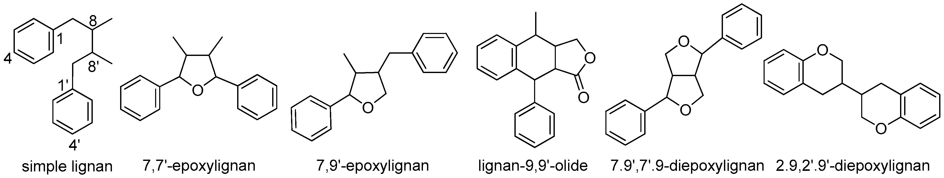
Figure 1.
Subtypes of classical lignans.

Figure 2.
Chemical structures of simple lignans.

Figure 3.
Chemical structures of 7,7’-epoxylignans.

Figure 4.
Chemical structures of 7,9’-epoxylignans, lignan-9,9’-olides, and 2.9,2’.9’-diepoxylignans.
Figure 4.
Chemical structures of 7,9’-epoxylignans, lignan-9,9’-olides, and 2.9,2’.9’-diepoxylignans.

Figure 5.
Chemical structures of 7.9’,7’.9-diepoxylignans.

Figure 6.
Chemical structures of 2,7’-cyclolignans.

Figure 7.
Subtypes of neolignans.

Figure 8.
Chemical structures of 8,1’-neolignans.

Figure 9.
Chemical structures of 8,3’-neolignans.

Figure 10.
Chemical structures of 7,1’-neolignans.

Figure 11.
Subtypes of cycloneolignans.

Figure 12.
Chemical structures of 7.3’,8.1’-cycloneolignans.

Figure 13.
Chemical structures of 7.3’,8.5’-cycloneolignans.

Figure 14.
Chemical structures of 7.1’,8.3’-cycloneolignans.

Figure 15.
Subtypes of oxyneolignans.

Figure 16.
Chemical structures of 7.3’,8.4’-dioxyneolignans.

Figure 17.
Chemical structures of 8,4’-oxyneolignans.

Figure 18.
Chemical structures of uncommon neolignans.

© 2018 by the authors. Licensee MDPI, Basel, Switzerland. This article is an open access article distributed under the terms and conditions of the Creative Commons Attribution (CC BY) license (http://creativecommons.org/licenses/by/4.0/).
Share and Cite
MDPI and ACS Style
Li, Y.; Xie, S.; Ying, J.; Wei, W.; Gao, K. Chemical Structures of Lignans and Neolignans Isolated from Lauraceae. Molecules 2018, 23, 3164. https://0-doi-org.brum.beds.ac.uk/10.3390/molecules23123164
AMA Style
Li Y, Xie S, Ying J, Wei W, Gao K. Chemical Structures of Lignans and Neolignans Isolated from Lauraceae. Molecules. 2018; 23(12):3164. https://0-doi-org.brum.beds.ac.uk/10.3390/molecules23123164
Chicago/Turabian StyleLi, Ya, Shuhan Xie, Jinchuan Ying, Wenjun Wei, and Kun Gao. 2018. "Chemical Structures of Lignans and Neolignans Isolated from Lauraceae" Molecules 23, no. 12: 3164. https://0-doi-org.brum.beds.ac.uk/10.3390/molecules23123164