Synthesis of 3-(Imidazo[2,1-b]thiazol-6-yl)-2H-chromen-2-one Derivatives and Study of Their Antiviral Activity against Parvovirus B19

 and
and
Abstract
:1. Introduction
2. Results
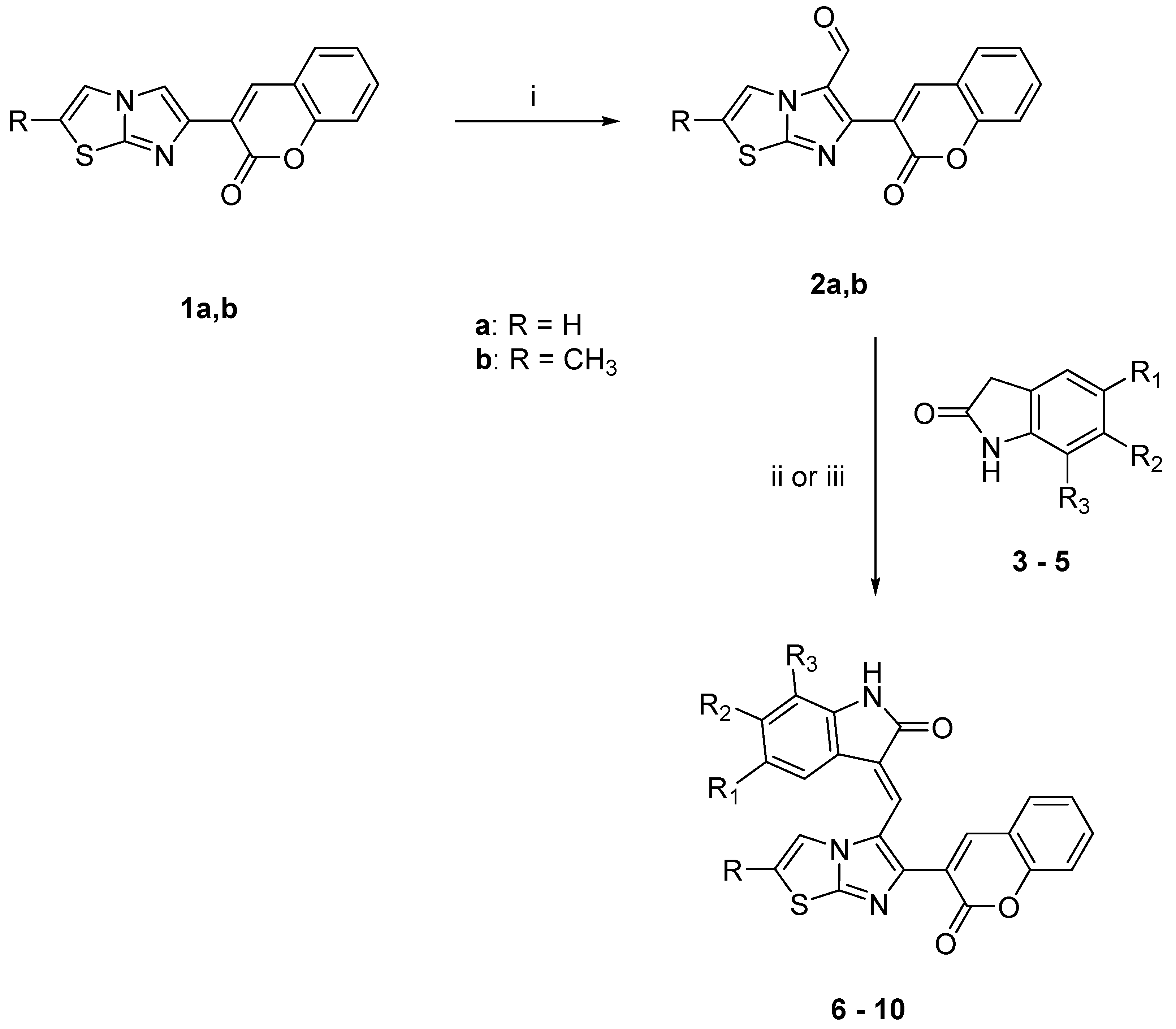
2.1. Chemistry
2.2. Biological Studies
2.2.1. Compound Solubility
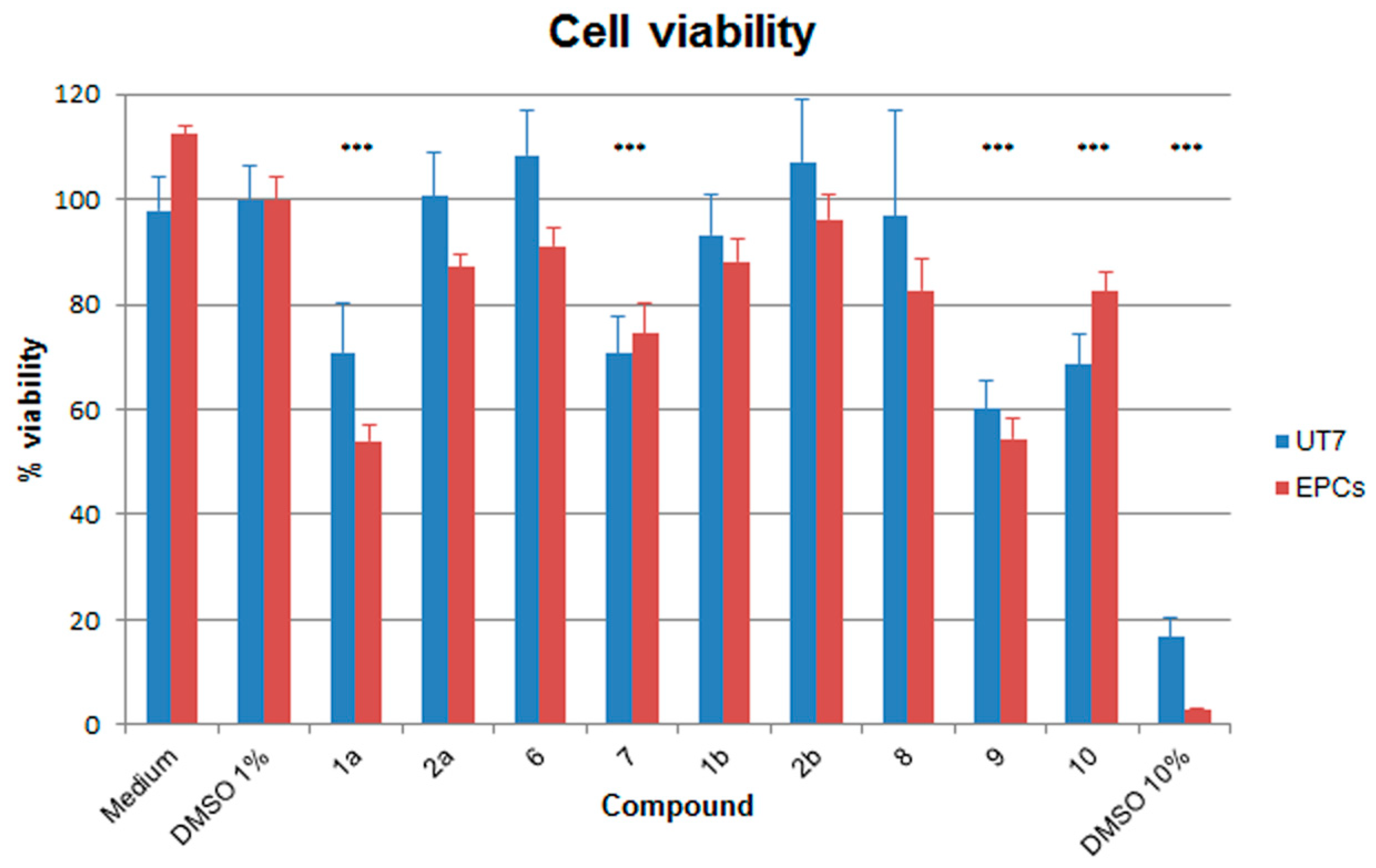
2.2.2. Effects on Cell Viability
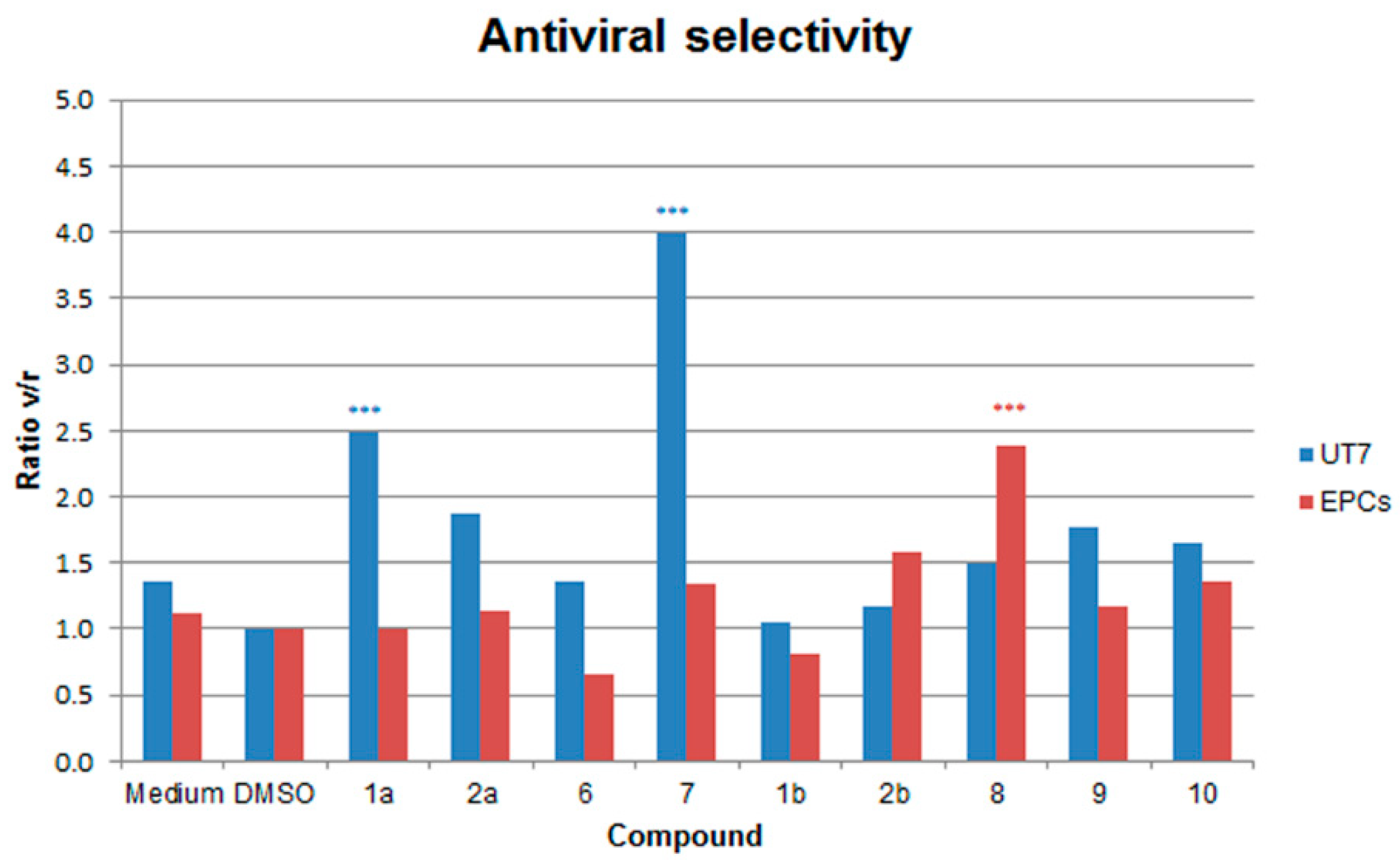
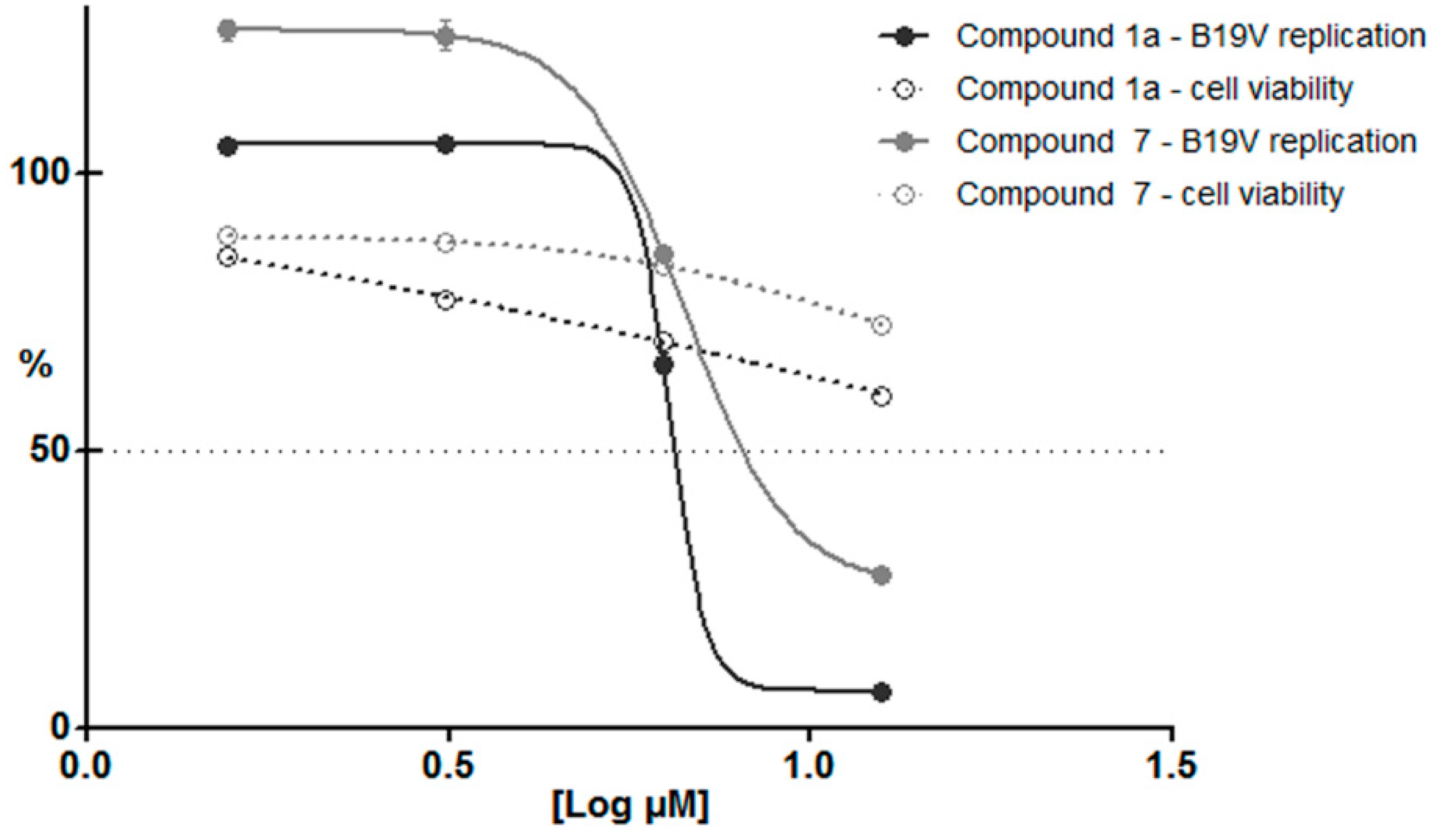
2.2.3. Antiviral Activity against B19V
3. Discussion
4. Materials and Methods
4.1. Chemistry
4.1.1. General Procedure for the Synthesis of the Aldehydes 2
4.1.2. General Procedure for the Synthesis of Compounds 6–10
4.2. Biological Activity
4.2.1. Cells
4.2.2. Cell Viability and Proliferation Assays
4.2.3. Infection
4.2.4. Nucleic Acid Purification and Analysis
4.2.5. Statistical Analysis
Author Contributions
Funding
Conflicts of Interest
References
- Penta, S. Advances in Structure and Activity Relationship of Coumarin Derivatives; Academic Press: Cambridge, MA, USA, 2015; pp. 1–182. [Google Scholar]
- Chen, Y.; Cheng, M.; Liu, F.Q.; Xia, P.; Qian, K.; Yu, D.; Xia, Y.; Yang, Z.Y.; Chen, C.H.; Morris-Natschke, S.L.; et al. Anti-AIDS agents 86. Synthesis and anti-HIV evaluation of 2′,3′-seco-3′-nor DCP and DCK analogues. Eur. J. Med. Chem. 2011, 46, 4924–4936. [Google Scholar] [CrossRef] [PubMed] [Green Version]
- Tang, J.; Qian, K.; Zhang, B.N.; Chen, Y.; Xia, P.; Yu, D.; Xia, Y.; Yang, Z.Y.; Chen, C.H.; Morris-Natschke, S.L.; et al. Anti-AIDS agents 82: Synthesis of seco-(3′R,4′R)-3′,4′-di-O-(S)-camphanoyl-(+)-cis-khellactone (DCK) derivatives as novel anti-HIV agents. Bioorg. Med. Chem. 2010, 18, 4363–4373. [Google Scholar] [CrossRef] [PubMed]
- Xie, L.; Yu, D.; Wild, C.; Allaway, G.; Turpin, J.; Smith, P.C.; Lee, K.H. Anti-AIDS agents. 52. Synthesis and anti-HIV activity of hydroxymethyl (3′R,4′R)-3′,4′-di-O-(S)-camphanoyl-(+)-cis-khellactone derivatives. J. Med. Chem. 2004, 47, 756–760. [Google Scholar] [CrossRef] [PubMed]
- Yu, D.; Chen, C.H.; Brossi, A.; Lee, K.H. Anti-AIDS agents. 60. Substituted 3′R,4′R-di-O-(-)-camphanoyl-2′,2′-dimethyldihydropyrano[2,3-f]chromone (DCP) analogues as potent anti-HIV agents. J. Med. Chem. 2004, 47, 4072–4082. [Google Scholar] [CrossRef] [PubMed]
- Zhou, T.; Shi, Q.; Chen, C.H.; Huang, L.; Ho, P.; Morris-Natschke, S.L.; Lee, K.H. Anti-AIDS agents 85. Design, synthesis, and evaluation of 1R,2R-dicamphanoyl-3,3-dimethyldihydropyrano-[2,3-c]xanthen-7(1H)-one (DCX) derivatives as novel anti-HIV agents. Eur. J. Med. Chem. 2012, 47, 86–96. [Google Scholar] [CrossRef] [PubMed]
- Hwu, J.R.; Lin, S.Y.; Tsay, S.C.; De Clercq, E.; Leyssen, P.; Neyts, J. Coumarin-purine ribofuranoside conjugates as new agents against hepatitis C virus. J. Med. Chem. 2011, 54, 2114–2126. [Google Scholar] [CrossRef] [PubMed]
- Neyts, J.; De Clercq, E.; Singha, R.; Chang, Y.H.; Das, A.R.; Chakraborty, S.K.; Hong, S.C.; Tsay, S.C.; Hsu, M.H.; Hwu, J.R. Structure-activity relationship of new anti-hepatitis C virus agents: Heterobicycle-coumarin conjugates. J. Med. Chem. 2009, 52, 1486–1490. [Google Scholar] [CrossRef] [PubMed]
- Tsay, S.C.; Lin, S.Y.; Huang, W.C.; Hsu, M.H.; Hwang, K.C.; Lin, C.C.; Horng, J.C.; Chen, I.C.; Hwu, J.R.; Shieh, F.K.; et al. Synthesis and Structure-Activity Relationships of Imidazole-Coumarin Conjugates against Hepatitis C Virus. Molecules 2016, 21, 228. [Google Scholar] [CrossRef] [PubMed]
- Hassan, M.Z.; Osman, H.; Ali, M.A.; Ahsan, M.J. Therapeutic potential of coumarins as antiviral agents. Eur. J. Med. Chem. 2016, 123, 236–255. [Google Scholar] [CrossRef] [PubMed]
- Gallinella, G. Parvovirus B19 Achievements and Challenges. ISRN Virol. 2013, 2013, 33. [Google Scholar] [CrossRef]
- Qiu, J.; Soderlund-Venermo, M.; Young, N.S. Human Parvoviruses. Clin. Microbiol. Rev. 2017, 30, 43–113. [Google Scholar] [CrossRef] [PubMed]
- Bua, G.; Manaresi, E.; Bonvicini, F.; Gallinella, G. Parvovirus B19 Replication and Expression in Differentiating Erythroid Progenitor Cells. PLoS ONE 2016, 11, e0148547. [Google Scholar] [CrossRef] [PubMed]
- Ganaie, S.S.; Qiu, J. Recent Advances in Replication and Infection of Human Parvovirus B19. Front. Cell Infect. Microbiol. 2018, 8, 166. [Google Scholar] [CrossRef] [PubMed]
- Zou, W.; Wang, Z.; Xiong, M.; Chen, A.Y.; Xu, P.; Ganaie, S.S.; Badawi, Y.; Kleiboeker, S.; Nishimune, H.; Ye, S.Q.; et al. Human Parvovirus B19 Utilizes Cellular DNA Replication Machinery for Viral DNA Replication. J. Virol. 2018, 92, e01881-17. [Google Scholar] [CrossRef] [PubMed]
- Gallinella, G. The clinical use of parvovirus B19 assays: Recent advances. Expert Rev. Mol. Diagn. 2018, 18, 821–832. [Google Scholar] [CrossRef] [PubMed]
- Bonvicini, F.; Bua, G.; Gallinella, G. Parvovirus B19 infection in pregnancy—Awareness and opportunities. Curr. Opin. Virol. 2017, 27, 8–14. [Google Scholar] [CrossRef] [PubMed]
- Chandramouli, S.; Medina-Selby, A.; Coit, D.; Schaefer, M.; Spencer, T.; Brito, L.A.; Zhang, P.; Otten, G.; Mandl, C.W.; Mason, P.W.; et al. Generation of a parvovirus B19 vaccine candidate. Vaccine 2013, 31, 3872–3878. [Google Scholar] [CrossRef] [PubMed]
- Crabol, Y.; Terrier, B.; Rozenberg, F.; Pestre, V.; Legendre, C.; Hermine, O.; Montagnier-Petrissans, C.; Guillevin, L.; Mouthon, L.; Montagnier-Petrissans, C.; Groupe d’experts de l’Assistance Publique-Hôpitaux de Paris. Intravenous immunoglobulin therapy for pure red cell aplasia related to human parvovirus b19 infection: A retrospective study of 10 patients and review of the literature. Clin. Infect. Dis. 2013, 56, 968–977. [Google Scholar] [CrossRef] [PubMed]
- Bonvicini, F.; Bua, G.; Conti, I.; Manaresi, E.; Gallinella, G. Hydroxyurea inhibits parvovirus B19 replication in erythroid progenitor cells. Biochem. Pharmacol. 2017, 136, 32–39. [Google Scholar] [CrossRef] [PubMed]
- Bonvicini, F.; Bua, G.; Manaresi, E.; Gallinella, G. Antiviral effect of cidofovir on parvovirus B19 replication. Antivir. Res. 2015, 113, 11–18. [Google Scholar] [CrossRef] [PubMed]
- Bonvicini, F.; Bua, G.; Manaresi, E.; Gallinella, G. Enhanced inhibition of parvovirus B19 replication by cidofovir in extendedly exposed erythroid progenitor cells. Virus Res. 2016, 220, 47–51. [Google Scholar] [CrossRef] [PubMed]
- Bua, G.; Conti, I.; Manaresi, E.; Sethna, P.; Foster, S.; Bonvicini, F.; Gallinella, G. Antiviral activity of brincidofovir on parvovirus B19. Antivir. Res. 2019, 162, 22–29. [Google Scholar] [CrossRef] [PubMed]
- Hostetler, K.Y. Alkoxyalkyl prodrugs of acyclic nucleoside phosphonates enhance oral antiviral activity and reduce toxicity: Current state of the art. Antivir. Res. 2009, 82, A84–A98. [Google Scholar] [CrossRef] [PubMed]
- Whitsett, J.; Maeda, Y. Methods and Composition to Treat Cancer. Patent WO2014205132A2, 24 December 2014. [Google Scholar]
- Andreani, A.; Granaiola, M.; Locatelli, A.; Morigi, R.; Rambaldi, M.; Varoli, L.; Calonghi, N.; Cappadone, C.; Farruggia, G.; Stefanelli, C.; et al. Substituted 3-(5-imidazo[2,1-b]thiazolylmethylene)-2-indolinones and analogues: Synthesis, cytotoxic activity, and study of the mechanism of action. J. Med. Chem. 2012, 55, 2078–2088. [Google Scholar] [CrossRef] [PubMed]
- Rao, V.R.; Reddy, V.R. Synthesis of some new types of 3-coumarinyl-substituted pyrazolopyrimidines and imidazothiazoles. Chem. Heterocycl. Compd. 2008, 44, 360–365. [Google Scholar]
- Srimanth, K.; Rao, V.R.; Krishna, D.R. Synthesis and evaluation of anticancer activity of some imidazothiazolyl, imidazobenzothiazolyl and dihydroimidazothiazolyl coumarins. Arzneimittelforschung 2002, 52, 388–392. [Google Scholar] [CrossRef]
- Koelsch, C.F. A Synthesis of Ethyl Quininate from m-Cresol. J. Am. Chem. Soc. 1944, 66, 2019–2020. [Google Scholar] [CrossRef]
- Mayer, F.; Oppenheimer, T. Über Naphthyl-essigsäuren. 3. Abhandlung: 1-Nitronaphthyl-2-brenztraubensäure und 1-Nitronaphthyl-2-essigsäure. Ber. Dtsch. Chem. Ges. 1918, 51, 1239–1245. [Google Scholar] [CrossRef]
- Bonvicini, F.; Manaresi, E.; Bua, G.; Venturoli, S.; Gallinella, G. Keeping pace with parvovirus B19 genetic variability: A multiplex genotype-specific quantitative pcr assay. J. Clin. Microbiol. 2013, 51, 3753–3759. [Google Scholar] [CrossRef] [PubMed]
- Bonvicini, F.; Filippone, C.; Manaresi, E.; Zerbini, M.; Musiani, M.; Gallinella, G. Functional analysis and quantitative determination of the expression profile of human parvovirus B19. Virology 2008, 381, 168–177. [Google Scholar] [CrossRef] [PubMed] [Green Version]
Sample Availability: Samples of the compounds 1a-b, 2a-b, 6-10 are available from the authors. |


| COMP | R | R1 | R2 | R3 |
|---|---|---|---|---|
| 3 | H | H | H | |
| 4 | OCH3 | H | H | |
| 5 | H |  | ||
| 6 | H | H | H | H |
| 7 | H | OCH3 | H | H |
| 8 | CH3 | H | H | H |
| 9 | CH3 | OCH3 | H | H |
| 10 | CH3 | H | | |
| Compound | Concentration (μM) |
|---|---|
| 1a | 12.50 |
| 2a | 6.25 |
| 6 | 0.78 |
| 7 | 12.50 |
| 1b | 1.56 |
| 2b | 0.78 |
| 8 | 3.13 |
| 9 | 12.50 |
| 10 | 6.25 |
| Cells | Compound | |||||||||||
|---|---|---|---|---|---|---|---|---|---|---|---|---|
| UT7 | 2hpi | 48hpi | DMSO | 1a | 2a | 6 | 7 | 1b | 2b | 8 | 9 | 10 |
| Log B19V Copies a | 5.03 | 7.02 | 7.16 | 6.61 | 6.89 | 7.06 | 6.41 | 7.11 | 7.13 | 6.97 | 6.69 | 6.78 |
| Delta Log b | 1.99 | 2.13 | 1.58 | 1.86 | 2.03 | 1.38 | 2.08 | 2.09 | 1.94 | 1.66 | 1.74 | |
| % viral replication c | 73.73 | 100.00 | 28.41 | 53.59 | 79.99 | 17.87 | 88.96 | 92.07 | 65.04 | 34.16 | 41.38 | |
| % cell viability c | 97.87 | 100.00 | 70.85 | 100.59 | 108.34 | 70.77 | 93.35 | 107.24 | 96.74 | 60.10 | 68.54 | |
| Ratio v/r d | 1.36 | 1.00 | 2.49 | 1.88 | 1.35 | 3.99 | 1.05 | 1.16 | 1.49 | 1.77 | 1.66 | |
| EPCs | 2hpi | 48hpi | DMSO | 1a | 2a | 6 | 7 | 1b | 2b | 8 | 9 | 10 |
| Log B19V Copies a | 5.02 | 7.37 | 7.37 | 7.10 | 7.26 | 7.51 | 7.12 | 7.40 | 7.16 | 6.91 | 7.04 | 7.15 |
| Delta Log b | 2.35 | 2.35 | 2.08 | 2.23 | 2.49 | 2.09 | 2.38 | 2.13 | 1.89 | 2.01 | 2.13 | |
| % viral replication c | 100.36 | 100.00 | 54.05 | 76.87 | 137.83 | 55.76 | 108.13 | 60.92 | 34.96 | 46.18 | 60.43 | |
| % cell viability c | 112.41 | 100.00 | 53.92 | 87.09 | 91.14 | 74.43 | 88.10 | 96.20 | 82.78 | 54.43 | 82.53 | |
| Ratio v/r d | 1.12 | 1.00 | 1.00 | 1.13 | 0.66 | 1.34 | 0.82 | 1.58 | 2.39 | 1.18 | 1.37 | |
© 2019 by the authors. Licensee MDPI, Basel, Switzerland. This article is an open access article distributed under the terms and conditions of the Creative Commons Attribution (CC BY) license (http://creativecommons.org/licenses/by/4.0/).
Share and Cite
Conti, I.; Morigi, R.; Locatelli, A.; Rambaldi, M.; Bua, G.; Gallinella, G.; Leoni, A. Synthesis of 3-(Imidazo[2,1-b]thiazol-6-yl)-2H-chromen-2-one Derivatives and Study of Their Antiviral Activity against Parvovirus B19. Molecules 2019, 24, 1037. https://0-doi-org.brum.beds.ac.uk/10.3390/molecules24061037
Conti I, Morigi R, Locatelli A, Rambaldi M, Bua G, Gallinella G, Leoni A. Synthesis of 3-(Imidazo[2,1-b]thiazol-6-yl)-2H-chromen-2-one Derivatives and Study of Their Antiviral Activity against Parvovirus B19. Molecules. 2019; 24(6):1037. https://0-doi-org.brum.beds.ac.uk/10.3390/molecules24061037
Chicago/Turabian StyleConti, Ilaria, Rita Morigi, Alessandra Locatelli, Mirella Rambaldi, Gloria Bua, Giorgio Gallinella, and Alberto Leoni. 2019. "Synthesis of 3-(Imidazo[2,1-b]thiazol-6-yl)-2H-chromen-2-one Derivatives and Study of Their Antiviral Activity against Parvovirus B19" Molecules 24, no. 6: 1037. https://0-doi-org.brum.beds.ac.uk/10.3390/molecules24061037