
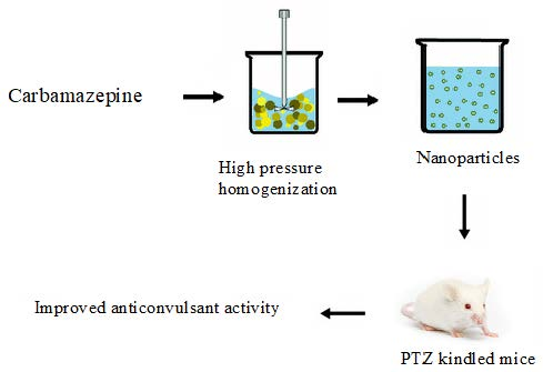
Preparation and Evaluation of Carbamazepine Solid Lipid Nanoparticle for Alleviating Seizure Activity in Pentylenetetrazole-Kindled Mice

 ,
,  , , and
Abstract
:
1. Introduction
2. The Experimental Part
2.1. Materials
2.2. Methods
2.2.1. The Preparation of the CBZ-SLN
2.2.2. The Determination of Entrapment Efficiency% (EE%) of CBZ in the Prepared CBZ-SLN
2.2.3. The Determination of Particle Size, Zeta Potential, and the Polydispersity Index of the Prepared CBZ-SLN
2.2.4. The In Vitro Release Study of CBZ from the Prepared CBZ-SLN
2.2.5. The Optimization of Formulation Variables
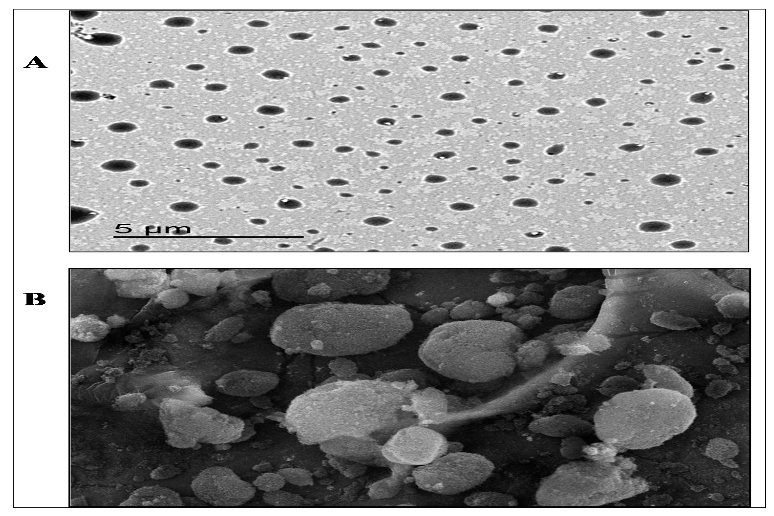
2.2.6. The Morphological Examination of the Best Formulation of the CBZ-SLN
Transmission Electron Microscope (TEM)
Scanning Electron Microscope (SEM)
2.2.7. The Fourier-Transform Infrared Spectroscopy (FT-IR)
2.2.8. The Differential Scanning Calorimetry (DSC)
2.2.9. Pharmacological Activity
Animals
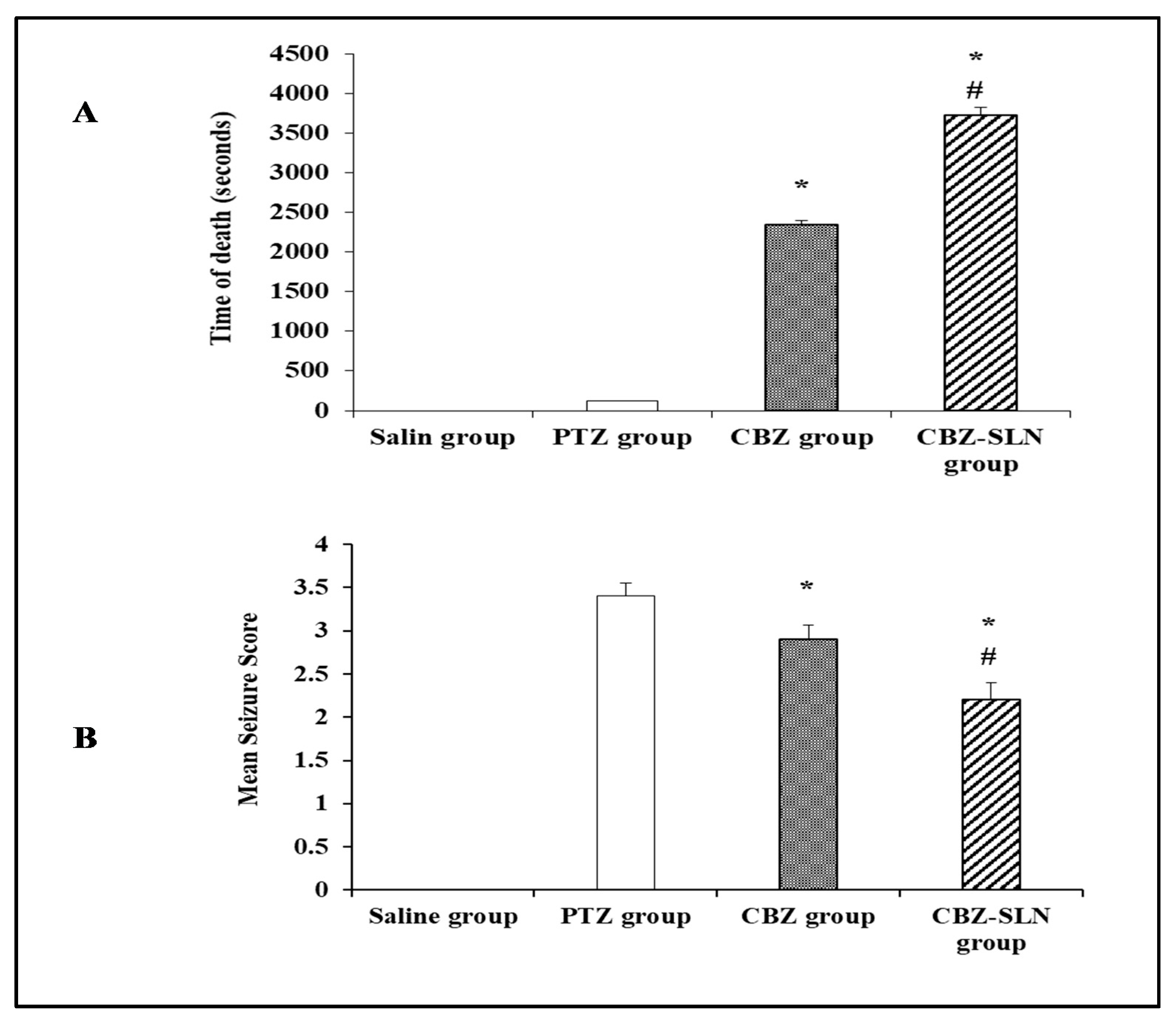
Time to Death Test
Seizure Activity Evaluation in PTZ-Kindled Mice
Tissue Preparation:
The Preparation of Histopathological Slides
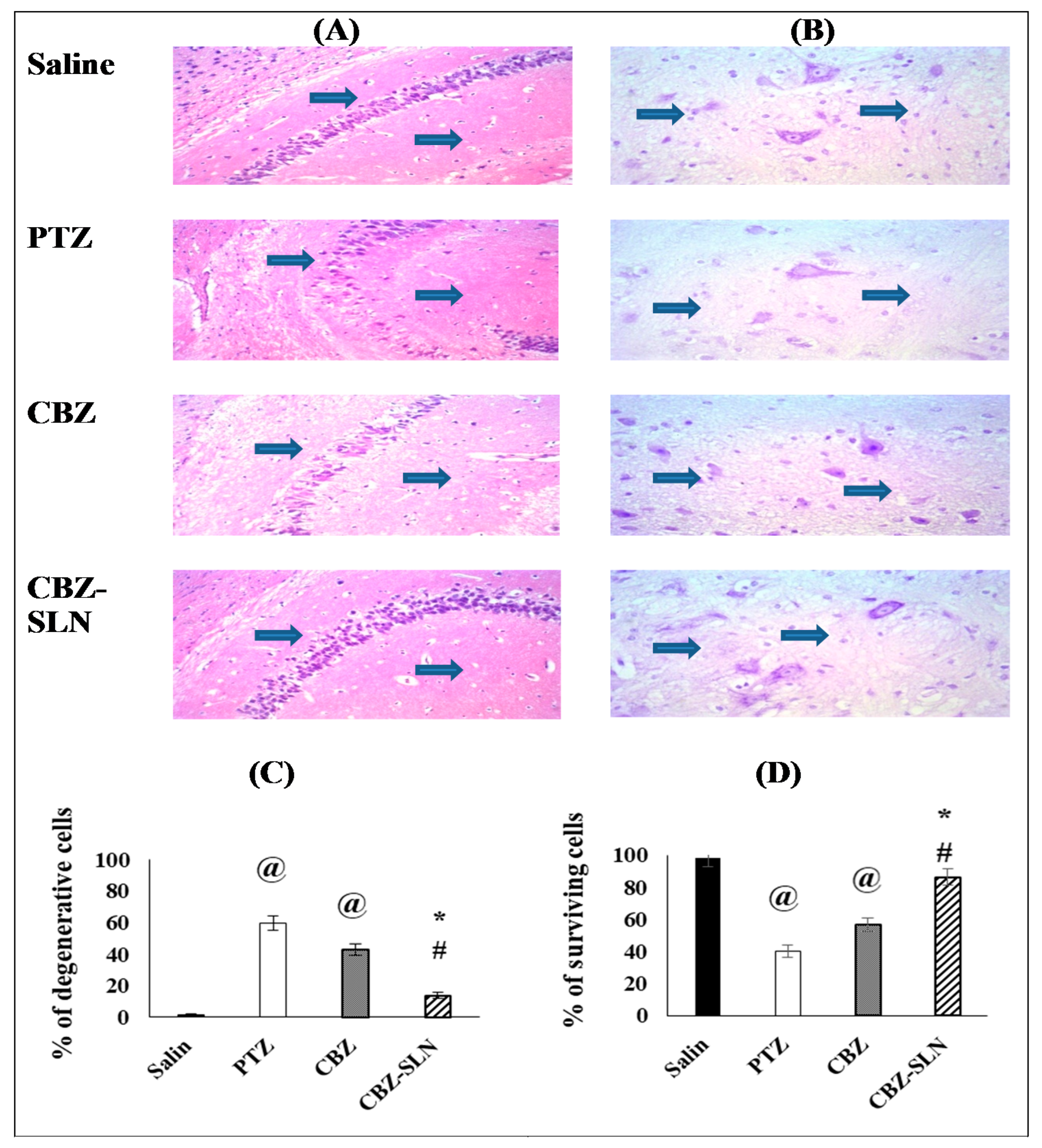
The Histopathological Examination of Cell Degeneration in the Hippocampus Region
The Histopathological Detection of Surviving Cell in Hippocampus by Cresyl Violet Staining
Statistical Analyses
3. Results and Discussion
3.1. The Preparation of the CBZ-SLN
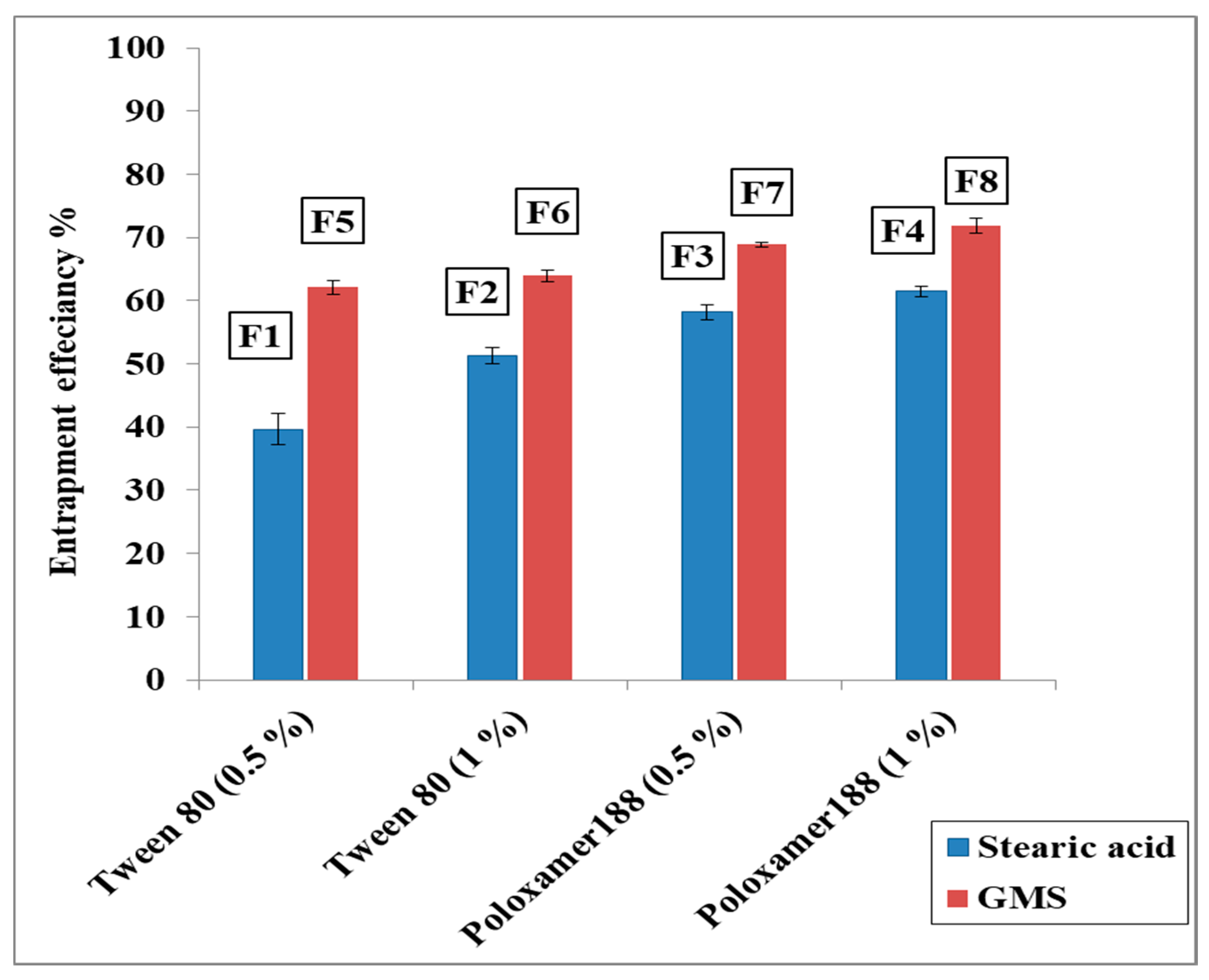
3.2. The Entrapment Efficiency% (EE%) of CBZ in the Prepared CBZ-SLN
3.2.1. The Effect of Lipid Type on the EE% of CBZ in the Prepared CBZ-SLN
3.2.2. The Effect of Surfactant Type and Concentration on the EE% of CBZ in the Prepared CBZ-SLN
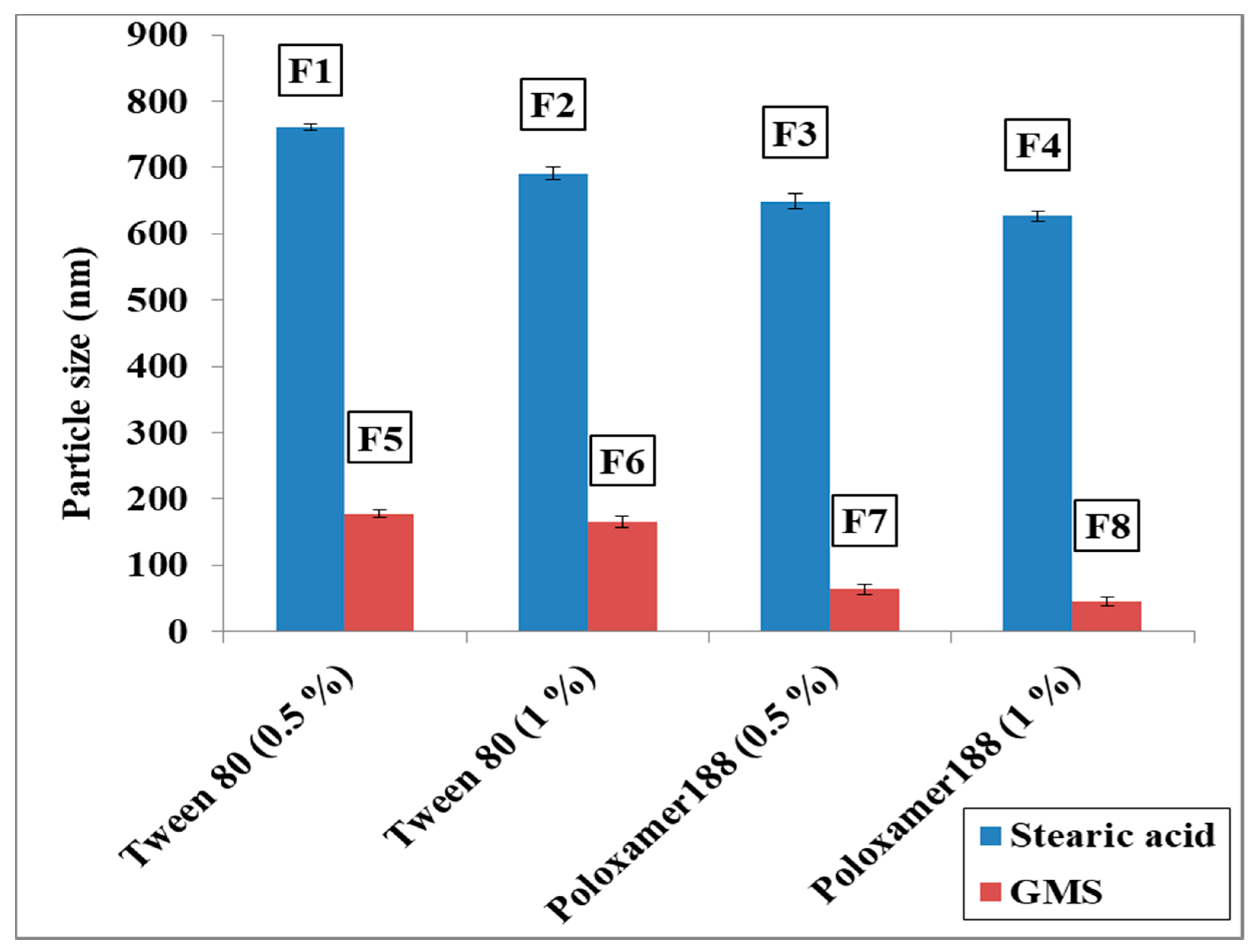
3.3. The Particle Size, Zeta potential, and Polydispersity Index of the Prepared CBZ-SLN
3.3.1. The Effect of Lipid Type on the Particle Size
3.3.2. The Effect of Surfactant Type and Concentration on the Particle Size
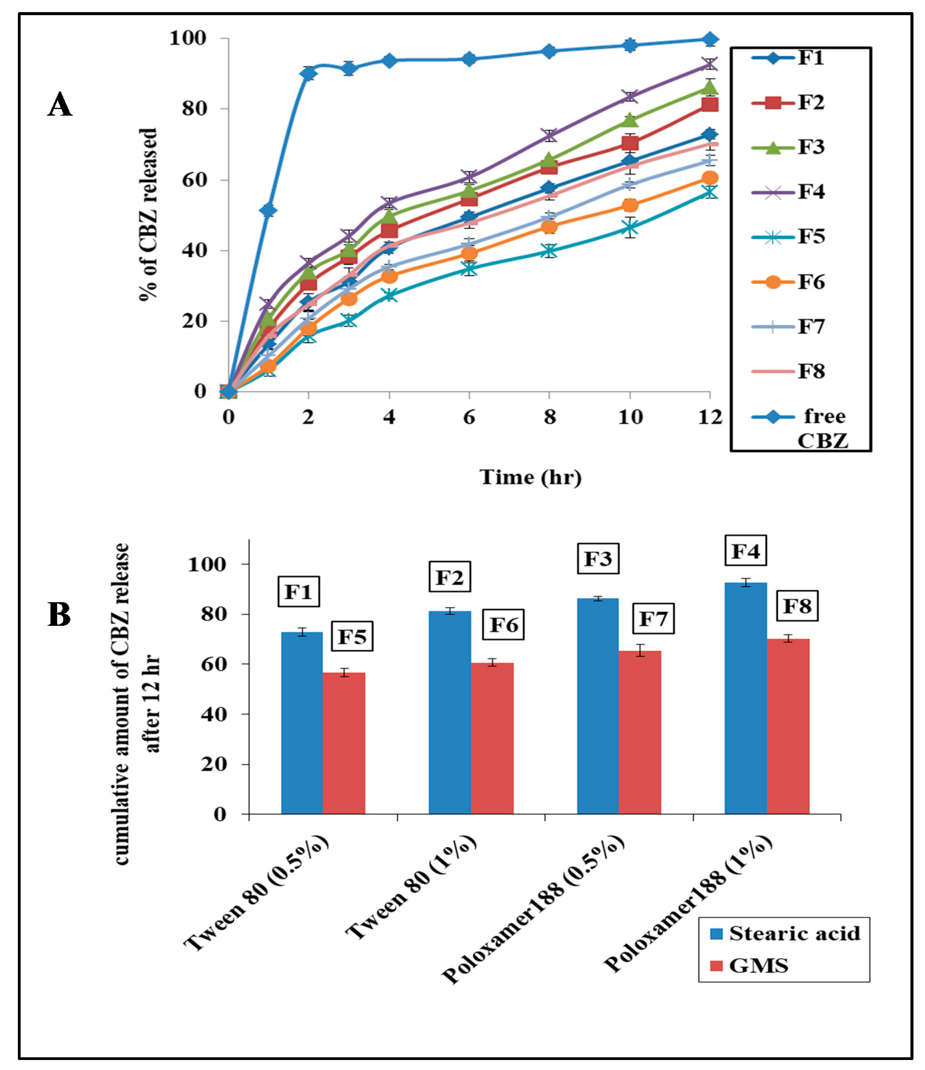
3.4. The In Vitro Release Study of CBZ from the Prepared CBZ-SLN
3.4.1. The Effect of Lipid Type on Drug Release
3.4.2. The Effect of Surfactant Type and Concentration on Drug Release
3.4.3. Release Kinetics
3.5. The Selection of the Optimized Formulation
3.6. The Surface Morphology of the CBZ-SLN
3.7. FT-IR Spectroscopy
3.8. The Thermal Analysis of the CBZ-SLN
3.9. The Pharmacological Activity of the CBZ-SLN
3.9.1. The Evaluation of the Anticonvulsant Activity of the CBZ-SLN by Time to Death Test
3.9.2. The Effect of the CBZ-SLN on Seizure Score in Mice Kindled with PTZ
3.9.3. The Effect of Repeated Administration of the CBZ-SLN on the Percentage of Degenerative Cells in the Hippocampal Sections Stained with H and E
3.9.4. The Effect of the CBZ-SLN on the Survival of Neurons in the Hippocampus of PTZ-Kindled Mice
4. Conclusions
Author Contributions
Funding
Acknowledgments
Conflicts of Interest
References
- Arya, R.K.K.; Juyal, V.; Kunwar, N. Preparation of carbamazepine chitosan nanoparticles for improving nasal absorption. J. Drug Deliv. Ther. 2016, 5, 101–108. [Google Scholar] [CrossRef]
- Nair, R.; Kumar, A.C.; Priya, V.K.; Yadav, C.M.; Raju, P.Y. Formulation and evaluation of chitosan solid lipid nanoparticles of carbamazepine. Lipids Health Dis. 2012, 11, 72. [Google Scholar] [CrossRef] [PubMed]
- Venkateswarlu, V.; Manjunath, K. Preparation, characterization and in vitro release kinetics of clozapine solid lipid nanoparticles. J. Control. Release 2004, 95, 627–638. [Google Scholar] [CrossRef] [PubMed]
- Abdelbary, G.; Fahmy, R.H. Diazepam-loaded solid lipid nanoparticles: Design and characterization. AAPS Pharmscitech. 2009, 10, 211–219. [Google Scholar] [CrossRef]
- Luo, Y.; Chen, D.; Ren, L.; Zhao, X.; Qin, J. Solid lipid nanoparticles for enhancing vinpocetine’s oral bioavailability. J. Control. Release 2006, 114, 53–59. [Google Scholar] [CrossRef] [PubMed]
- Ekambaram, P.; Sathali, A.A.H.; Priyanka, K. Solid lipid nanoparticles: A review. Sci. Rev. Chem. Commun. 2012, 2, 80–102. [Google Scholar]
- Lopalco, A.; Ali, H.; Denora, N.; Rytting, E. Oxcarbazepine-loaded polymeric nanoparticles: Development and permeability studies across in vitro models of the blood-brain barrier and human placental trophoblast. Int. J. Nanomed. 2015, 10, 1985–1996. [Google Scholar]
- Gavini, E.; Hegge, A.B.; Rassu, G.; Sanna, V.; Testa, C.; Pirisino, G.; Karlsen, J.; Giunchedi, P. Nasal administration of carbamazepine using chitosan microspheres: In vitro/in vivo studies. Int. J. Pharm. 2006, 307, 9–15. [Google Scholar] [CrossRef]
- Das, S.; Chaudhury, A. Recent advances in lipid nanoparticle formulations with solid matrix for oral drug delivery. Aaps. Pharmscitech. 2011, 12, 62–76. [Google Scholar] [CrossRef]
- Esfandyari-Manesh, M.; Javanbakht, M.; Dinarvand, R.; Atyabi, F. Molecularly imprinted nanoparticles prepared by miniemulsion polymerization as selective receptors and new carriers for the sustained release of carbamazepine. J. Mater. Sci. Mater. Med. 2012, 23, 963–972. [Google Scholar] [CrossRef]
- Leyva-Gómez, G.; Gonzalez-Trujano, M.E.; Lopez-Ruiz, E.; Couraud, P.O.; Wekslerg, B.; Romero, I.; Miller, F.; Delie, F.; Allemann, E.; Quintanar-Guerrero, D. Nanoparticle formulation improves the anticonvulsant effect of clonazepam on the pentylenetetrazole-induced seizures: Behavior and electroencephalogram. J. Pharm. Sci. 2014, 103, 2509–2519. [Google Scholar] [CrossRef] [PubMed]
- Jain, A.S.; Nagarsenker, M.S. Design, characterization and evaluation of anti-epileptic activity of nanoprecipitating preconcentrate of Carbamazepine. Drug Deliv. Lett. 2013, 3, 61–69. [Google Scholar] [CrossRef]
- Ruiz, M.E.; Castro, G.R. Nanoformulations of Antiepileptic Drugs: In Vitro and In Vivo Studies. Antiepileptic Drug Discov. 2016, 299–326. [Google Scholar] [CrossRef]
- Gangurde, P.K.; Kumar, L. Lamotrigine lipid nanoparticles for effective treatment of epilepsy: A focus on brain targeting via nasal route. J. Pharm. Innov. 2019, 14, 91–111. [Google Scholar] [CrossRef]
- Kumar, P.; Sharma, G.; Gupta, V.; Kaur, R.; Thakur, K.; Malik, R.; Kumar, A.; Kaushal, N.; Katare, O.P.; Raza, K. Oral Delivery of Methylthioadenosine to the Brain Employing Solid Lipid Nanoparticles: Pharmacokinetic, Behavioral, and Histopathological Evidences. AAPS Pharm. Sci. Tech. 2019, 20, 74. [Google Scholar] [CrossRef]
- Swidan, S.A.; Ghonaim, H.M.; Samy, A.M.; Ghorab, M.M. Efficacy and in vitro cytotoxicity of nanostructured lipid carriers for paclitaxel delivery. J. Appl. Pharm. Sci. 2016, 6, 18–26. [Google Scholar] [CrossRef]
- Kelidari, H.R.; Saeedi, M.; Akbari, J.; Morteza-Semnani, K.; Gill, P.; Valizadeh, H.; Nokhodchi, A. Formulation optimization and in vitro skin penetration of spironolactone loaded solid lipid nanoparticles. Colloids Surf. B Biointerfaces 2015, 128, 473–479. [Google Scholar] [CrossRef]
- Vivek, K.; Reddy, H.; Murthy, R.S. Investigations of the effect of the lipid matrix on drug entrapment, in vitro release, and physical stability of olanzapine-loaded solid lipid nanoparticles. Aaps. Pharm. Sci. Tech. 2007, 8, 16–24. [Google Scholar] [CrossRef] [Green Version]
- Ahmed, A.; Ghourab, M.; Shedid, S.; Qushawy, M. Optimization of piroxicam niosomes using central composite design. Int. J. Pharm. Pharm. Sci. 2013, 5, 229–236. [Google Scholar]
- Qushawy, M.; Nasr, A.; Abd-Alhaseeb, M.; Swidan, S. Design, Optimization and Characterization of a Transfersomal Gel Using Miconazole Nitrate for the Treatment of Candida Skin Infections. Pharmaceutics 2018, 10, 26. [Google Scholar] [CrossRef]
- Priyanka, K.; Sathali, A.A. Preparation and evaluation of montelukast sodium loaded solid lipid nanoparticles. J. Young Pharm. 2012, 4, 129–137. [Google Scholar] [CrossRef] [PubMed]
- Dhawan, S.; Kapil, R.; Singh, B. Formulation development and systematic optimization of solid lipid nanoparticles of quercetin for improved brain delivery. J. Pharm. Pharmacol. 2011, 63, 342–351. [Google Scholar] [CrossRef] [PubMed]
- Gad, S.; Ahmed, A.M.; Ghourab, M.M.; Queshawy, M.K. Design, formulation, and evaluation of Piroxicam niosomal gel. Int. J. Pharmtech. Res. 2014, 6, 185–195. [Google Scholar]
- Neves, A.R.; Queiroz, J.F.; Reis, S. Brain-targeted delivery of resveratrol using solid lipid nanoparticles functionalized with apolipoprotein E. J. Nanobiotechnol. 2016, 14, 27. [Google Scholar] [CrossRef]
- Yassin, A.E.; Anwer, M.K.; Mowafy, H.A.; El-Bagory, I.M.; Bayomi, M.A.; Alsarra, I.A. Optimization of 5-flurouracil solid-lipid nanoparticles: A preliminary study to treat colon cancer. Int. J. Med. Sci. 2010, 7, 398–408. [Google Scholar] [CrossRef]
- Eltahawy, N.A.; Ibrahim, A.K.; Radwan, M.M.; Zaitone, S.A.; Gomaa, M.; ElSohly, M.A.; Hassanean, H.A.; Ahmed, S.A. Mechanism of action of antiepileptic ceramide from Red Sea soft coral Sarcophyton auritum. Bioorg. Med. Chem. Lett. 2015, 25, 5819–5824. [Google Scholar] [CrossRef]
- Yum, M.S.; Ko, T.S. β-Hydroxybutyrate increases the pilocarpineinduced seizure threshold in young mice. Brain Dev. 2012, 34, 181–184. [Google Scholar] [CrossRef]
- Racine, R.J. Modification of seizure activity by electrical stimulation. II. Motor seizure. Electroencephalogr. Clin. Neurophysiol. 1972, 32, 281–294. [Google Scholar] [CrossRef]
- Alhaj, M.W.; Zaitone, S.A.; Moustafa, Y.M. Fluvoxamine alleviates seizure activity and downregulates hippocampal GAP-43 expression in pentylenetetrazole-kindled mice: Role of 5-HT3 receptors. Behav. Pharmacol. 2015, 26, 369–382. [Google Scholar] [CrossRef]
- Abd-Elghafour, B.A.; El-Sayed, N.M.; Ahmed, A.A.; Zaitone, S.A.; Moustafa, Y.M. Aspirin and (or) omega-3 polyunsaturated fatty acids protect against corticohippocampal neurodegeneration and downregulate lipoxin A4 production and formyl peptide receptor-like 1 expression in pentylenetetrazole-kindled rats. Can. J. Physiol. Pharmacol. 2017, 95, 340–348. [Google Scholar] [CrossRef]
- Aldawsari, H.M.; Eid, B.G.; Neamatallah, T.; Zaitone, S.A.; Badr, J.M. Anticonvulsant and Neuroprotective Activities of Phragmanthera austroarabica Extract in Pentylenetetrazole-Kindled Mice. Evid. Based Complement. Altern. Med. 2017, 2017, 5148219. [Google Scholar] [CrossRef] [PubMed]
- Tran, T.D.; Kelly, S.J. Critical periods for ethanol-induced cell loss in the hippocampal formation. Neurotoxicol. Teratol. 2003, 25, 519–528. [Google Scholar] [CrossRef]
- Gad, E.S.; Zaitone, S.A.; Moustafa, Y.M. Pioglitazone and exenatide enhance cognition and downregulate hippocampal beta amyloid oligomer and microglia expression in insulin-resistant rats. Can. J. Physiol. Pharmacol. 2016, 94, 819–828. [Google Scholar] [CrossRef] [PubMed]
- Swarnkar, S.; Singh, S.; Sharma, S.; Mathur, R.; Patro, I.K.; Nath, C. Rotenone induced neurotoxicity in rat brain areas: A histopathological study. Neurosci Lett. 2011, 501, 123–127. [Google Scholar] [CrossRef] [PubMed]
- Bancroft, J.D.; Gamble, M. Theory and practice of histological technique, 6th edition. J. Neuropathol. Exp. Neurol. 2008, 67, 633. [Google Scholar] [CrossRef]
- Arafat, E.A.G.; Khalaf, H.A. Effect of aqueous extract of Hibiscus sabdariffa on hyperthyroidism-induced changes in the renal cortex of rats: A histological study. Egypt. J. Histol. 2014, 37, 603–614. [Google Scholar] [CrossRef]
- Ekambaram, P.; Abdul, H.S. Formulation and evaluation of solid lipid nanoparticles of ramipril. J. Young Pharm. 2011, 3, 216–220. [Google Scholar] [CrossRef]
- Joseph, E.; Reddi, S.; Rinwa, V.; Balwani, G.; Saha, R. Design and in vivo evaluation of solid lipid nanoparticulate systems of Olanzapine for acute phase schizophrenia treatment: Investigations on antipsychotic potential and adverse effects. Eur. J. Pharm. Sci. 2017, 104, 315–325. [Google Scholar] [CrossRef]
- Zur Muhlen, A.; Schwarz, C.; Mehnert, W. Solid lipid nanoparticles (SLN) for controlled drug delivery--drug release and release mechanism. Eur. J. Pharm. Biopharm. 1998, 45, 149–155. [Google Scholar] [CrossRef]
- Scioli Montoto, S.; Sbaraglini, M.L.; Talevi, A.; Couyoupetrou, M.; Di Ianni, M.; Pesce, G.O.; Alvarez, V.A.; Bruno-Blanch, L.E.; Castro, G.R.; Ruiz, M.E.; et al. Carbamazepine-loaded solid lipid nanoparticles and nanostructured lipid carriers: Physicochemical characterization and in vitro/in vivo evaluation. Colloids Surf. B Biointerfaces 2018, 167, 73–81. [Google Scholar] [CrossRef]
- Muller, R.H.; Mader, K.; Gohla, S. Solid lipid nanoparticles (SLN) for controlled drug delivery—A review of the state of the art. Eur. J. Pharm. Biopharm. 2000, 50, 161–177. [Google Scholar] [CrossRef]
- Kaur, I.P.; Bhandari, R.; Bhandari, S.; Kakkar, V. Potential of solid lipid nanoparticles in brain targeting. J. Control. Release. 2008, 127, 97–109. [Google Scholar] [CrossRef] [PubMed]
- Garg, A.; Bhalala, K.; Tomar, D.S.; Wahajuddin. In-situ single pass intestinal permeability and pharmacokinetic study of developed Lumefantrine loaded solid lipid nanoparticles. Int. J. Pharm. 2017, 516, 120–130. [Google Scholar] [CrossRef] [PubMed]
- Rustichelli, C.; Gamberini, G.; Ferioli, V.; Gamberini, M.C.; Ficarra, R.; Tommasini, S. Solid-state study of polymorphic drugs: Carbamazepine. J. Pharm. Biomed. Anal. 2000, 23, 41–54. [Google Scholar] [CrossRef]
- Jahangiri, L.; Kesmati, M.; Najafzadeh, H. Evaluation of Anticonvulsive Effect of Magnesium Oxide Nanoparticles in Comparison with Conventional MgO in Diabetic and Non-diabetic Male Mice. Basic Clin. Neurosci. 2014, 5, 156–161. [Google Scholar] [PubMed]
- Kreuter, J.; Alyautdin, R.N.; Kharkevich, D.A.; Ivanov, A.A. Passage of peptides through the blood-brain barrier with colloidal polymer particles (nanoparticles). Brain Res. 1995, 674, 171–174. [Google Scholar] [CrossRef]
- Wang, J.X.; Sun, X.; Zhang, Z.R. Enhanced brain targeting by synthesis of 3′, 5′-dioctanoyl-5-fluoro-2′-deoxyuridine and incorporation into solid lipid nanoparticles. Eur. J. Pharm. Biopharm. 2002, 54, 285–290. [Google Scholar] [CrossRef]
- Minagawa, T.; Sakanaka, K.; Inaba, S.; Sai, Y.; Tamai, I.; Suwa, T.; Tsuji, A. Blood-brain-barrier transport of lipid microspheres containing clinprost, a prostaglandin I2 analogue. J. Pharm. Pharmacol. 1996, 48, 1016–1022. [Google Scholar] [CrossRef]
- Yang, S.C.; Lu, L.F.; Cai, Y.; Zhu, J.B.; Liang, B.W.; Yang, C.Z. Body distribution in mice of intravenously injected camptothecin solid lipid nanoparticles and targeting effect on brain. J. Control. Release 1999, 59, 299–307. [Google Scholar] [CrossRef]
- Kreuter, J. Nanoparticulate systems for brain delivery of drugs. Adv. Drug Deliv. Rev. 2001, 47, 65–81. [Google Scholar] [CrossRef]
Sample Availability: Samples of the compound are not available from the authors. |


| Formulation No. | Drug (mg) | Type of Lipid | Type of Surfactant | Surfactant% (w/v) | Total Volume (mL) |
|---|---|---|---|---|---|
| F1 | 10 | Stearic acid | Tween 80 | 0.5% (100 mg) | 20 |
| F2 | 10 | Stearic acid | Tween 80 | 1% (200 mg) | 20 |
| F3 | 10 | Stearic acid | Poloxamer 188 | 0.5% (100 mg) | 20 |
| F4 | 10 | Stearic acid | Poloxamer 188 | 1% (200 mg) | 20 |
| F5 | 10 | GMS | Tween 80 | 0.5% (100 mg) | 20 |
| F6 | 10 | GMS | Tween 80 | 1% (200 mg) | 20 |
| F7 | 10 | GMS | Poloxamer 188 | 0.5% (100 mg) | 20 |
| F8 | 10 | GMS | Poloxamer 188 | 1% (200 mg) | 20 |
| Formulation No. | Entrapment Efficiency% EE (%) | Particle Size (nm) | Zeta Potential (mv) | Polydispersity Index (PDI) |
|---|---|---|---|---|
| F1 | 39.66 ± 2.42 | 760.7 ± 5.25 | −25.1 ± 0.98 | 0.234 ± 0.08 |
| F2 | 51.22 ± 1.28 | 691.1 ± 9.02 | −27.6 ± 0.75 | 0.196 ± 0.05 |
| F3 | 58.16 ± 1.15 | 648.7 ± 11.54 | −27.7 ± 0.85 | 0.277 ± 0.01 |
| F4 | 61.49 ± 0.85 | 626.5 ± 7.25 | −21.5 ± 1.02 | 0.217 ± 0.04 |
| F5 | 62.08 ± 1.05 | 177.8 ± 6.24 | −32.5 ± 1.14 | 0.394 ± 0.07 |
| F6 | 63.95 ± 0.89 | 165.0 ± 8.21 | −38.4 ± 1.32 | 0.419 ± 0.02 |
| F7 | 68.88 ± 0.37 | 63.30 ± 8.27 | −30.1 ± 0.58 | 0.318 ± 0.04 |
| F8 | 71.91 ± 1.20 | 45.11 ± 6.72 | −33.3 ± 1.45 | 0.277 ± 0.03 |
| Formulation No. | Correlation Coefficient (r) | |||||
|---|---|---|---|---|---|---|
| Zero | First | Second | Diffusion | H-C | B-L | |
| F1 | 0.983 | −0.998 | 0.985 | 0.999 | 0.996 | 0.994 |
| F2 | 0.984 | −0.991 | 0.945 | 0.998 | 0.995 | 0.984 |
| F3 | 0.987 | −0.985 | 0.919 | 0.997 | 0.993 | 0.978 |
| F4 | 0.991 | −0.973 | 0.861 | 0.998 | 0.991 | 0.974 |
| F5 | 0.987 | −0.993 | 0.983 | 0.996 | 0.993 | 0.976 |
| F6 | 0.977 | −0.994 | 0.993 | 0.996 | 0.990 | 0.992 |
| F7 | 0.983 | −0.996 | 0.988 | 0.997 | 0.994 | 0.989 |
| F8 | 0.982 | −0.997 | 0.988 | 0.998 | 0.995 | 0.994 |
| Free CBZ | 0.652 | −0.910 | 0.679 | 0.737 | 0.880 | 0.866 |
© 2019 by the authors. Licensee MDPI, Basel, Switzerland. This article is an open access article distributed under the terms and conditions of the Creative Commons Attribution (CC BY) license (http://creativecommons.org/licenses/by/4.0/).
Share and Cite
Qushawy, M.; Prabahar, K.; Abd-Alhaseeb, M.; Swidan, S.; Nasr, A. Preparation and Evaluation of Carbamazepine Solid Lipid Nanoparticle for Alleviating Seizure Activity in Pentylenetetrazole-Kindled Mice. Molecules 2019, 24, 3971. https://0-doi-org.brum.beds.ac.uk/10.3390/molecules24213971
Qushawy M, Prabahar K, Abd-Alhaseeb M, Swidan S, Nasr A. Preparation and Evaluation of Carbamazepine Solid Lipid Nanoparticle for Alleviating Seizure Activity in Pentylenetetrazole-Kindled Mice. Molecules. 2019; 24(21):3971. https://0-doi-org.brum.beds.ac.uk/10.3390/molecules24213971
Chicago/Turabian StyleQushawy, Mona, Kousalya Prabahar, Mohammed Abd-Alhaseeb, Shady Swidan, and Ali Nasr. 2019. "Preparation and Evaluation of Carbamazepine Solid Lipid Nanoparticle for Alleviating Seizure Activity in Pentylenetetrazole-Kindled Mice" Molecules 24, no. 21: 3971. https://0-doi-org.brum.beds.ac.uk/10.3390/molecules24213971