
Determination of Asymmetric and Symmetric Dimethylarginine in Serum from Patients with Chronic Kidney Disease: UPLC-MS/MS versus ELISA

 , , ,
, , ,
Abstract
:
1. Introduction
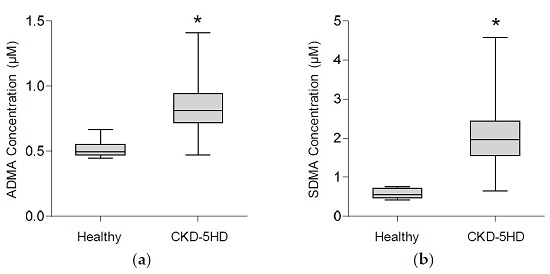
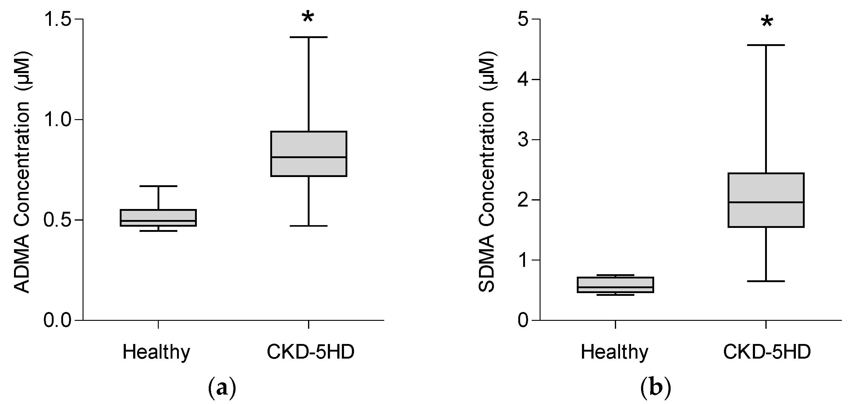
2. Results
3. Conclusions
4. Materials and Methods
4.1. Chemicals
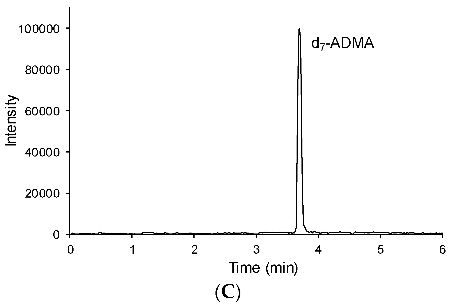
4.2. UPLC-MS/MS Assay
4.2.1. Instrumentation
4.2.2. Preparation of Stock Solutions, Calibration Standards and Quality Control (QC) Samples
4.2.3. Sample Preparation
4.2.4. Validation
4.2.5. Application to Biological Samples
4.3. ELISA Assay
4.4. Nephelometric Assay
4.5. Statistical Analysis
Acknowledgments
Author Contributions
Conflicts of Interest
Abbreviations
| ADMA | asymmetric dimethylarginine |
| SDMA | symmetric dimethylarginine |
| CKD | chronic kidney disease |
| CKD5HD | CKD patients on dialysis |
| HPLC | high performance liquid chromatography |
| UPLC | Ultra performance liquid chromatography |
| ELISA | enzyme-linked immunosorbent assay |
| GC | gas chromatography |
| MS | mass spectrometry |
| FLD | fluorence detection |
| CE | capillary electrophoresis |
| UV | ultraviolet |
| m/z | mass-to-charge ratio |
| LOD | limit of detection |
| LOQ | limit of quantification |
| QC | quality control |
| AAG | a1-acid glycoprotein |
| OPA | orthophtaldialdehyde |
| S/N | signal to noise |
| RSD | relative standard deviation |
References
- Foley, R.N.; Parfrey, P.S.; Sarnak, M.J. Clinical epidemiology of cardiovascular disease in chronic renal disease. Am. J. Kidney Dis. 1998, 32, S112–S119. [Google Scholar] [CrossRef] [PubMed]
- Vanholder, R.; Massy, Z.; Argiles, A.; Spasovski, G.; Verbeke, F.; Lameire, N. Chronic kidney disease as cause of cardiovascular morbidity and mortality. Nephrol. Dial. Transplant. 2005, 20, 1048–1056. [Google Scholar] [CrossRef] [PubMed]
- Kielstein, J.T.; Fliser, D.; Veldink, H. Asymmetric dimethylarginine and symmetric dimethylarginine: Axis of evil or useful alliance? Semin. Dial. 2009, 22, 346–350. [Google Scholar] [CrossRef] [PubMed]
- Boger, R.H. The emerging role of asymmetric dimethylarginine as a novel cardiovascular risk factor. Cardiovasc. Res. 2003, 59, 824–833. [Google Scholar] [CrossRef]
- Vallance, P.; Leone, A.; Calver, A.; Collier, J.; Moncada, S. Accumulation of an endogenous inhibitor of nitric oxide synthesis in chronic renal failure. Lancet 1992, 339, 572–575. [Google Scholar] [PubMed]
- Boger, R.H.; Bode-Boger, S.M.; Szuba, A.; Tsao, P.S.; Chan, J.R.; Tangphao, O.; Blaschke, T.F.; Cooke, J.P. Asymmetric dimethylarginine (ADMA): A novel risk factor for endothelial dysfunction: Its role in hypercholesterolemia. Circulation 1998, 98, 1842–1847. [Google Scholar] [CrossRef] [PubMed]
- Caglar, K.; Yilmaz, M.I.; Sonmez, A.; Cakir, E.; Kaya, A.; Acikel, C.; Eyileten, T.; Yenicesu, M.; Oguz, Y.; Bilgi, C.; et al. ADMA, proteinuria, and insulin resistance in non-diabetic stage I chronic kidney disease. Kidney Int. 2006, 70, 781–787. [Google Scholar] [CrossRef] [PubMed]
- Kielstein, J.T.; Boger, R.H.; Bode-Boger, S.M.; Frolich, J.C.; Haller, H.; Ritz, E.; Fliser, D. Marked increase of asymmetric dimethylarginine in patients with incipient primary chronic renal disease. J. Am. Soc. Nephrol. 2002, 13, 170–176. [Google Scholar] [PubMed]
- Fliser, D.; Kronenberg, F.; Kielstein, J.T.; Morath, C.; Bode-Boger, S.M.; Haller, H.; Ritz, E. Asymmetric dimethylarginine and progression of chronic kidney disease: The mild to moderate kidney disease study. J. Am. Soc. Nephrol. 2005, 16, 2456–2461. [Google Scholar] [CrossRef] [PubMed]
- Ravani, P.; Tripepi, G.; Malberti, F.; Testa, S.; Mallamaci, F.; Zoccali, C. Asymmetrical dimethylarginine predicts progression to dialysis and death in patients with chronic kidney disease: A competing risks modeling approach. J. Am. Soc. Nephrol. 2005, 16, 2449–2455. [Google Scholar] [CrossRef] [PubMed]
- Meinitzer, A.; Seelhorst, U.; Wellnitz, B.; Halwachs-Baumann, G.; Boehm, B.O.; Winkelmann, B.R.; Marz, W. Asymmetrical dimethylarginine independently predicts total and cardiovascular mortality in individuals with angiographic coronary artery disease (the Ludwigshafen Risk and Cardiovascular Health study). Clin. Chem. 2007, 53, 273–283. [Google Scholar] [CrossRef] [PubMed]
- Schnabel, R.; Blankenberg, S.; Lubos, E.; Lackner, K.J.; Rupprecht, H.J.; Espinola-Klein, C.; Jachmann, N.; Post, F.; Peetz, D.; Bickel, C.; et al. Asymmetric dimethylarginine and the risk of cardiovascular events and death in patients with coronary artery disease: Results from the AtheroGene Study. Circ. Res. 2005, 97, e53–e59. [Google Scholar] [CrossRef] [PubMed]
- Aucella, F.; Maas, R.; Vigilante, M.; Tripepi, G.; Schwedhelm, E.; Margaglione, M.; Gesualdo, L.; Boeger, R.; Zoccali, C. Methylarginines and mortality in patients with end stage renal disease: A prospective cohort study. Atherosclerosis 2009, 207, 541–545. [Google Scholar] [CrossRef] [PubMed]
- Zoccali, C.; Bode-Boger, S.; Mallamaci, F.; Benedetto, F.; Tripepi, G.; Malatino, L.; Cataliotti, A.; Bellanuova, I.; Fermo, I.; Frolich, J.; et al. Plasma concentration of asymmetrical dimethylarginine and mortality in patients with end-stage renal disease: A prospective study. Lancet 2001, 358, 2113–2117. [Google Scholar] [CrossRef]
- Bode-Boger, S.M.; Scalera, F.; Kielstein, J.T.; Martens-Lobenhoffer, J.; Breithardt, G.; Fobker, M.; Reinecke, H. Symmetrical dimethylarginine: A new combined parameter for renal function and extent of coronary artery disease. J. Am. Soc. Nephrol. 2006, 17, 1128–1134. [Google Scholar] [CrossRef] [PubMed]
- Schepers, E.; Glorieux, G.; Dhondt, A.; Leybaert, L.; Vanholder, R. Role of symmetric dimethylarginine in vascular damage by increasing ros via store-operated calcium influx in monocytes. Nephrol. Dial. Transplant. 2009, 24, 1429–1435. [Google Scholar] [CrossRef] [PubMed]
- Schepers, E.; Barreto, D.V.; Liabeuf, S.; Glorieux, G.; Eloot, S.; Barreto, F.C.; Massy, Z.; Vanholder, R. Symmetric dimethylarginine as a proinflammatory agent in chronic kidney disease. Clin. J. Am. Soc. Nephrol. 2011, 6, 2374–2383. [Google Scholar] [CrossRef] [PubMed]
- Schwedhelm, E.; Boger, R.H. The role of asymmetric and symmetric dimethylarginines in renal disease. Nat. Rev. Nephrol. 2011, 7, 275–285. [Google Scholar] [CrossRef] [PubMed]
- Tutarel, O.; Denecke, A.; Bode-Boger, S.M.; Martens-Lobenhoffer, J.; Schieffer, B.; Westhoff-Bleck, M.; Kielstein, J.T. Symmetrical dimethylarginine outperforms CKD-EPI and MDRD-derived eGFR for the assessment of renal function in patients with adult congenital heart disease. Kidney Blood Press. Res. 2011, 34, 41–45. [Google Scholar] [CrossRef] [PubMed]
- Kielstein, J.T.; Boger, R.H.; Bode-Boger, S.M.; Martens-Lobenhoffer, J.; Lonnemann, G.; Frolich, J.C.; Haller, H.; Fliser, D. Low dialysance of asymmetric dimethylarginine (ADMA)—In vivo and in vitro evidence of significant protein binding. Clin. Nephrol. 2004, 62, 295–300. [Google Scholar] [CrossRef] [PubMed]
- Grooteman, M.P.C.; Wauters, I.M.P.M.J.; Teerlink, T.; Twisk, J.W.R.; Nubé, M.J. Plasma dimethylarginine levels in chronic hemodialysis patients are independent of the type of dialyzer applied. Blood Purif. 2007, 25, 281–289. [Google Scholar] [CrossRef] [PubMed]
- Kalousová, M.; Kielstein, J.T.; Hodková, M.; Zima, T.; Dusilová-Sulková, S.; Martens-Lobenhoffer, J.; Bode-Boger, S.M. No benefit of hemodiafiltration over hemodialysis in lowering elevated levels of asymmetric dimethylarginine in ESRD patients. Blood Purif. 2006, 24, 439–444. [Google Scholar] [PubMed]
- Albsmeier, J.; Schwedhelm, E.; Schulze, F.; Kastner, M.; Böger, R.H. Determination of NG,NG-dimethyl-l-arginine, an endogenous NO synthase inhibitor, by gas chromatography-mass spectrometry. J. Chromatogr. B 2004, 809, 59–65. [Google Scholar] [CrossRef] [PubMed]
- Tsikas, D.; Beckmann, B.; Gutzki, F.M.; Jordan, J. Simultaneous gas chromatography-tandem mass spectrometry quantification of symmetric and asymmetric dimethylarginine in human urine. Anal. Biochem. 2011, 413, 60–62. [Google Scholar] [CrossRef] [PubMed]
- Blackwell, S.; O’Reilly, D.S.J.; Talwar, D.K. HPLC analysis of asymmetric dimethylarginine (ADMA) and related arginine metabolites in human plasma using a novel non-endogenous internal standard. Clin. Chim. Acta 2009, 401, 14–19. [Google Scholar] [CrossRef] [PubMed]
- Heresztyn, T.; Worthley, M.I.; Horowitz, J.D. Determination of l-arginine and NG,NG- and NG,NG'-dimethyl-l-arginine in plasma by liquid chromatography as AccQ-Fluor fluorescent derivatives. J. Chromatogr. B Anal. Technol. Biomed. Life Sci. 2004, 805, 325–329. [Google Scholar] [CrossRef] [PubMed]
- Ivanova, M.; Artusi, C.; Boffa, G.M.; Zaninotto, M.; Plebani, M. HPLC determination of plasma dimethylarginines: Method validation and preliminary clinical application. Clin. Chim. Acta 2010, 411, 1632–1636. [Google Scholar] [CrossRef] [PubMed]
- Jones, C.E.; Darcy, C.J.; Woodberry, T.; Anstey, N.M.; McNeil, Y.R. HPLC analysis of asymmetric dimethylarginine, symmetric dimethylarginine, homoarginine and arginine in small plasma volumes using a Gemini-NX column at high pH. J. Chromatogr. B Anal. Technol. Biomed. Life Sci. 2010, 878, 8–12. [Google Scholar] [CrossRef] [PubMed]
- Markowski, P.; Baranowska, I.; Baranowski, J. Simultaneous determination of l-arginine and 12 molecules participating in its metabolic cycle by gradient RP-HPLC method: Application to human urine samples. Anal. Chim. Acta 2007, 605, 205–217. [Google Scholar] [CrossRef] [PubMed]
- Marra, M.; Bonfigli, A.R.; Testa, R.; Testa, I.; Gambini, A.; Coppa, G. High-performance liquid chromatographic assay of asymmetric dimethylarginine, symmetric dimethylarginine, and arginine in human plasma by derivatization with naphthalene-2,3-dicarboxaldehyde. Anal. Biochem. 2003, 318, 13–17. [Google Scholar] [CrossRef]
- Teerlink, T.; Nijveldt, R.J.; de Jong, S.; van Leeuwen, P.A.M. Determination of arginine, asymmetric dimethylarginine, and symmetric dimethylarginine in human plasma and other biological samples by high-performance liquid chromatography. Anal. Biochem. 2002, 303, 131–137. [Google Scholar] [CrossRef] [PubMed]
- Tsunoda, M.; Nonaka, S.; Funatsu, T. Determination of methylated arginines by column-switching high-performance liquid chromatography-fluorescence detection. Analyst 2005, 130, 1410–1413. [Google Scholar] [CrossRef] [PubMed]
- Brown, C.M.; Becker, J.O.; Wise, P.M.; Hoofnagle, A.N. Simultaneous determination of 6 l-arginine metabolites in human and mouse plasma by using hydrophilic-interaction chromatography and electrospray tandem mass spectrometry. Clin. Chem. 2011, 57, 701–709. [Google Scholar] [CrossRef] [PubMed]
- D’Apolito, O.; Paglia, G.; Tricarico, F.; Garofalo, D.; Pilotti, A.; Lamacchia, O.; Cignarelli, M.; Corso, G. Development and validation of a fast quantitative method for plasma dimethylarginines analysis using liquid chromatography-tandem mass spectrometry. Clin. Biochem. 2008, 41, 1391–1395. [Google Scholar] [CrossRef] [PubMed]
- El-Khoury, J.M.; Bunch, D.R.; Reineks, E.; Jackson, R.; Steinle, R.; Wang, S. A simple and fast liquid chromatography-tandem mass spectrometry method for measurement of underivatized l-arginine, symmetric dimethylarginine, and asymmetric dimethylarginine and establishment of the reference ranges. Anal. Bioanal. Chem. 2012, 402, 771–779. [Google Scholar] [CrossRef] [PubMed]
- Huang, L.F.; Guo, F.Q.; Liang, Y.Z.; Li, B.Y.; Cheng, B.M. Simultaneous determination of l-arginine and its mono- and dimethylated metabolites in human plasma by high-performance liquid chromatography-mass spectrometry. Anal. Bioanal. Chem. 2004, 380, 643–649. [Google Scholar] [CrossRef] [PubMed]
- Martens-Lobenhoffer, J.; Bode-Boger, S.M. Fast and efficient determination of arginine, symmetric dimethylarginine, and asymmetric dimethylarginine in biological fluids by hydrophilic-interaction liquid chromatography-electrospray tandem mass spectrometry. Clin. Chem. 2006, 52, 488–493. [Google Scholar] [CrossRef] [PubMed]
- Martens-Lobenhoffer, J.; Bode-Boger, S.M. Quantification of l-arginine, asymmetric dimethylarginine and symmetric dimethylarginine in human plasma: A step improvement in precision by stable isotope dilution mass spectrometry. J. Chromatogr. B Anal. Technol. Biomed. Life Sci. 2012, 904, 140–143. [Google Scholar] [CrossRef] [PubMed]
- Shin, S.; Fung, S.-M.; Mohan, S.; Fung, H.-L. Simultaneous bioanalysis of l-arginine, l-citrulline, and dimethylarginines by LC-MS/MS. J. Chromatogr. B Anal. Technol. Biomed. Life Sci. 2011, 879, 467–474. [Google Scholar] [CrossRef] [PubMed]
- Vishwanathan, K.; Tackett, R.L.; Stewart, J.T.; Bartlett, M.G. Determination of arginine and methylated arginines in human plasma by liquid chromatography-tandem mass spectrometry. J. Chromatogr. B Biomed. Life Sci. 2000, 748, 157–166. [Google Scholar] [CrossRef]
- Wang, H.-Y.; Hu, P.; Jiang, J. Rapid determination of underivatized arginine, ornithine, citrulline and symmetric/asymmetric dimethylarginine in human plasma by LC-MS. Chromatography 2010, 71, 933–939. [Google Scholar] [CrossRef]
- Davids, M.; Swieringa, E.; Palm, F.; Smith, D.E.; Smulders, Y.M.; Scheffer, P.G.; Blom, H.J.; Teerlink, T. Simultaneous determination of asymmetric and symmetric dimethylarginine, l-monomethylarginine, l-arginine, and l-homoarginine in biological samples using stable isotope dilution liquid chromatography tandem mass spectrometry. J. Chromatogr. B Anal. Technol. Biomed. Life Sci. 2012, 900, 38–47. [Google Scholar] [CrossRef] [PubMed]
- Di Gangi, I.M.; Chiandetti, L.; Gucciardi, A.; Moret, V.; Naturale, M.; Giordano, G. Simultaneous quantitative determination of NG,NG-dimethyl-l-arginine or asymmetric dimethylarginine and related pathway’s metabolites in biological fluids by ultrahigh-performance liquid chromatography/electrospray ionization-tandem mass spectrometry. Anal. Chim. Acta 2010, 677, 140–148. [Google Scholar] [CrossRef] [PubMed]
- Hui, Y.; Wong, M.; Kim, J.-O.; Love, J.; Ansley, D.M.; Chen, D.D.Y. A new derivatization method coupled with LC-MS/MS to enable baseline separation and quantification of dimethylarginines in human plasma from patients to receive on-pump CABG surgery. Electrophoresis 2012, 33, 1911–1920. [Google Scholar] [CrossRef] [PubMed]
- Martens-Lobenhoffer, J.; Krug, O.; Bode-Boger, S.M. Determination of arginine and asymmetric dimethylarginine (ADMA) in human plasma by liquid chromatography/mass spectrometry with the isotope dilution technique. J. Mass Spectrom. 2004, 39, 1287–1294. [Google Scholar] [CrossRef] [PubMed]
- Schwedhelm, E.; Maas, R.; Tan-Andresen, J.; Schulze, F.; Riederer, U.; Boger, R.H. High-throughput liquid chromatographic-tandem mass spectrometric determination of arginine and dimethylated arginine derivatives in human and mouse plasma. J. Chromatogr. B Anal. Technol. Biomed. Life Sci. 2007, 851, 211–219. [Google Scholar] [CrossRef] [PubMed]
- Zinellu, A.; Sotgia, S.; Usai, M.F.; Pintus, G.; Deiana, L.; Carru, C. Improved method for plasma ADMA, SDMA, and arginine quantification by field-amplified sample injection capillary electrophoresis UV detection. Anal. Bioanal. Chem. 2011, 399, 1815–1821. [Google Scholar] [CrossRef] [PubMed]
- Desiderio, C.; Rossetti, D.V.; Messana, I.; Giardina, B.; Castagnola, M. Analysis of arginine and methylated metabolites in human plasma by field amplified sample injection capillary electrophoresis tandem mass spectrometry. Electrophoresis 2010, 31, 1894–1902. [Google Scholar] [CrossRef] [PubMed]
- Schulze, F.; Wesemann, R.; Schwedhelm, E.; Sydow, K.; Albsmeier, J.; Cooke, J.P.; Boger, R.H. Determination of asymmetric dimethylarginine (ADMA) using a novel ELISA assay. Clin. Chem. Lab. Med. 2004, 42, 1377–1383. [Google Scholar] [CrossRef] [PubMed]
- Horowitz, J.D.; Heresztyn, T. An overview of plasma concentrations of asymmetric dimethylarginine (ADMA) in health and disease and in clinical studies: Methodological considerations. J. Chromatogr. B Anal. Technol. Biomed. Life Sci. 2007, 851, 42–50. [Google Scholar] [CrossRef] [PubMed]
- Martens-Lobenhoffer, J.; Westphal, S.; Awiszus, F.; Bode-Boger, S.M.; Luley, C. Determination of asymmetric dimethylarginine: Liquid chromatography-mass spectrometry or ELISA? Clin. Chem. 2005, 51, 2188–2189. [Google Scholar] [CrossRef] [PubMed]
- Pecchini, P.; Malberti, F.; Mieth, M.; Quinn, R.; Tripepi, G.; Mallamaci, F.; Maas, R.; Zoccali, C.; Ravani, P. Measuring asymmetric dimethylarginine (ADMA) in CKD: A comparison between enzyme-linked immunosorbent assay and liquid chromatography-electrospray tandem mass spectrometry. J. Nephrol. 2012, 25, 1016–1022. [Google Scholar] [CrossRef]
- Schulze, F.; Schwedhelm, E.; Maas, R.; Boger, R.H. Comparison of HPLC method and commercial ELISA assay for asymmetric dimethylarginine (ADMA) determination in human serum. J. Chromatogr. B Anal. Technol. Biomed. Life Sci. 2006, 831, 333. [Google Scholar] [CrossRef] [PubMed]
- Siroka, R.; Trefil, L.; Rajdl, D.; Racek, J.; Cibulka, R. Asymmetric dimethylarginine—Comparison of HPLC and ELISA methods. J. Chromatogr. B Anal. Technol. Biomed. Life Sci. 2007, 850, 586–587. [Google Scholar] [CrossRef] [PubMed]
- Valtonen, P.; Karppi, J.; Nyyssonen, K.; Valkonen, V.P.; Halonen, T.; Punnonen, K. Comparison of HPLC method and commercial ELISA assay for asymmetric dimethylarginine (ADMA) determination in human serum. J. Chromatogr. B Anal. Technol. Biomed. Life Sci. 2005, 828, 97–102. [Google Scholar] [CrossRef] [PubMed]
- Fleck, C.; Schweitzer, F.; Karge, E.; Busch, M.; Stein, G. Serum concentrations of asymmetric (ADMA) and symmetric (SDMA) dimethylarginine in patients with chronic kidney diseases. Clin. Chim. Acta 2003, 336, 1–12. [Google Scholar] [CrossRef]
- Kremer, J.M.; Wilting, J.; Janssen, L.H. Drug binding to human alpha-1-acid glycoprotein in health and disease. Pharmacol. Rev. 1988, 40, 1–47. [Google Scholar] [PubMed]
- Pacifici, G.M.; Viani, A.; Taddeucci-Brunelli, G.; Rizzo, G.; Carrai, M.; Schulz, H.U. Effects of development, aging, and renal and hepatic insufficiency as well as hemodialysis on the plasma concentrations of albumin and alpha 1-acid glycoprotein: Implications for binding of drugs. Ther. Drug Monit. 1986, 8, 259–263. [Google Scholar] [CrossRef] [PubMed]
- Schepers, E.; Speer, T.; Bode-Boger, S.M.; Fliser, D.; Kielstein, J.T. Dimethylarginines ADMA and SDMA: The real water-soluble small toxins? Semin. Nephrol. 2014, 34, 97–105. [Google Scholar] [CrossRef] [PubMed]
- Sitar, M.E.; Kayacelebi, A.A.; Beckmann, B.; Kielstein, J.T.; Tsikas, D. Asymmetric dimethylarginine (ADMA) in human blood: Effects of extended haemodialysis in the critically ill patient with acute kidney injury, protein binding to human serum albumin and proteolysis by thermolysin. Amino Acids 2015, 47, 1983–1993. [Google Scholar] [CrossRef] [PubMed]
- Chambers, E.; Wagrowski-Diehl, D.M.; Lu, Z.; Mazzeo, J.R. Systematic and comprehensive strategy for reducing matrix effects in LC/MS/MS analyses. J. Chromatogr. B 2007, 852, 22–34. [Google Scholar] [CrossRef] [PubMed]
- Meert, N.; Schepers, E.; Glorieux, G.; van Landschoot, M.; Goeman, J.L.; Waterloos, M.-A.; Dhondt, A.; van der Eycken, J.; Vanholder, R. Novel method for simultaneous determination of p-cresylsulphate and p-cresylglucuronide: Clinical data and pathophysiological implications. Nephrol. Dial. Transplant. 2012, 27, 2388–2396. [Google Scholar] [CrossRef] [PubMed]
- Definition and Procedure for the Determination of the Method Detection Limit-Revision 1.11.40 CFR, Appendix B to Part 136. Available online: https://www.gpo.gov/fdsys/pkg/CFR-2015-title40-vol23/pdf/CFR-2015-title40-vol23-part136-appB.pdf (accessed on 11 May 2016).


| Compound | Structure | tR (min) | Transition (after Derivatization) | Cone (V) | Collision Energy (eV) |
|---|---|---|---|---|---|
| ADMA |  | 3.7 | 259 > 214 | 27 | 15 |
| SDMA |  | 3.9 | 259 > 228 | 27 | 15 |
| 2,3,3,4,4,5,5-d7-ADMA |  | 3.7 | 266 > 221 | 27 | 15 |
| Uremic Toxin | LOD (nM) | LOQ (nM) | QC Concentration Added (µM) | Accuracy (%) | Within-Day Precision (%) | Between-Day Precision (%) | Recovery (%) |
|---|---|---|---|---|---|---|---|
| ADMA | 7.9 | 23.7 | 0.25 | −9.65 | 2.00 | 7.79 | 100.5 ± 3.3 |
| 0.49 | −8.14 | 3.48 | 6.13 | 102.2 ± 3.2 | |||
| 0.99 | −4.87 | 2.56 | 5.25 | 98.7 ± 2.5 | |||
| SDMA | 6.4 | 19.2 | 0.25 | −12.35 | 3.34 | 9.92 | 96.2 ± 4.8 |
| 1.24 | −6.83 | 2.31 | 10.93 | 94.6 ± 5.5 | |||
| 3.71 | 11.69 | 1.93 | 8.61 | 97.4 ± 4.1 |
| Characteristics | Healthy Controls | CKD5HD Patients |
|---|---|---|
| Number | 10 | 77 |
| Female | 6 | 32 |
| Age | 61.4 ± 11.2 | 69.6 ± 12.4 |
| Body weight (kg) | 69.2 ± 15.9 | 71.2 ± 16.3 |
| Dialysis vintage (months) | n.a. | 39.2 ± 23.1 |
| Diabetes | 0 | 31 |
© 2016 by the authors; licensee MDPI, Basel, Switzerland. This article is an open access article distributed under the terms and conditions of the Creative Commons Attribution (CC-BY) license (http://creativecommons.org/licenses/by/4.0/).
Share and Cite
Boelaert, J.; Schepers, E.; Glorieux, G.; Eloot, S.; Vanholder, R.; Lynen, F. Determination of Asymmetric and Symmetric Dimethylarginine in Serum from Patients with Chronic Kidney Disease: UPLC-MS/MS versus ELISA. Toxins 2016, 8, 149. https://0-doi-org.brum.beds.ac.uk/10.3390/toxins8050149
Boelaert J, Schepers E, Glorieux G, Eloot S, Vanholder R, Lynen F. Determination of Asymmetric and Symmetric Dimethylarginine in Serum from Patients with Chronic Kidney Disease: UPLC-MS/MS versus ELISA. Toxins. 2016; 8(5):149. https://0-doi-org.brum.beds.ac.uk/10.3390/toxins8050149
Chicago/Turabian StyleBoelaert, Jente, Eva Schepers, Griet Glorieux, Sunny Eloot, Raymond Vanholder, and Frédéric Lynen. 2016. "Determination of Asymmetric and Symmetric Dimethylarginine in Serum from Patients with Chronic Kidney Disease: UPLC-MS/MS versus ELISA" Toxins 8, no. 5: 149. https://0-doi-org.brum.beds.ac.uk/10.3390/toxins8050149